Copyright
©The Author(s) 2017.
World J Gastroenterol. Sep 21, 2017; 23(35): 6540-6545
Published online Sep 21, 2017. doi: 10.3748/wjg.v23.i35.6540
Published online Sep 21, 2017. doi: 10.3748/wjg.v23.i35.6540
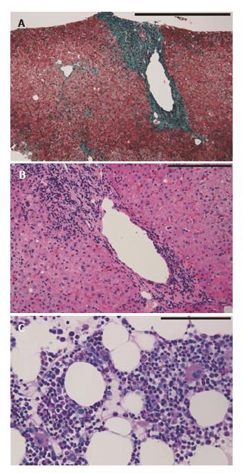
Figure 1 Histopathological features of liver biopsy specimen and clot section of bone marrow aspirate.
A: The liver biopsy specimen shows fibrous portal expansion. There is no fibrous bridging (Elastica-Goldner staining, scale bar; 500 μm); B: Mild piecemeal necrosis, mild intralobular degeneration and focal necrosis, and moderate portal inflammation are observed (H and E staining, scale bar; 200 μm); C: The clot section of bone marrow aspirate shows normal numbers of megakaryocytes and other cell lineages are preserved (Periodic Acid Schiff staining, scale bar; 100 μm).
- Citation: Ichimata S, Kobayashi M, Honda K, Shibata S, Matsumoto A, Kanno H. Acquired amegakaryocytic thrombocytopenia previously diagnosed as idiopathic thrombocytopenic purpura in a patient with hepatitis C virus infection. World J Gastroenterol 2017; 23(35): 6540-6545
- URL: https://www.wjgnet.com/1007-9327/full/v23/i35/6540.htm
- DOI: https://dx.doi.org/10.3748/wjg.v23.i35.6540


