Copyright
©The Author(s) 2017.
World J Gastroenterol. Sep 21, 2017; 23(35): 6540-6545
Published online Sep 21, 2017. doi: 10.3748/wjg.v23.i35.6540
Published online Sep 21, 2017. doi: 10.3748/wjg.v23.i35.6540
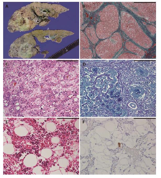
Figure 2 Macroscopic and histopathological features of autopsy specimens.
A: The cut surface of the liver shows diffuse micronodular cirrhosis with a yellow-green lesion in the right lobe; B: The non-tumorous liver shows diffuse small regenerative nodules with fibrous septum (Elastica-Goldner staining, scale bar; 1000 μm); C and D: Histopathological findings of combined hepatocellular-cholangiocarcinoma; C: hepatocellular carcinoma component (H and E staining) and D: adenocarcinoma component (Alcian Blue-Periodic Acid Schiff staining) (C and D, scale bar; 200 μm); E: In the bone marrow, no megakaryocytes are observed (H and E staining); F: A small megakaryocyte is identified through immunostaining for CD41 (E and F, scale bar; 100 μm).
- Citation: Ichimata S, Kobayashi M, Honda K, Shibata S, Matsumoto A, Kanno H. Acquired amegakaryocytic thrombocytopenia previously diagnosed as idiopathic thrombocytopenic purpura in a patient with hepatitis C virus infection. World J Gastroenterol 2017; 23(35): 6540-6545
- URL: https://www.wjgnet.com/1007-9327/full/v23/i35/6540.htm
- DOI: https://dx.doi.org/10.3748/wjg.v23.i35.6540


