Copyright
©2014 Baishideng Publishing Group Inc.
World J Gastroenterol. Dec 7, 2014; 20(45): 17247-17253
Published online Dec 7, 2014. doi: 10.3748/wjg.v20.i45.17247
Published online Dec 7, 2014. doi: 10.3748/wjg.v20.i45.17247
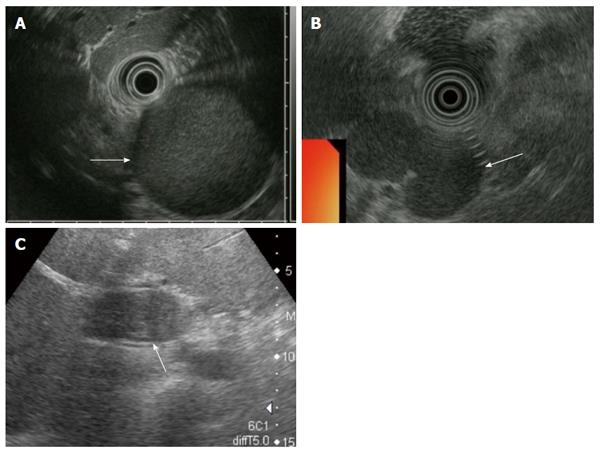
Figure 1 Ultrasonography and endoscopic ultrasonography findings.
A: Findings of Patient #2, the cystic lesion had a hyperechoic appearance (arrow); B: Findings of Patient #3, the cystic lesion had a homogenous hypoechoic appearance (arrow); C: Findings of Patient #4, the cystic lesion displayed a mosaic pattern (arrow).
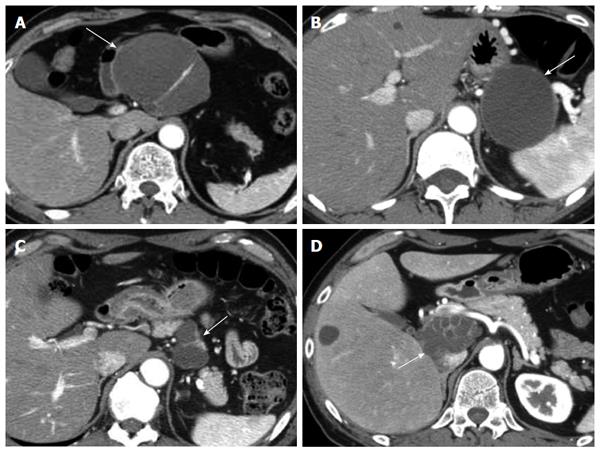
Figure 2 Computed tomography findings.
All lesions were well-defined and were exophytic off the pancreatic parenchyma. The wall and septum of the cysts were enhanced. A: Findings of Patient #1, the lesion was localized in the body of the pancreas and had a multilocular cystic appearance (arrow); B: Findings of Patient #2, the lesion was localized in the tail of the pancreas and had a unilocular cystic appearance (arrow); C: Findings of Patient #3, the lesion was localized in the body of the pancreas and had a multilocular cystic appearance (arrow); D: Findings of Patient #4, the lesion was localized in the head of the pancreas and had a multilocular cystic appearance (arrow).
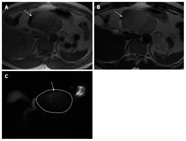
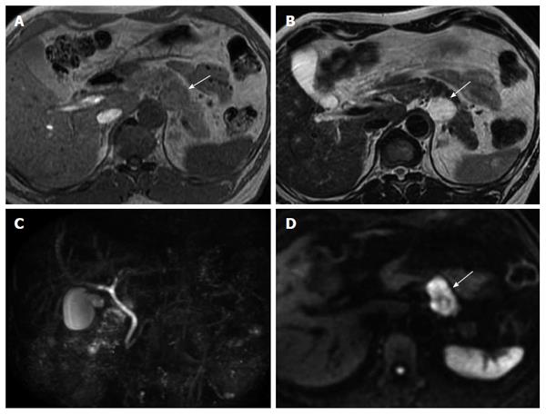
Figure 3 Findings of patient #1.
A: Findings of T1-weighted imaging on magnetic resonance imaging (MRI). T1-weighted imaging of patient showed a higher intensity than free water (arrow), B: Findings of T2-weighted imaging on MRI. T2-weighted imaging of patient showed a lower intensity than free water (arrow); C: Findings of magnetic resonance cholangiopancreatography (MRCP). MRCP of patient showed a lower intensity than free water (arrow).
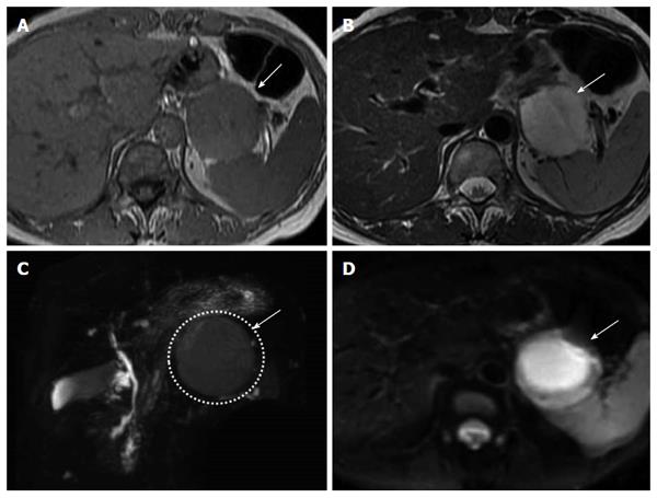
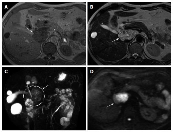
Figure 4 Findings of patient #2.
A: Findings of T1-weighted imaging on magnetic resonance imaging (MRI). T1-weighted imaging of patient showed a higher intensity than free water (arrow); B: Findings of T2-weighted imaging on MRI. T2-weighted imaging of patient showed a lower intensity than free water (arrow); C: Findings of magnetic resonance cholangiopancreatography (MRCP). MRCP of patient showed a lower intensity than free water (arrow); D: Findings of diffusion-weighted imaging on MRI (arrow). Diffusion-weighted imaging of three patients showed a higher intensity than free water. The cystic lesions showed high intense signal in the central part and iso-intense in the periphery.
Figure 5 Findings of patient #3.
A: Findings of T1-weighted imaging on magnetic resonance imaging (MRI). T1-weighted imaging of patient showed a higher intensity than free water (arrow); B: Findings of T2-weighted imaging on MRI. T2-weighted imaging of patient showed a lower intensity than free water (arrow); C: Findings of magnetic resonance cholangiopancreatography (MRCP). MRCP of patient showed a lower intensity than free water; D: Findings of diffusion-weighted imaging on MRI. Diffusion-weighted imaging of three patients showed a higher intensity than free water (arrow). The cystic lesions showed high intense signal in the central part and iso-intense in the periphery.
Figure 6 Findings of patient #4.
A: Findings of T1-weighted imaging on magnetic resonance imaging (MRI). T1-weighted imaging of patient showed a higher intensity than free water (arrow); B: Findings of T2-weighted imaging on MRI. T2-weighted imaging of patient showed a lower intensity than free water (arrow); C: Findings of magnetic resonance cholangiopancreatography (MRCP). MRCP of patient showed a lower intensity than free water (arrow); D: Findings of diffusion-weighted imaging on MRI (arrow). Diffusion-weighted imaging of three patients showed a higher intensity than free water. The cystic lesions showed high intense signal in the central part and iso-intense in the periphery.
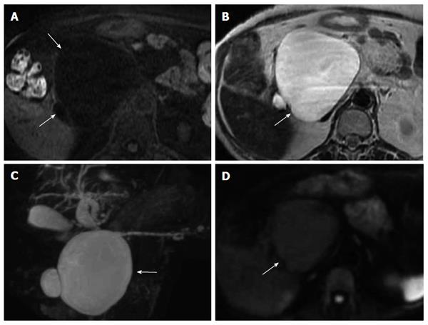
Figure 7 Findings of a case of serous cystic neoplasm.
A: Findings of T1-weighted imaging on magnetic resonance imaging (MRI), the cystic lesion showed low intensity (arrow); B: Findings of T2-weighted imaging on MRI, the cystic lesion showed high intensity (arrow); C: Findings of magnetic resonance cholangiopancreatography, the cystic lesion showed high intensity (arrow); D: Findings of diffusion-weighted imaging on MRI, the cystic lesion showed high intensity (arrow).
- Citation: Terakawa H, Makino I, Nakagawara H, Miyashita T, Tajima H, Kitagawa H, Fujimura T, Inoue D, Kozaka K, Gabata T, Ohta T. Clinical and radiological feature of lymphoepithelial cyst of the pancreas. World J Gastroenterol 2014; 20(45): 17247-17253
- URL: https://www.wjgnet.com/1007-9327/full/v20/i45/17247.htm
- DOI: https://dx.doi.org/10.3748/wjg.v20.i45.17247


