Copyright
©2014 Baishideng Publishing Group Inc.
World J Gastroenterol. Dec 7, 2014; 20(45): 17247-17253
Published online Dec 7, 2014. doi: 10.3748/wjg.v20.i45.17247
Published online Dec 7, 2014. doi: 10.3748/wjg.v20.i45.17247
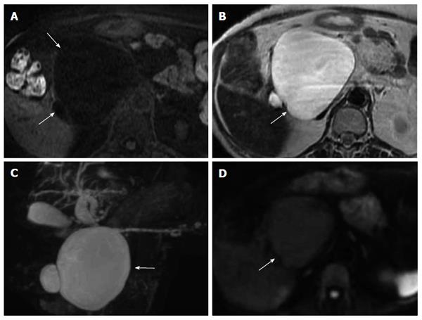
Figure 7 Findings of a case of serous cystic neoplasm.
A: Findings of T1-weighted imaging on magnetic resonance imaging (MRI), the cystic lesion showed low intensity (arrow); B: Findings of T2-weighted imaging on MRI, the cystic lesion showed high intensity (arrow); C: Findings of magnetic resonance cholangiopancreatography, the cystic lesion showed high intensity (arrow); D: Findings of diffusion-weighted imaging on MRI, the cystic lesion showed high intensity (arrow).
- Citation: Terakawa H, Makino I, Nakagawara H, Miyashita T, Tajima H, Kitagawa H, Fujimura T, Inoue D, Kozaka K, Gabata T, Ohta T. Clinical and radiological feature of lymphoepithelial cyst of the pancreas. World J Gastroenterol 2014; 20(45): 17247-17253
- URL: https://www.wjgnet.com/1007-9327/full/v20/i45/17247.htm
- DOI: https://dx.doi.org/10.3748/wjg.v20.i45.17247


