Copyright
©2014 Baishideng Publishing Group Inc.
World J Gastroenterol. Dec 7, 2014; 20(45): 17247-17253
Published online Dec 7, 2014. doi: 10.3748/wjg.v20.i45.17247
Published online Dec 7, 2014. doi: 10.3748/wjg.v20.i45.17247
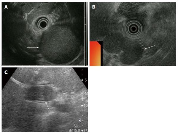
Figure 1 Ultrasonography and endoscopic ultrasonography findings.
A: Findings of Patient #2, the cystic lesion had a hyperechoic appearance (arrow); B: Findings of Patient #3, the cystic lesion had a homogenous hypoechoic appearance (arrow); C: Findings of Patient #4, the cystic lesion displayed a mosaic pattern (arrow).
- Citation: Terakawa H, Makino I, Nakagawara H, Miyashita T, Tajima H, Kitagawa H, Fujimura T, Inoue D, Kozaka K, Gabata T, Ohta T. Clinical and radiological feature of lymphoepithelial cyst of the pancreas. World J Gastroenterol 2014; 20(45): 17247-17253
- URL: https://www.wjgnet.com/1007-9327/full/v20/i45/17247.htm
- DOI: https://dx.doi.org/10.3748/wjg.v20.i45.17247


