Copyright
©2009 The WJG Press and Baishideng.
World J Gastroenterol. Dec 14, 2009; 15(46): 5813-5820
Published online Dec 14, 2009. doi: 10.3748/wjg.15.5813
Published online Dec 14, 2009. doi: 10.3748/wjg.15.5813
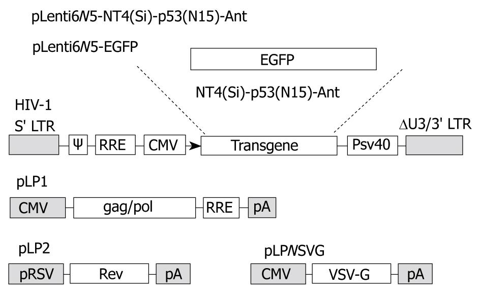
Figure 1 Packaging system of LV-NT4(Si)-p53(N15)-Ant and LV-EGFP.
The packaging system: lentiviral vector, pLenti6/V5-D-TOPO, contains HIV-1 5'LTR, packaging signal (ψ), RRE sequence, CMV promoter and 3'LTR from which some regulatory sequences are deleted, resulting in “self-inactivation” of lentivirus after transduction of the target cells. pLP1, pLP2 and pLP/VSVG are three helper plasmids, which provide structural and replication proteins to produce lentivirus.
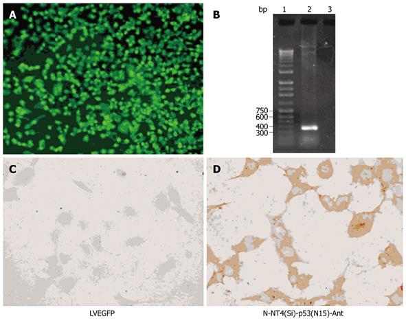
Figure 2 Fluorescent microscopic analysis and immunohistochemical staining.
Fluorescence microscopy analysis showing EGFP expression in HepG2 cells (A), agarose gel electrophoresis of RT-PCR products from HepG2 cells (B) [lane 1: 1.0 kb DNA ladder; lane 2: RT-PCR products from cells infected with LV-NT4(Si)-p53(N15)-Ant; lane 3: negative controls infected with LV-EGFP], immunohistochemical staining showing no p53 protein (C), and p53 protein expression in cytoplasm of HepG2 cells (D) 48 h after infection with LV-NT4(Si)-p53(N15)-Ant.
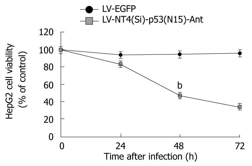
Figure 3 Cytotoxicity of LV-NT4(Si)-p53(N15)-Ant to HepG2 cells at different time points (24, 48 and 72 h) after infection with lentivirus.
bP < 0.01 vs LV-EGFP.
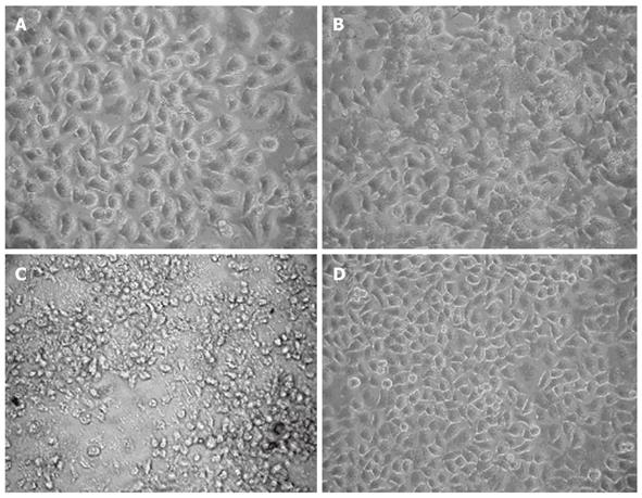
Figure 4 Morphological changes in HepG2 cells 24 h (A), 48 h (B), 72 h (C) after infection with LV-NT4(Si)-p53(N15)-Ant and 72 h (D) after infection with LV-EGFP (× 400).
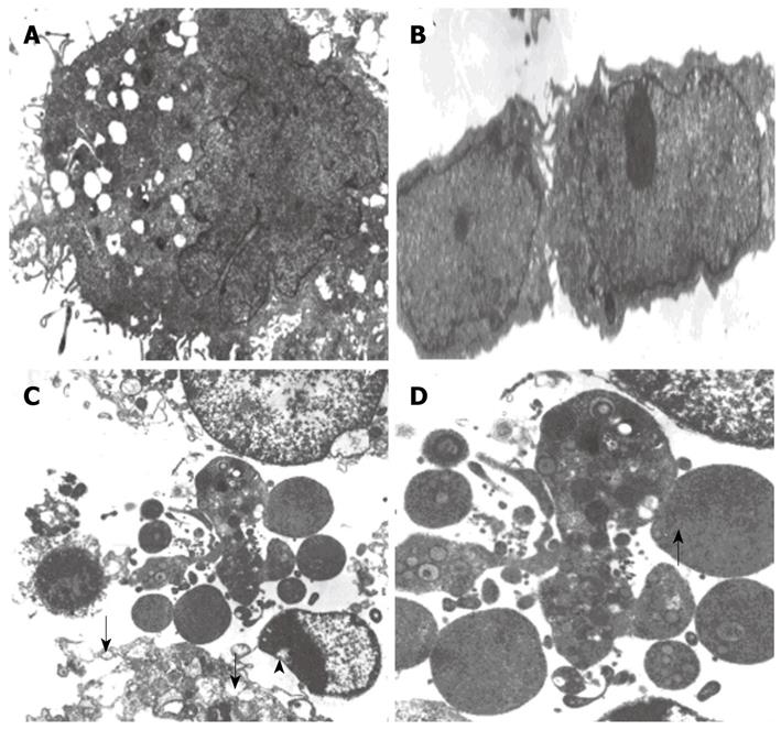
Figure 5 Ultra-structure of HepG2 cells 48 h after infection with LV-EGFP (A, B) and LV-NT4(Si)-p53 (N15)-Ant (C, D) (× 5000).
A: Forty-eight hours after LV-EGFP infection, the cell membranes and nuclear membranes of HepG2 cells were intact; there were some crimples in the nuclear membranes and some microvilli on the surface of cells. The structure of mitochondria and ER was normal; B: Forty-eight hours after LV-EGFP infection, there were two HepG2 cells which had just completed division and this phenomenon suggested that LV-EGFP did not have a significant effect on cell growth and division; C: Forty-eight hours after LV-NT4(Si)-p53(N15)-Ant infection, it can be seen that cell membranes were incomplete and several fractures (arrows) were seen. Cell contents also appeared to be leaking. Mitochondria were swollen and showed vacuolization. Vacuoles were also seen under the nuclear membrane (arrowhead); D: Forty-eight hours after LV-NT4(Si)-p53(N15)-Ant infection, the remaining bare nuclei (arrow), after cytoplasm collapse can be seen.
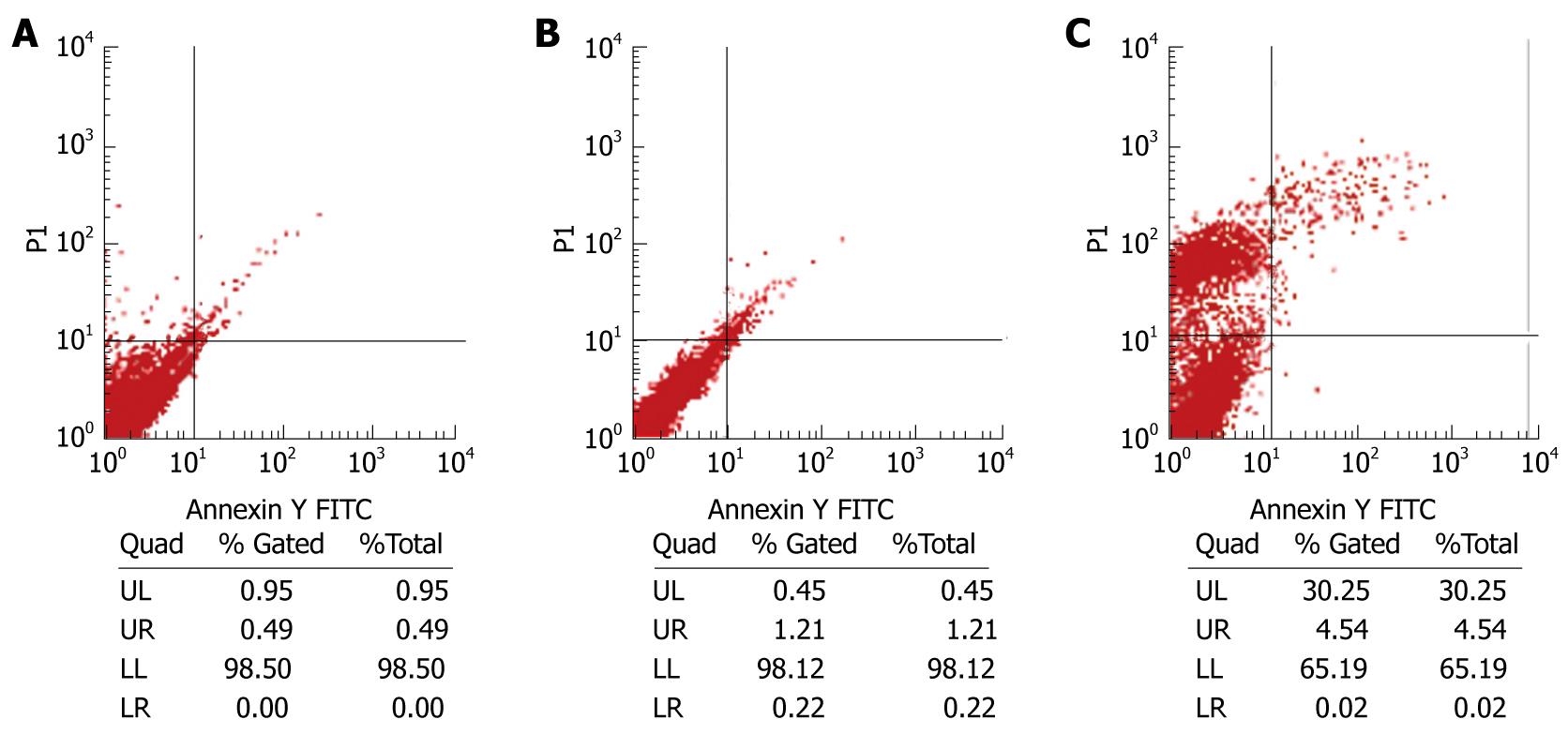
Figure 6 Flow cytometry analysis of HepG2 cells without treatment (A), 48 h after infection with LV-EGFP (B) and LV-NT4(Si)-p53(N15)-Ant (C) after stained with Annexin V and propidium iodide.
The left lower quadrant represents normal cells (An-PI-), the right lower quadrant represents early apoptotic cells (An+ PI-), the right upper quadrant represents apoptotic cells and necrotic cells (An + PI +), the upper left quadrant represents early necrotic cells (An-PI +).
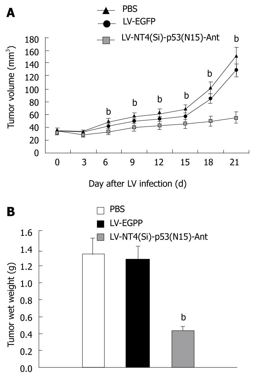
Figure 7 Tumor growth curve (A) and tumor wet weight (B) after treatment with LV-NT4(Si)-p53(N15)-Ant.
A: Tumor growth curve after treatment with LV-NT4(Si)-p53(N15)-Ant. Tumors grew rapidly in the PBS and LV-EGFP groups, while the tumor growth was significantly inhibited in the LV-NT4(Si)-p53(N15)-Ant group 3 wk after infection. There was no statistical significance between the PBS group (151.0 ± 50.1 mm3) and the LV-EGFP group (128.5 ± 45.3 mm3) in terms of tumor volume (P > 0.05). But the tumor volume in the LV-NT4(Si)-p53(N15)-Ant group (55.1 ± 18.7 mm3) was significantly less than that of the PBS and LV-EGFP groups (bP < 0.01); B: Tumor wet weight after treatment with LV-NT4(Si)-p53(N15)-Ant. There were no significant differences in the wet weight of tumors between the two control groups: PBS group 1.33 ± 0.93 g vs LV-EGFP group 1.27 ± 1.04 g (P > 0.05). But the wet weight of the LV-NT4(Si)-p53(N15)-Ant treatment group (0.43 ± 0.77 g) was significantly less than that of the two control groups (bP < 0.01).
- Citation: Song LP, Li YP, Wang N, Li WW, Ren J, Qiu SD, Wang QY, Yang GX. NT4(Si)-p53(N15)-antennapedia induces cell death in a human hepatocellular carcinoma cell line. World J Gastroenterol 2009; 15(46): 5813-5820
- URL: https://www.wjgnet.com/1007-9327/full/v15/i46/5813.htm
- DOI: https://dx.doi.org/10.3748/wjg.15.5813


