Copyright
©2009 The WJG Press and Baishideng.
World J Gastroenterol. Dec 14, 2009; 15(46): 5813-5820
Published online Dec 14, 2009. doi: 10.3748/wjg.15.5813
Published online Dec 14, 2009. doi: 10.3748/wjg.15.5813
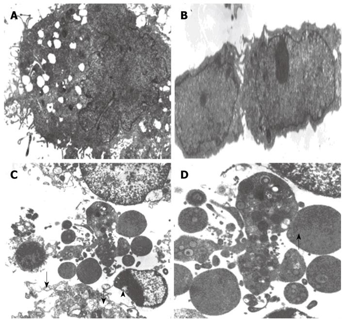
Figure 5 Ultra-structure of HepG2 cells 48 h after infection with LV-EGFP (A, B) and LV-NT4(Si)-p53 (N15)-Ant (C, D) (× 5000).
A: Forty-eight hours after LV-EGFP infection, the cell membranes and nuclear membranes of HepG2 cells were intact; there were some crimples in the nuclear membranes and some microvilli on the surface of cells. The structure of mitochondria and ER was normal; B: Forty-eight hours after LV-EGFP infection, there were two HepG2 cells which had just completed division and this phenomenon suggested that LV-EGFP did not have a significant effect on cell growth and division; C: Forty-eight hours after LV-NT4(Si)-p53(N15)-Ant infection, it can be seen that cell membranes were incomplete and several fractures (arrows) were seen. Cell contents also appeared to be leaking. Mitochondria were swollen and showed vacuolization. Vacuoles were also seen under the nuclear membrane (arrowhead); D: Forty-eight hours after LV-NT4(Si)-p53(N15)-Ant infection, the remaining bare nuclei (arrow), after cytoplasm collapse can be seen.
- Citation: Song LP, Li YP, Wang N, Li WW, Ren J, Qiu SD, Wang QY, Yang GX. NT4(Si)-p53(N15)-antennapedia induces cell death in a human hepatocellular carcinoma cell line. World J Gastroenterol 2009; 15(46): 5813-5820
- URL: https://www.wjgnet.com/1007-9327/full/v15/i46/5813.htm
- DOI: https://dx.doi.org/10.3748/wjg.15.5813


