Copyright
©The Author(s) 2021.
World J Clin Cases. Jun 26, 2021; 9(18): 4844-4851
Published online Jun 26, 2021. doi: 10.12998/wjcc.v9.i18.4844
Published online Jun 26, 2021. doi: 10.12998/wjcc.v9.i18.4844
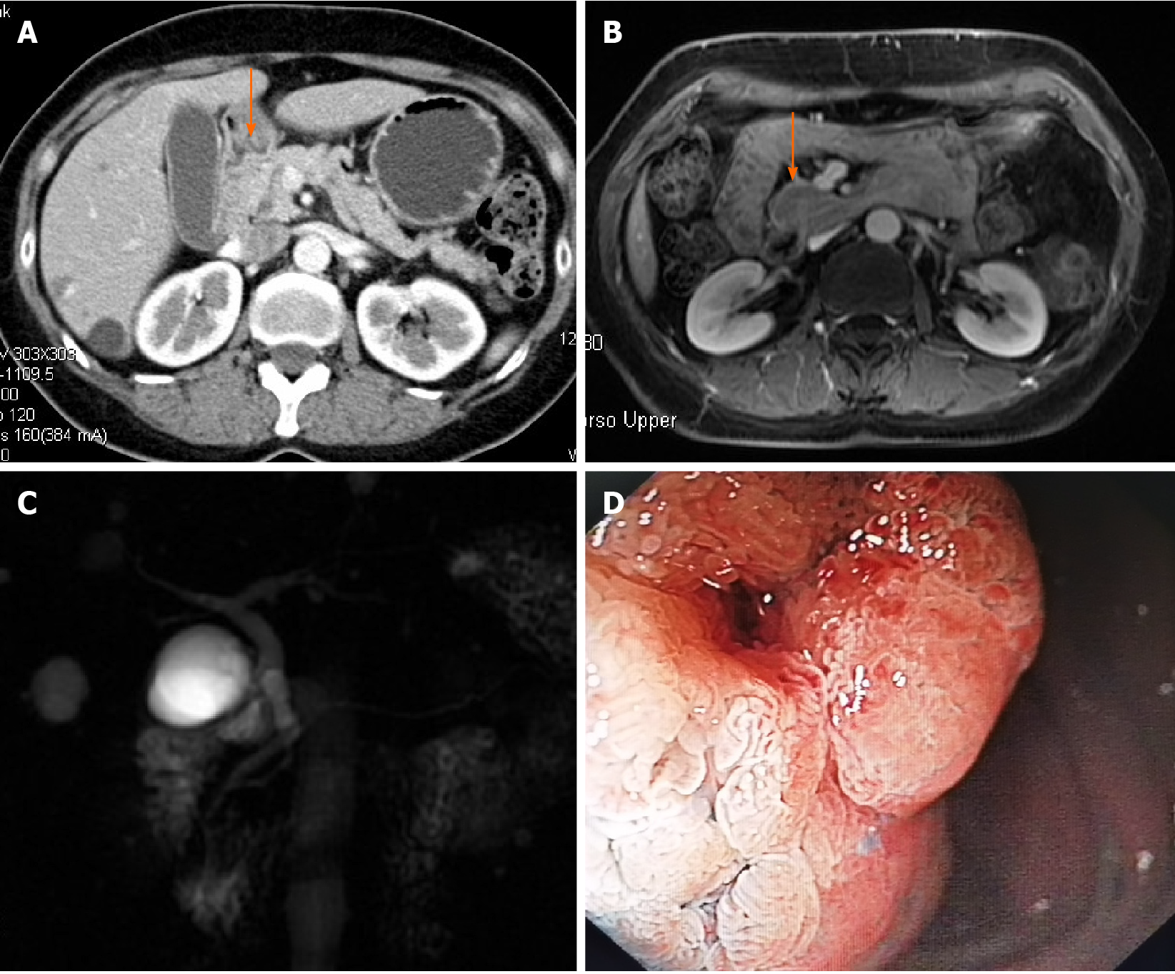
Figure 1 Preoperative examination of the ampullary tumor.
A and B: Contrast-enhanced computed tomography and magnetic resonance imaging showing an enhanced lesion (arrow) located in the descending part of the duodenum; C: Magnetic resonance cholangiography revealing a mild dilation of the common bile duct, and no cut-off sign or stricture of either the bile duct or pancreatic duct; D: Endoscopic view of the ampullary adenoma.
- Citation: Zheng X, Sun QJ, Zhou B, Jin M, Yan S. Microscopic transduodenal excision of an ampullary adenoma: A case report and review of the literature. World J Clin Cases 2021; 9(18): 4844-4851
- URL: https://www.wjgnet.com/2307-8960/full/v9/i18/4844.htm
- DOI: https://dx.doi.org/10.12998/wjcc.v9.i18.4844


