Copyright
©The Author(s) 2020.
World J Clin Cases. Mar 6, 2020; 8(5): 939-945
Published online Mar 6, 2020. doi: 10.12998/wjcc.v8.i5.939
Published online Mar 6, 2020. doi: 10.12998/wjcc.v8.i5.939
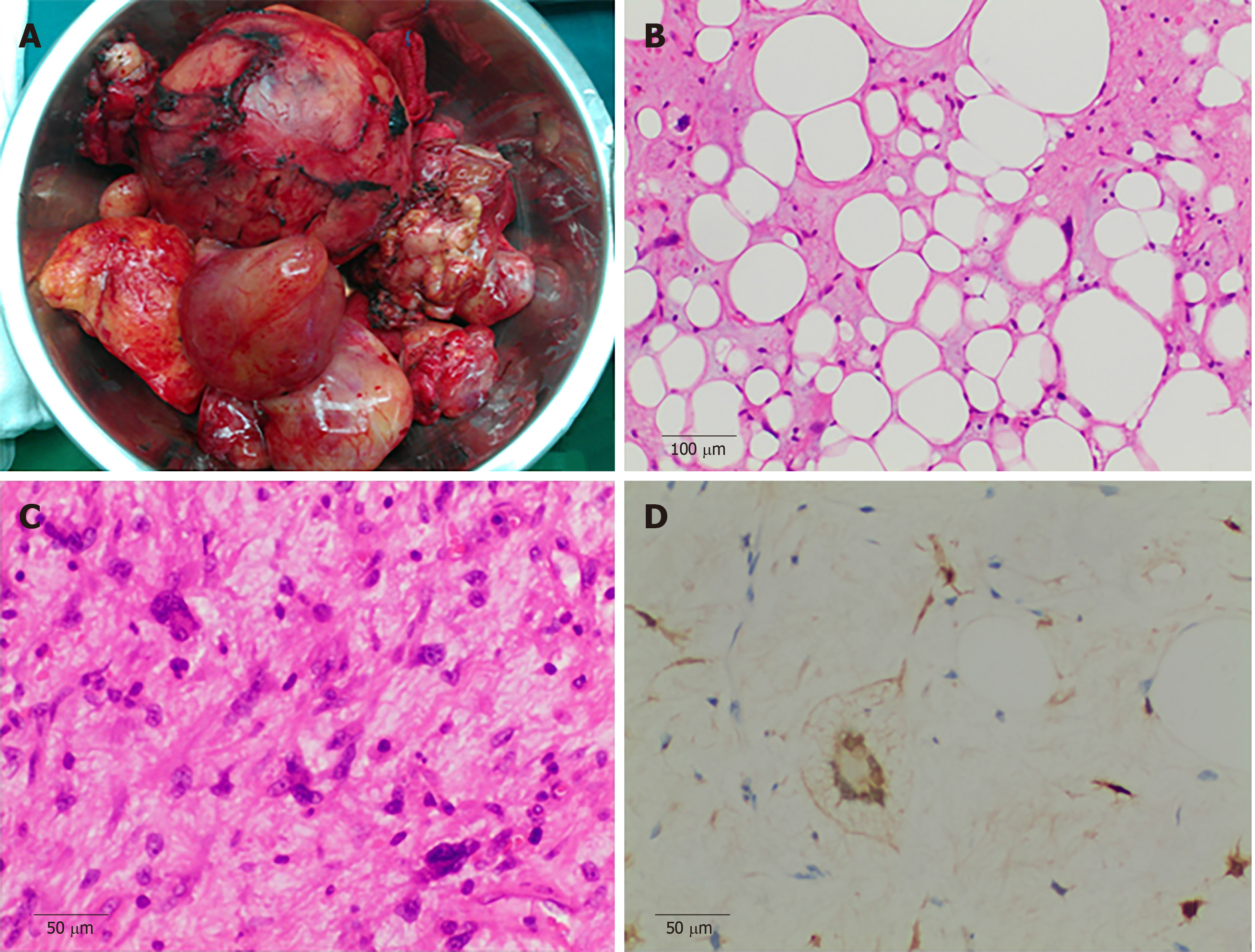
Figure 2 Pathological findings.
A: Gross appearance showed the largest tumor was about 105 mm × 94 mm × 88 mm in size, and the smallest was about 20 mm in diameter; the tumors had integrated capsules; B and C: H&E staining showed a well-differentiated liposarcoma component and spindle cell component (× 100 and × 200, respectively); C: Immunohistochemical analysis showed the tumor was positive for S-100 protein in scattered cells (× 200).
- Citation: Chen HG, Zhang K, Wu WB, Wu YH, Zhang J, Gu LJ, Li XJ. Combining surgery with 125I brachytherapy for recurrent mediastinal dedifferentiated liposarcoma: A case report and review of literature. World J Clin Cases 2020; 8(5): 939-945
- URL: https://www.wjgnet.com/2307-8960/full/v8/i5/939.htm
- DOI: https://dx.doi.org/10.12998/wjcc.v8.i5.939


