Published online Feb 26, 2020. doi: 10.12998/wjcc.v8.i4.820
Peer-review started: November 21, 2019
First decision: December 12, 2019
Revised: January 5, 2020
Accepted: January 11, 2020
Article in press: January 11, 2020
Published online: February 26, 2020
Processing time: 97 Days and 2.7 Hours
A diverticulum is the medical or biological term for outpouching of a hollow structure in the body. It particularly occurs in the digestive system, but rarely occurs in the oral mucosa.
This report describes a rare case of diverticulum presenting in the buccal mucosa in a 44-year-old woman that was initially diagnosed in Stomatology Hospital, Wuhan University. We made our diagnosis under the guidance of imaging data, and the patient underwent surgical resection.
This report is the first confirmed case of buccal mucosal diverticulum. in addition, we elucidated that in general, idiopathic developmental abnormalities caused by succinate muscle defects are responsible for diverticulum development.
Core tip: Oral mucosal diverticulum is rare, and diverticulum of digestive tract has not been previously reported. Partial defects of the muscle layer around the diverticulum were also observed during the operation, and the diverticulum was located at the fragile edge of the anatomy of the muscle layer. After the operation, the patient’s food impaction symptoms were resolved, and have not relapsed.
- Citation: Zhang Y, Wang L, Liu K. Diverticulum of the buccal mucosa: A case report. World J Clin Cases 2020; 8(4): 820-824
- URL: https://www.wjgnet.com/2307-8960/full/v8/i4/820.htm
- DOI: https://dx.doi.org/10.12998/wjcc.v8.i4.820
The expansion of buccal mucosa is very rare. The first case of buccal mucosal diverticulum was reported by Bailey, who reported that large numbers of diverticula arise from the buccal sulcus and extend to the neck. Subsequently, only nine such cases have been reported in the literature. A buccal mucosal diverticulum is a small compartment formed through defects in the muscle layer through protrusions of the mucosa and submucosa[1]. In published reports, the diverticulum occurs in various organs including the esophagus, stomach, duodenum[3], bladder[5], urethra, vagina, and circulatory system[2-6]. Here, we present a rare case of diverticulum in the oral buccal mucosa, with discussion of the presentation, investigation, and management of this case.
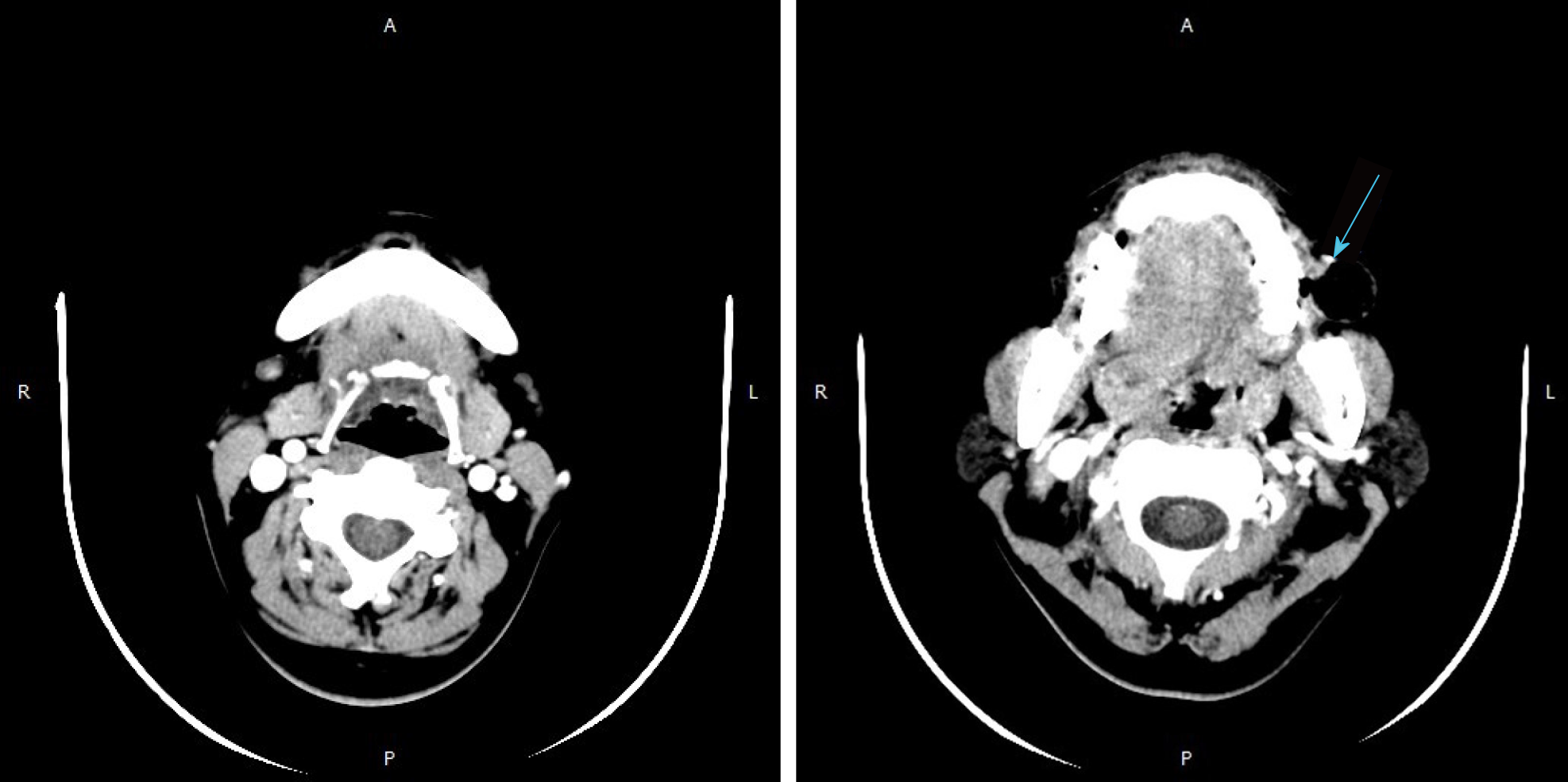
A 37-year-old female patient was referred to Stomatology Hospital, Wuhan University (Wuhan, China) for investigation and treatment, complaining of a progressing mass with pain in the left buccal mucosa perceived 4 mo before consultation. The mass was ulcerated with clear liquid 2 mo prior. The patient had no history of facial trauma, infection, tooth pain, or restriction of the mouth. At initial diagnosis, a hard, fixed, round lump of 1.5 cm × 1.0 cm in size was seen in the left cheek mucosa, with food residue. After cleaning up the food residue, a palpable sinus tract measuring about 1.5 cm in diameter and 2 cm in depth was visible in that area. The skin on the left buccal area appeared normal. There was a slight inflammatory reaction in the sinus mucosa. Dental hard tissues and gingiva did not show obvious abnormality. Computed tomography (CT) showed a 2.1 cm × 2 cm × 2.2 cm sinus cavity in the left buccal region (Figure 1), which provided radiographic evidence that the sinus cavity had no connection with the teeth, jaw bone, or parotid gland.
Diverticulum of the buccal mucosa.
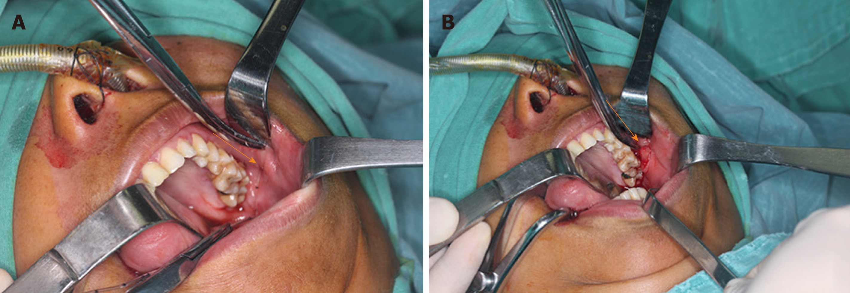
The sinus tract was removed by surgery (Figure 2). The surgical procedure was conventional disinfection and drapes. The inner volume of the inner volume was pulled out and gradually cut out with a vascular clamp. The fat pad was pulled out and an adjacent flap to close the wound was designed.
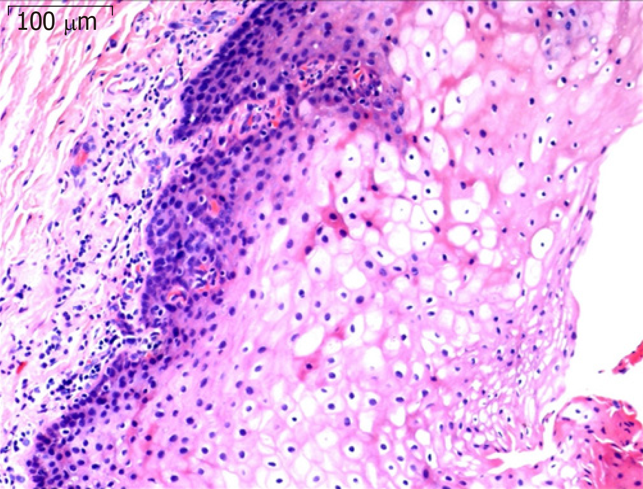
Pathological examination reported that squamous epithelium was normal, and infiltration of chronic inflammation cell was discovered in the lamina propria. According to the clinical and pathological examinations, the sinus tract showed a diverticulum in the oral mucosa (Figure 3). The patient came to our hospital 1, 3, 6, and 12 mo after operation and showed good healing with no recurrence.
Diverticulum is a “pocket” or “tube” structure in the mucosa. Typically, diverticula protrude from the mucosa and submucosa and form through defects in the muscle layers[1]. The cheeks are mainly composed of three groups of muscle fibers. It has been suggested that diverticulum lesions are a developmental anomaly, which is formed by the relatively weak partial dissection along these muscle fibers. According to published literature, diverticulum is most common in the digestive tract, including Meckel’s diverticulum, gastric diverticulum, colonic diverticulum, and duodenal diverticulum. Diverticulum also occurs in the genitourinary system (bladder diverticulum, vagina diverticulum, and uterus diverticulum), circulatory system (Kommerell’s diverticulum and cardiac diverticulum)[3-7], and pharyngeal region (Zenker’s diverticulum)[2]. The true or false nature of the diverticulum depends on the level it involves. True diverticulum involves all levels of the anatomical structure of the site, including propria muscle and adventitia such as Meckel’s diverticulum. Pseudo diverticulum does not involve the muscle layer or outer membrane of the gastrointestinal tract, but only the submucous and mucosal layers[8].
Usually a “tube” structure in the oral mucosa is diagnosed as sinus tract or fistula. The sinus tract originating in the dental region is usually caused by a periapical infection around the root apices because of necrotic pulp. Pulp sensibility test and panoramic views are used to located involved teeth. In addition, radiological examination has revealed that the sinus tract originates from the apical periodontitis[9]. However, computed tomography examination in this case has shown that the sinus tract is irrelevant to the dental hard tissues, and there is no abnormal bone resorption or bone defection. Because the sinus tract is in the upper of the buccal mucosa, it was irrelevant with Stensen’s duct or parotid gland. According to clinical and radiological examinations, and based on literature about the diverticulum, this case reported a rare diverticulum in the oral buccal mucosa.
The mouth is the beginning of the digestive tract, composed of both the epithelium and lamina propria. The cause of diverticulum of the digestive tract is unknown. Some theories are proposed including genetics, diet, motility, microbiome, and inflammation. One leading theory is that pressure in the areas of weakened walls may lead to the occurrence of diverticulosis[1]. Zenker’s diverticulum occurs in the oropharynx, although traction and propulsion mechanisms have long been identified as major drivers of diverticulum development. The current industry consensus suggests that occlusal mechanisms are the most important: increased distal pharyngeal pressure due to uncoordinated swallowing, impaired relaxation, and cyclopharyngeal muscle cramps is the leading cause of diverticulum. In addition, degeneration of the mucosal wall of the digestive tract with age results in the development of diverticula[8]. So the occurrence and development of diverticula in the buccal mucosa still needs more research and discussion.
Diverticulum of the digestive tract shows a variety of clinical symptoms and signs. Symptomatic uncomplicated diverticular disease, infection complications, and bleeding are common symptoms of diverticulum of the digestive tract. Other symptoms include nausea, vomiting, urinary symptoms, ulceration, diverticulitis, and perforation. There was no obvious symptom in our case beside food impaction. In addition, partial defects of the muscle layer around the diverticulum were also observed during the operation, and the diverticulum was located at the fragile edge of the anatomy of the muscle layer. After the operation, the patient’s food impaction symptoms were resolved, and have not relapsed.
This report is our first confirmed buccal mucosal diverticulum and we provide substantial clinical, pathological, and imaging evidence. In addition, we elucidated that in general, idiopathic developmental abnormalities caused by succinate muscle defects are responsible for diverticulum development.
Manuscript source: Unsolicited manuscript
Specialty type: Medicine, research and experimental
Country of origin: China
Peer-review report classification
Grade A (Excellent): 0
Grade B (Very good): 0
Grade C (Good): C
Grade D (Fair): D
Grade E (Poor): 0
P-Reviewer: Kuwai T, de Melo FF S-Editor: Wang YQ L-Editor: Filipodia E-Editor: Xing YX
| 1. | Bortz JH, Bortz JH, Ramlaul A, Munro L. Diverticular Disease. Bortz JH, Ramlaul A, Munro L. CT Colonography for Radiographers. Switzerland: Springer International Publishing 2016; 221-232. [DOI] [Full Text] |
| 2. | Groher ME. Esophageal disorders. Groher ME, Crary MA. Amsterdam: Elsevier 2016; 97-114. [RCA] [DOI] [Full Text] [Cited by in Crossref: 1] [Cited by in RCA: 1] [Article Influence: 0.1] [Reference Citation Analysis (0)] |
| 3. | Palmer ED. Gastric diverticulosis. Am Fam Physician. 1973;7:114-117. [PubMed] |
| 4. | Tamas EF, Stephenson AJ, Campbell SC, Montague DK, Trusty DC, Hansel DE. Histopathologic features and clinical outcomes in 71 cases of bladder diverticula. Arch Pathol Lab Med. 2009;133:791-796. [RCA] [PubMed] [DOI] [Full Text] [Cited by in RCA: 3] [Reference Citation Analysis (0)] |
| 5. | Bayne AP, Jones, EA, Taneja SS. Complications of hypospadias repair. Complications of Urologic Surgery. Taneja SS. Amsterdam: Elsevier 2010; 713-722. [DOI] [Full Text] |
| 6. | van Son JA, Konstantinov IE. Burckhard F. Kommerell and Kommerell's diverticulum. Tex Heart Inst J. 2002;29:109-112. [PubMed] |
| 7. | Sagar J, Kumar V, Shah DK. Meckel's diverticulum: a systematic review. J R Soc Med. 2006;99:501-505. [RCA] [PubMed] [DOI] [Full Text] [Cited by in Crossref: 374] [Cited by in RCA: 196] [Article Influence: 10.3] [Reference Citation Analysis (0)] |
| 8. | Marianne C, Ur Rehman M, Min Hoe C. A case report of large gastric diverticulum with literature review. Int J Surg Case Rep. 2018;44:82-84. [RCA] [PubMed] [DOI] [Full Text] [Full Text (PDF)] [Cited by in Crossref: 4] [Cited by in RCA: 3] [Article Influence: 0.4] [Reference Citation Analysis (0)] |
| 9. | Lee SH, Yun SJ. Odontogenic cutaneous sinus tract presenting as a growing cheek mass in the emergency department. Am J Emerg Med. 2017;35:808.e5-808.e7. [RCA] [PubMed] [DOI] [Full Text] [Cited by in Crossref: 1] [Cited by in RCA: 1] [Article Influence: 0.1] [Reference Citation Analysis (0)] |


