Published online Feb 6, 2020. doi: 10.12998/wjcc.v8.i3.638
Peer-review started: October 28, 2019
First decision: December 4, 2019
Revised: December 13, 2019
Accepted: December 22, 2019
Article in press: December 22, 2019
Published online: February 6, 2020
Processing time: 100 Days and 20.2 Hours
Sternal tumors are difficult to diagnose, and usually need to be differentiated from other diseases such as tuberculosis, osteosarcoma, intrathoracic thyroid and thymoma. The sternum is a rare site of Hodgkin’s lymphoma, which is often misdiagnosed as tuberculosis on routine histopathology.
We reported a 47-year-old female patient with chest pain in the upper sternum for 1 mo. Chest computed tomography found a mass in the upper sternum. Pathology and immunohistochemistry of the biopsy confirmed the diagnosis of typical Hodgkin’s lymphoma (mixed cellularity subtype). Patient was diagnosed with primary sternal Hodgkin’s lymphoma and administered 6 cycles of adriamycin, bleomycin, vinblastine, and dacarbazine chemotherapy. Patient had no tumor recurrence and progression at a follow-up visit 2 years later.
This study highlights the rarity of primary sternal Hodgkin’s lymphoma and the challenges of its diagnosis. A PubMed and Web of Science search revealed 10 reported cases of sternal involvement in Hodgkin’s lymphoma.
Core tip: Sternal tumors are difficult to diagnose, and usually need to be differentiated from other diseases. The sternum is a rare site of Hodgkin’s lymphoma, which is usually misdiagnosed as tuberculosis on routine histopathology. We here report a 47-year-old female patient with chest pain in the upper sternum for 1 mo. The patient was diagnosed with primary sternal Hodgkin’s lymphoma and administered 6 cycles of adriamycin, bleomycin, vinblastine, and dacarbazine chemotherapy. No tumor recurrence and progression at a follow-up visit 2 years later. This is a first case report about diagnosis at initial presentation with a primary lesion.
- Citation: Yin YY, Zhao N, Yang B, Xin H. Sternal Hodgkin’s lymphoma: A case report and review of literature. World J Clin Cases 2020; 8(3): 638-644
- URL: https://www.wjgnet.com/2307-8960/full/v8/i3/638.htm
- DOI: https://dx.doi.org/10.12998/wjcc.v8.i3.638
Lymphomas, both Hodgkin lymphomas (HL) and non-HL, comprise approximately 5% to 6% of all malignancies, approximately half of all newly diagnosed hematological tumors[1,2]. HL is a type of lymphoma that originates from B lymphocytes and characteristically presents in a group of lymph nodes, most commonly in the cervical and supraclavicular lymph nodes (LN)[3]. Extranodal HL, especially in sternum, is extremely rare. This study highlights the rarity of primary sternal Hodgkin’s lymphoma and the challenges of its diagnosis.
Chest pain in the upper sternum for 1 mo in a 47-year-old woman.
The patient presented to our department with complaints of paroxysmal chest pain, radiating to the left side, and associated with fatigue, fever of 37.8 °C, and significant weight loss (6.5 kg over 3 mo), these symptoms gradually aggravated within 1 mo. Patient denied experiencing night sweats or other symptoms.
The patient had no history of other diseases.
She had no history of cigarette smoking or alcohol use, and there were no similar cases in the family.
Physical examination after admission revealed the following: blood pressure of 110/70 mmHg, pulse of 80 beats/min and temperature of 37.5 °C. The general examination was unremarkable, and no palpable lymph nodules were found. Chest examination revealed a solid mass in the upper sternal segment. On palpation, the swelling was fixed, firm, and non-tender with smooth edges without redness and ulceration, and no obvious abnormality was found in other systemic examinations.
Blood test results were as follows: Differential counts of neutrophils, 72.9%; lymphocytes, 19.8%; platelet counts, 257 × 109/L; lactate dehydrogenase level, 169.69 IU/L and human immunodeficiency virus was negative.
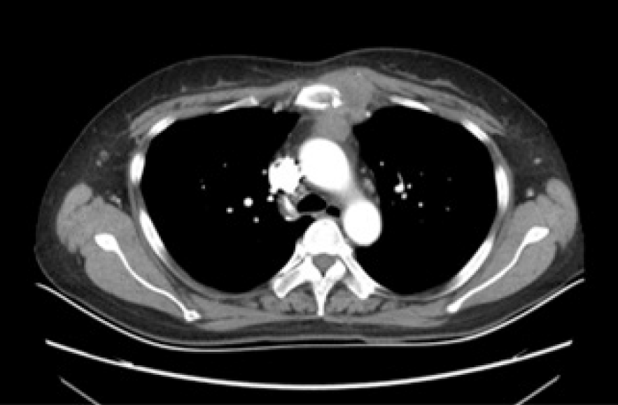
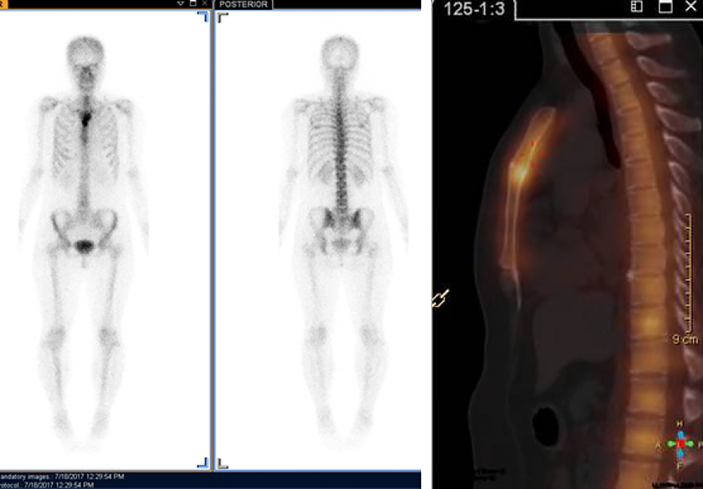
Chest contrast-enhanced computed tomography (CT) (Figure 1) showed a patellar low-density shadow in the upper sternum, surrounding a soft tissue mass measuring 3.9 cm × 2.8 cm with mild enhancement. The initial diagnosis was a mediastinal mass. We performed a technetium 99m-methyl diphosphonate (Tc99MDP) bone scintigraphy scan (Figure 2) to exclude other skeletal system tumors in the patient. The results revealed apparent fixation of the radiopharmaceuticals in the sternal manubrium and upper segment of the sternal body, and no involvement of other sites in the skeleton was found.
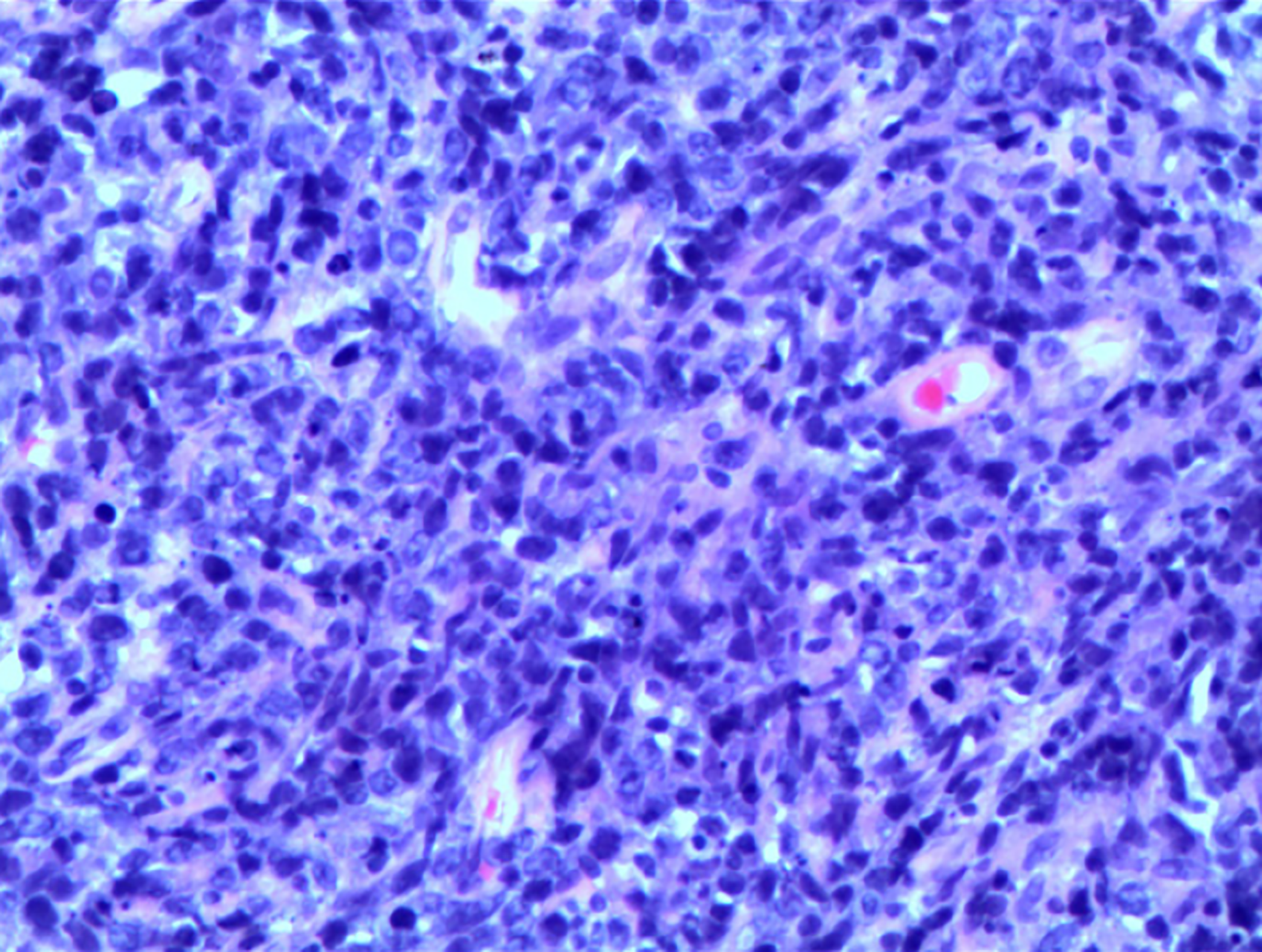
A histopathological biopsy (Figure 3) from the swelling revealed that most tissues were necrotic and contained inflammatory granulation, blood vessels, lymphocytes, plasma cells and a few large heterotypic cells (suspected to be Reed Sternberg cells). On immunohistochemistry (Figure 4): CD30 (+), Pax-5 (+), Ki67 (60%+), CD20 (-), and CD5 (-) were revealed, indicating a pattern typical of classical Hodgkin’s lymphoma (mixed cellularity, MC).
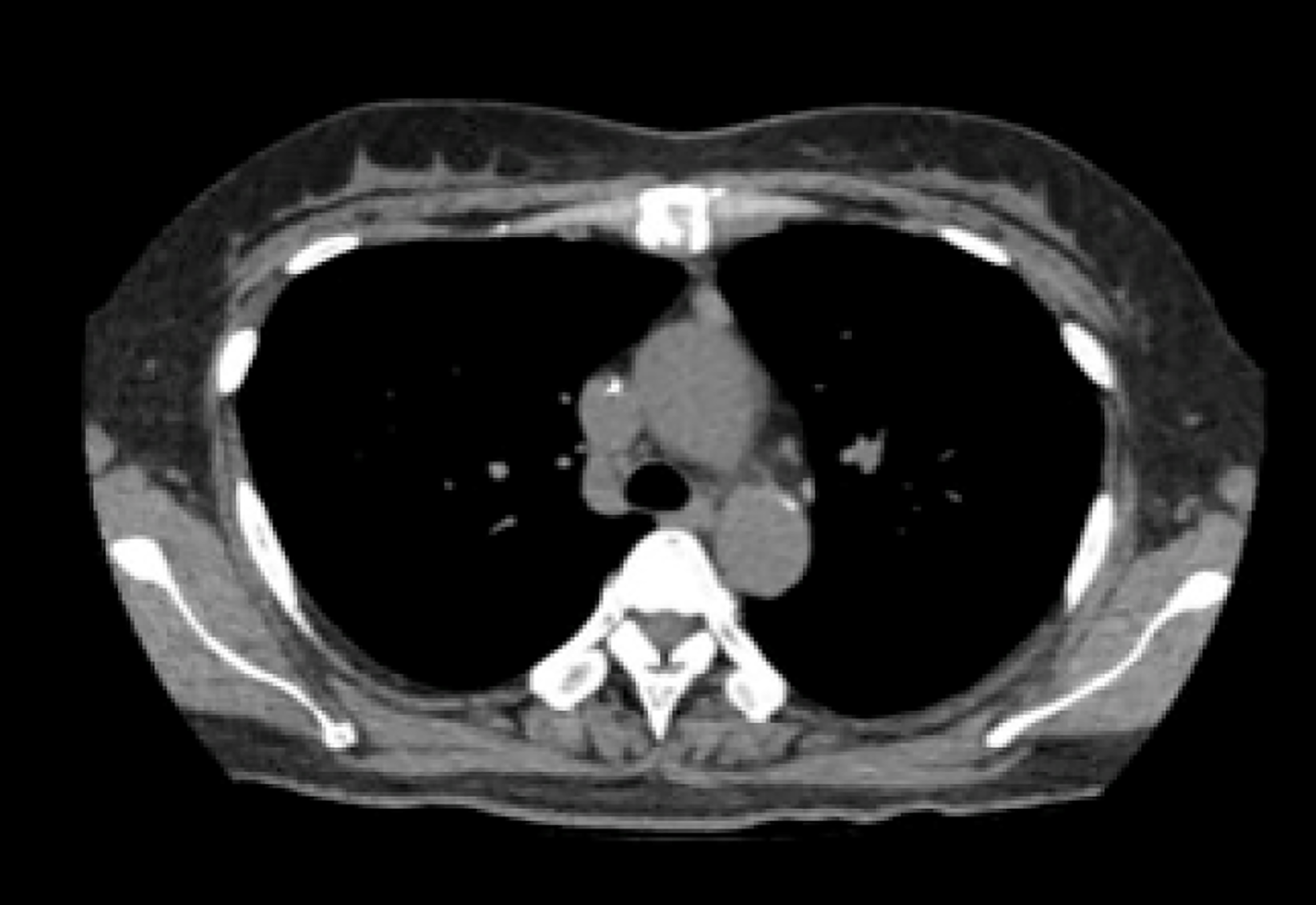
To prevent disease progression and relieve symptoms, patient was administered 6 cycles of the ABVD regimen (Adriamycin 25 mg/m2, Bleomycin 10 mg/m2, Vinblastine 6 mg/m2, and Dacarbazine 375 mg/m2 every 28 d). Patient’s symptoms improved significantly after the treatment, and chest CT examination (Figure 5) revealed a 90% reduction in the size of the mass lesion.
Patient has achieved complete remission after treatment and followed up for 2 years without recurrence of the tumor or clinically detectable lymph nodes enlargement.
HL is a type of lymphoma that originates in B lymphocytes, which characteristically presents in a group of lymph nodes (LN), most commonly in the cervical and supraclavicular LN. Four typical subtypes of HL are defined: Lymphocytic predominance, nodular sclerosis, MC, and lymphocytic depletion[3]. Extranodal HL is extremely rare and most commonly presents in the gastrointestinal tract, respiratory system, thyroid gland, skin, urogenital tract and central nervous system[4]. HL initially manifesting in the skeleton is rare because bone involvement is a peculiarity of advanced HL and occurs in about 10%-20% of cases at some stages of the disease course [5]. Although some cases of HL may initially appear with skeletal symptoms, other findings may eventually indicate the involvement of other areas, such as the mammary[6], the supraclavicular[7], the axillary[8], and the inguinal LNs[9]. Primary HL of the skeleton is extremely rare with an incidence of only 1%-4%.[7] Biswas et al[10] reported that the spine, pelvis, ribs, femur and clavicle are the most common sites of bone involvement in HL, while sternum involvement is distinctly uncommon. Primary bone lymphoma is defined as being limited to bone with or without an associated soft tissue component and no regional lymphadenopathy at diagnosis and during the 6-mo follow-up. Bone involvement is classified as stage IV HL, but primary solitary osseous HL is defined as stage IE according to the Ann Arbor staging[4].
Sternal tumors confer certain difficulties in clinical diagnosis, and usually need to be differentiated from other diseases such as tuberculosis, eosinophilic granuloma, osteosarcoma, intrathoracic thyroid and thymoma. Therefore, patients often delay treatment or even die because of misdiagnosis. Hence, in such patients with sternal pain, HL should be cautiously considered, although HL involving the sternum is very rare. In our case, the patient was admitted to the hospital with complaints of chest pain for 1 mo, in view of the patients’ age, symptoms, physical signs and imaging examination results, a mediastinal mass was considered as the primary diagnosis. For further diagnosis and treatment and to prevent misdiagnosis, the patient underwent a tissue biopsy and was finally diagnosed with sternal HL (MC) based on the pathological results. Following 6 cycles of chemotherapy, the patient achieved complete remission. Although CT, magnetic resonance imaging and positron emission tomography can be used for the diagnosis of chest wall or sternal tumors, careful clinical and comprehensive diagnostic workup is still needed in the treatment of such cases, which can improve the diagnosis rate, and avoid ultimate death due to misdiagnosis.
We conducted a search of PubMed and Web of Science using the key words “Hodgkin’s, sternum” for papers published from 2000 to 2019 and summarized 11 articles in Table 1. Nine patients had involvements in the LNs, 3 had involvements at other skeletal sites, and 2 patients had isolated and primary sternal involvement. Men had a higher prevalence than women (6, 54.55%) and the mean age was 29 years (ranged from 7-82, R software 3.5.1 was used). Histological results were available in 7 articles, NS was the most common subtype, which accounted for 71.43%, and MC accounted for 28.57%. Five cases (45.45%) were initially misdiagnosed (most commonly with tuberculosis). Chemoradiotherapy was the most common treatment (5, 45.45%), followed by chemotherapy (4, 36.36%) and one patient was treated by stem cell transplantation. Based on the review, about half of the patients were misdiagnosed initially, and only 2 patients were diagnosed with primary sternal HL, but they both experienced relapses; thus, our case is the first reported case diagnosed at initial presentation with a primary lesion.
| Ref. | Country | Age (yr) | Sex | Initial diagnosis | Pathological type | Extra-sternal involvement regions | Stage | Treatment |
| Priola et al[9], 2006 | Italy | 20 | M | EG | NS | Supraclavicular LN | NA | CT and RT |
| Petkov et al[11], 2006 | Bulgaria | 33 | M | HL | NA | NA | IIB | CT and SCT |
| Karimi et al[12], 2007 | Iran | 82 | F | HL | NA | Chest wall, Thoracic inlet | NA | No treatment |
| Langley et al[13], 2008 | UK | 7 | M | TB | NA | L1 vertebra, left sacroiliac joint, right acetabulum | IVB | CT |
| Biswas et al[10], 2008 | India | 21 | M | TB | NS | Left cervical LN, liver, spleen, abdominal LN | IVB | CT and RT |
| Oshikawa et al[14], 2009 | Japan | 28 | M | HL | MC | Right ilium, left scapula, rib | IVB | CT and RT |
| Goyal et al[7], 2015 | India | 25 | F | LCH | NS | Axillary, supraclavicular, anterior diaphragmatic LN | IIBS | CT and RT |
| Singh et al[15], 2015 | India | 30 | F | TB | MC | Cervical LN, pre aortic LN | NA | CT |
| Jain et al[16], 2016 | India | 30 | F | HL | NS | NA | IEB | CT and RT |
| Li et al[17], 2018 | China | 25 | F | HL | NS | Right clavicle, axillary, mediastinal SN | NA | CT |
This study highlights the rarity of primary sternal Hodgkin’s lymphoma and the challenges of its diagnosis. Because of the rarity of such tumors, we report this case to raise awareness of this disease.
Manuscript source: Unsolicited manuscript
Specialty type: Medicine, research and experimental
Country of origin: China
Peer-review report classification
Grade A (Excellent): A
Grade B (Very good): B
Grade C (Good): 0
Grade D (Fair): 0
Grade E (Poor): 0
P-Reviewer: El-Razek AA, Yildiz K S-Editor: Zhang L L-Editor: MedE-Ma JY E-Editor: Liu JH
| 1. | Siegel RL, Miller KD, Jemal A. Cancer statistics, 2015. CA Cancer J Clin. 2015;65:5-29. [RCA] [PubMed] [DOI] [Full Text] [Cited by in Crossref: 9172] [Cited by in RCA: 9956] [Article Influence: 995.6] [Reference Citation Analysis (0)] |
| 2. | Razek AAKA, Shamaa S, Lattif MA, Yousef HH. Inter-Observer Agreement of Whole-Body Computed Tomography in Staging and Response Assessment in Lymphoma: The Lugano Classification. Pol J Radiol. 2017;82:441-447. [RCA] [PubMed] [DOI] [Full Text] [Full Text (PDF)] [Cited by in Crossref: 12] [Cited by in RCA: 14] [Article Influence: 1.8] [Reference Citation Analysis (0)] |
| 3. | Zhu XZ, Li XQ. Interpretation of WHO classification of malignant lymphoma in 2008 – Hodgkin’s lymphoma and others. Linchuang Yu Shiyan Binglixue Zazhi. 2010;26:10-13. |
| 4. | Li Y, Wang XB, Tian XY, Li B, Li Z. Unusual primary osseous Hodgkin lymphoma in rib with associated soft tissue mass: a case report and review of literature. Diagn Pathol. 2012;7:64. [RCA] [PubMed] [DOI] [Full Text] [Full Text (PDF)] [Cited by in Crossref: 15] [Cited by in RCA: 19] [Article Influence: 1.5] [Reference Citation Analysis (0)] |
| 5. | Ostrowski ML, Inwards CY, Strickler JG, Witzig TE, Wenger DE, Unni KK. Osseous Hodgkin disease. Cancer. 1999;85:1166-1178. [RCA] [PubMed] [DOI] [Full Text] [Cited by in RCA: 2] [Reference Citation Analysis (0)] |
| 6. | Arnold HS, Meese EH, D'Amato NA, Maughon JS. Localized Hodgkin's disease presenting as a sternal tumor and treated by total sternectomy. Ann Thorac Surg. 1966;2:87-93. [RCA] [PubMed] [DOI] [Full Text] [Cited by in Crossref: 17] [Cited by in RCA: 18] [Article Influence: 0.3] [Reference Citation Analysis (0)] |
| 7. | Goyal S, Biswas A, Puri T, Gupta R, Julka PK. Osseous Hodgkin’s lymphoma with sternal involvement at presentation: Diagnostic challenges. Clin Cancer Investig J. 2015;4:447-450. [RCA] [DOI] [Full Text] [Cited by in Crossref: 1] [Cited by in RCA: 1] [Article Influence: 0.1] [Reference Citation Analysis (0)] |
| 8. | Miano C, Lombardi A, Russo LA, Ceci A, Baronci C, Bonaldi U, Rosati D. [Hodgkin's lymphoma with nodular sclerosis. A report of a case with an unusual sternal location at the onset]. Pediatr Med Chir. 1991;13:639-640. [PubMed] |
| 9. | Priola SM, Priola AM, Cataldi A, Fava C. Nodular sclerosing Hodgkin disease presenting as a sternal mass. Br J Haematol. 2006;135:594. [RCA] [PubMed] [DOI] [Full Text] [Cited by in Crossref: 5] [Cited by in RCA: 6] [Article Influence: 0.3] [Reference Citation Analysis (0)] |
| 10. | Biswas A, Puri T, Goyal S, Haresh KP, Gupta R, Julka PK, Rath GK. Osseous Hodgkin's lymphoma-review of literature and report of an unusual case presenting as a large ulcerofungating sternal mass. Bone. 2008;43:636-640. [RCA] [PubMed] [DOI] [Full Text] [Cited by in Crossref: 12] [Cited by in RCA: 11] [Article Influence: 0.6] [Reference Citation Analysis (0)] |
| 11. | Petkov R, Nossikoff AV, Alexandrova K, Stanchev A, Ousheva R, Hadgiev E. A case of sternal involvement in an early relapse of hodgkin disease verified with ultrasound guided core needle biopsy. Eur J Radiol Extra. 2006;60:75-78. [RCA] [DOI] [Full Text] [Cited by in Crossref: 1] [Cited by in RCA: 1] [Article Influence: 0.1] [Reference Citation Analysis (0)] |
| 12. | Karimi S, Mohammadi F, Pejhan S, Zahirifard S, Azari PA. An unusual presentation of Hodgkin’s lymphoma as a chest wall abscess in association with old tuberculosis. Tanaffos. 2007;6:71-74. |
| 13. | Langley CR, Garrett SJ, Urand J, Kohler J, Clarke NM. Primary multifocal osseous Hodgkin's lymphoma. World J Surg Oncol. 2008;6:34. [RCA] [PubMed] [DOI] [Full Text] [Full Text (PDF)] [Cited by in Crossref: 25] [Cited by in RCA: 26] [Article Influence: 1.5] [Reference Citation Analysis (0)] |
| 14. | Oshikawa G, Arai A, Sasaki K, Ichinohasama R, Miura O. [Primary multifocal osseous Hodgkin lymphoma]. Rinsho Ketsueki. 2009;50:92-96. [PubMed] |
| 15. | Singh S, Jenaw RK, Jindal A, Bhandari C. Hodgkin's lymphoma presenting as lytic sternal swelling. Lung India. 2015;32:410-412. [RCA] [PubMed] [DOI] [Full Text] [Full Text (PDF)] [Cited by in Crossref: 2] [Cited by in RCA: 3] [Article Influence: 0.3] [Reference Citation Analysis (0)] |
| 16. | Jain A, Gupta N. Primary Hodgkin's Lymphoma of the Sternum: Report of a Case and Review of the Literature. J Clin Diagn Res. 2016;10:XE07-XE10. [RCA] [PubMed] [DOI] [Full Text] [Cited by in Crossref: 2] [Cited by in RCA: 4] [Article Influence: 0.4] [Reference Citation Analysis (0)] |
| 17. | Li Y, Qin Y, Zheng L, Liu H. Extranodal presentation of Hodgkin's lymphoma of the sternum: A case report and review of the literature. Oncol Lett. 2018;15:2079-2084. [RCA] [PubMed] [DOI] [Full Text] [Full Text (PDF)] [Cited by in Crossref: 1] [Cited by in RCA: 2] [Article Influence: 0.3] [Reference Citation Analysis (0)] |


