Published online Nov 26, 2020. doi: 10.12998/wjcc.v8.i22.5802
Peer-review started: August 22, 2020
First decision: September 24, 2020
Revised: September 26, 2020
Accepted: October 26, 2020
Article in press: October 26, 2020
Published online: November 26, 2020
Processing time: 95 Days and 5.9 Hours
Ureteral reconstruction is a highly technical type of laparoscopic or open surgery. The incidence of ureteral injury is low; however, ureteral injuries tend to be overtreated. Robotic surgery for urinary reconstructive surgery is growing in popularity, which has made procedures such as pyeloplasty, ureteroureterostomy, and ureteroneocystostomy possible, with minimal damage to the patient. To the best of our knowledge, this is the first report of robot-assisted laparoscopic pyeloureterostomy in Korea, in a 17-year-old female patient with a ureteral injury.
The patient, a 17-year-old girl without previous medical history, was presented at the emergency room and complained of abdominal and back pain. Tenderness in the right upper quadrant was observed on physical examination. Hemorrhage in the right perirenal space was observed without abdominal organ injuries on the initial enhanced abdomen computed tomography (CT) scan. Ureteral injury was not suspected at this time. The patient was stabilized via conservative treatment, but complained of right flank pain 3 wk later and revisited the emergency room. An enhanced abdominal CT scan revealed a huge urinoma in the right perirenal space with hydronephrosis of the right kidney. Retrograde and antegrade pyelography were performed. Extravasation and discontinuity of the ureter were found. A rupture of the ureteropelvic junction was diagnosed and reconstructive surgery was performed. After 3 mo, the patient did not complain of any symptoms without any abnormal radiologic findings.
This case report discusses the safety and effectiveness of this minimal invasive procedure as an alternative to conventional open or laparoscopic surgery.
Core Tip: The incidence of ureteral injuries is lower than other urinary tract injuries due to their anatomical features, located deep and surrounded by muscles or other organs. However, the small size, mobility, and location of the ureter made the operation difficult, and laparoscopic surgery was difficult to overcome the learning curve. Robotic surgery is a tool that can compensate for these shortcomings and treat patients less-invasively. This report might introduce the surgical methods and benefits of robot-assisted laparoscopic pyeloureterostomy, the first reported in Korea, and suggest that these methods can be used more widely.
- Citation: Kim SH, Kim WB, Kim JH, Lee SW. Robot-assisted laparoscopic pyeloureterostomy for ureteropelvic junction rupture sustained in a traffic accident: A case report. World J Clin Cases 2020; 8(22): 5802-5808
- URL: https://www.wjgnet.com/2307-8960/full/v8/i22/5802.htm
- DOI: https://dx.doi.org/10.12998/wjcc.v8.i22.5802
Ureteral reconstruction remains a challenge for reconstructive urology[1] because the ureter is small, delicate, and vulnerable. Tension and the blood supply are two factors that must be considered, and the difficulty of the surgery is relatively high. Many techniques have been introduced as interest in minimally invasive surgery has increased.
The first laparoscopic ureteral reconstruction was performed in the early 1990s, and many successful cases have been reported since. The success rate of laparoscopic procedures parallels that of open procedures. However, laparoscopic ureteral reconstruction surgery is technically difficult. The limitations of conventional laparoscopic reconstruction surgery have been overcome by robotic-assisted technologies[2]. Robotic surgery is currently used in a wide range of reconstructive urology procedures. For the first time in Korea, we report a case of ureteropelvic junction rupture, caused by a traffic accident, treated by robot-assisted laparoscopic pyeloureterostomy in a 17-year-old girl.
A 17-year-old Asian girl, involved in a traffic accident, was entered the emergency room shortly after the accident by ambulance and complained of abdominal and back pain.
The patient presented with the symptom of abdominal pain and back pain. The patient did not complain about any other symptoms.
The patient had no special previous medical history.
The patient had no special personal and family history.
Vital sign of the patient was stable. Tenderness in the right upper quadrant was observed on physical examination. There was no costovertebral angle tenderness.
Alanine aminotransferase and aspartate aminotransferase were slightly elevated, but other blood test results were within the normal ranges.
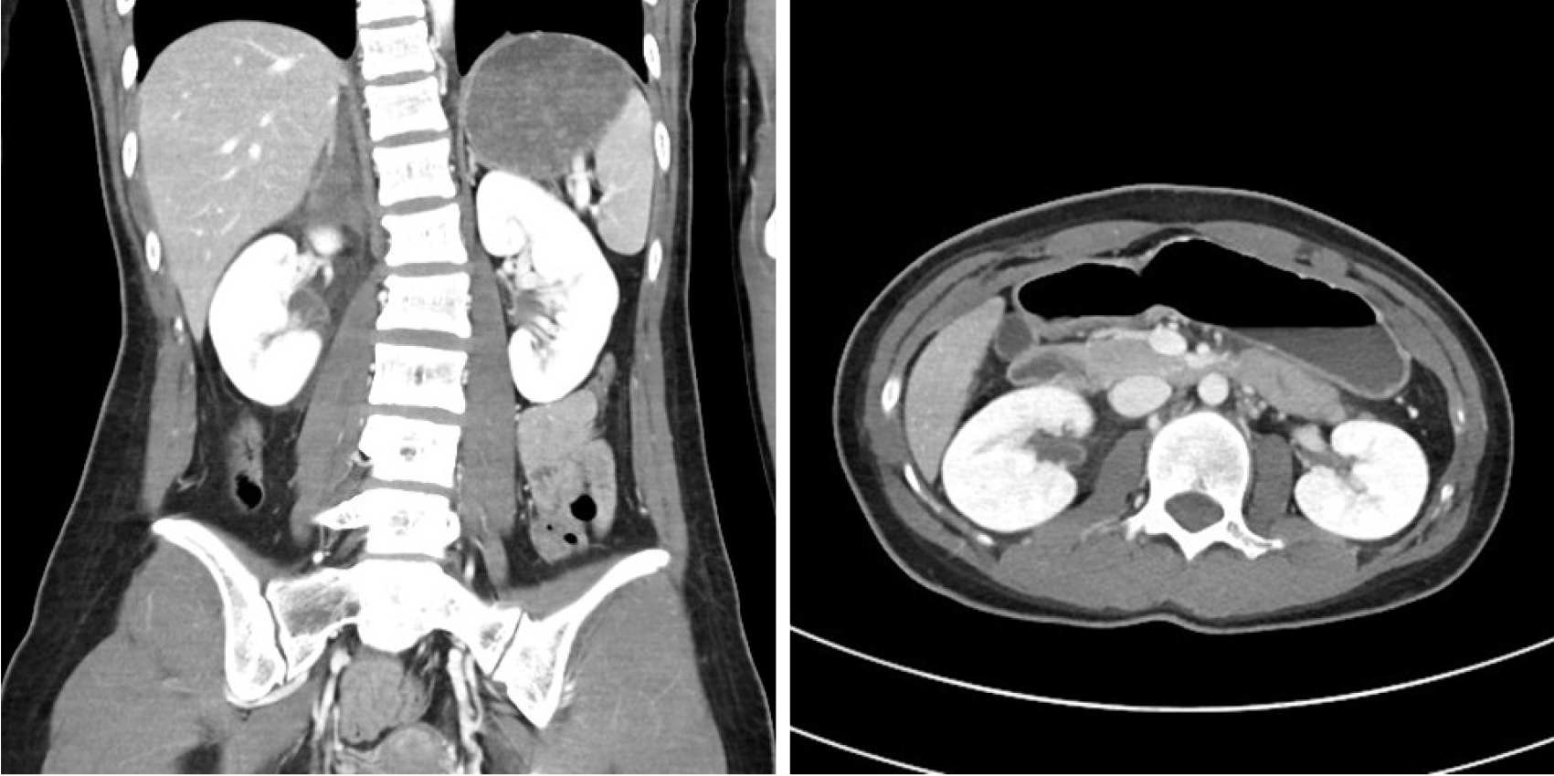
Hemorrhage in the right perirenal space was observed without abdominal organ injuries on the initial enhanced abdomen computed tomography (CT) scan (Figure 1). Ureteral injury was not suspected at this time. An orthosis was used for back pain and compression fracture of the first lumbar vertebra. The patient was stabilized via conservative treatment.
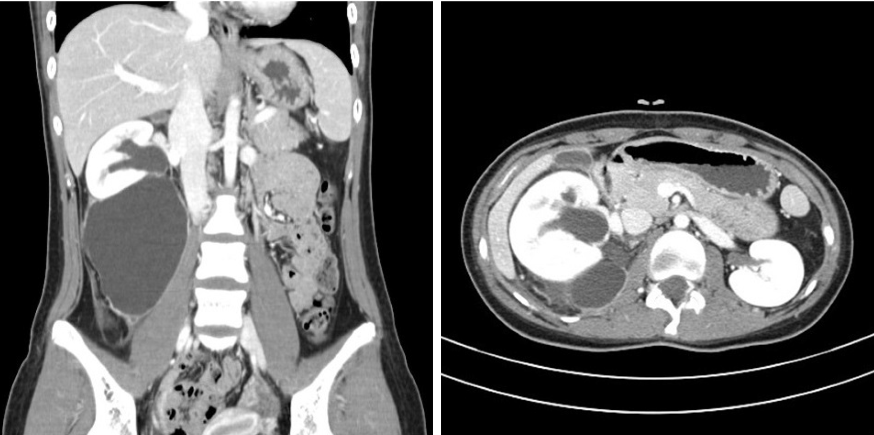
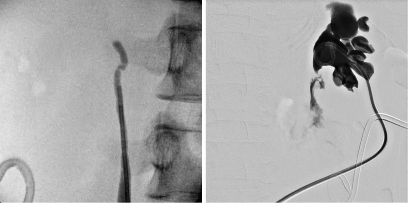
The patient complained of right flank pain 3 wk later and revisited the emergency room. An enhanced abdominal CT scan revealed a huge urinoma in the right perirenal space with hydronephrosis of the right kidney (Figure 2). A percutaneous catheter was inserted into the urinoma. Retrograde and antegrade pyelography were performed. Extravasation and discontinuity of the ureter were found (Figure 3), corresponding to an American Association for the Surgery of Trauma Organ Injury Scale (AAST-OIS) grade of 5. Percutaneous nephrostomy was performed for pain relief and urinary diversion. We decided on reconstructive surgery 1 wk later.
The final diagnosis of the presented case is ureteropelvic junction rupture.
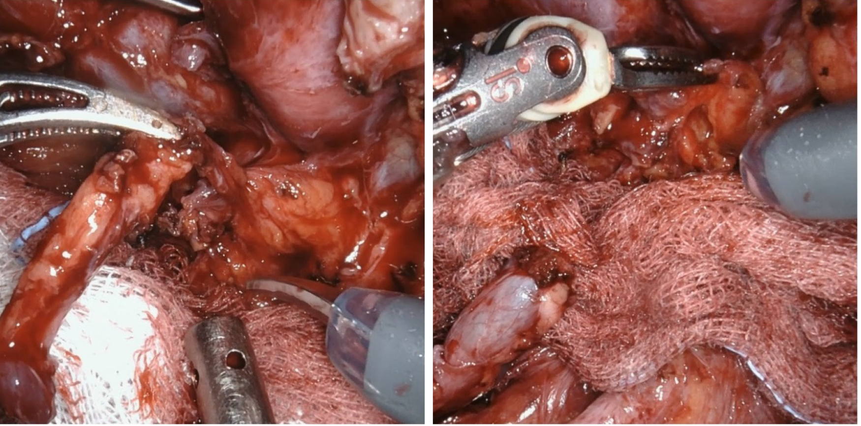
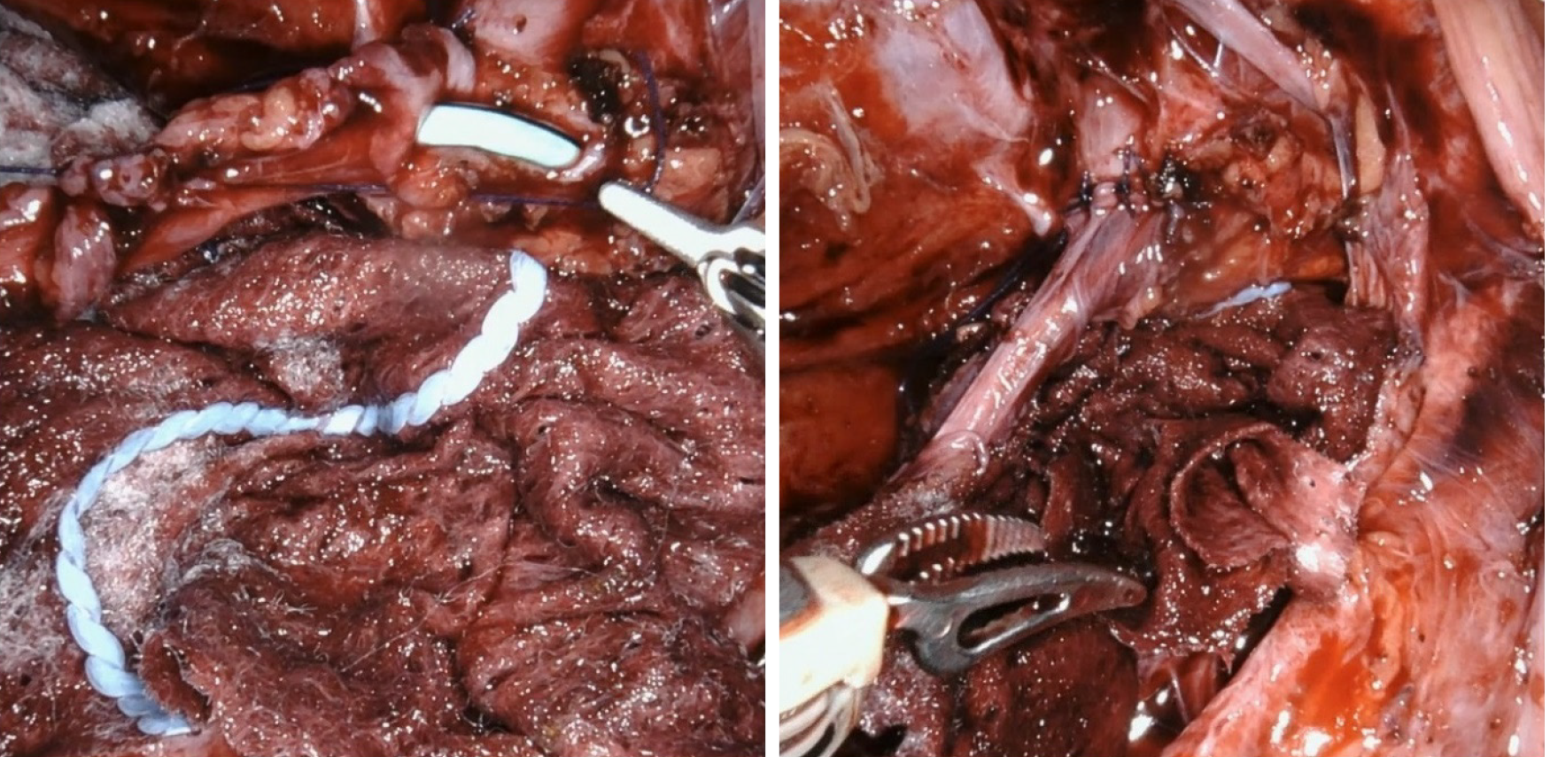
The surgeon was experienced in robot-assisted laparoscopic radical prostatectomy and nephrectomy. The da Vinci® Xi system (Intuitive Surgical, Inc., Sunnyvale, CA, Untied States) was used for the operation, which was performed under general anesthesia. A urethral catheter and nasogastric tube were placed before positioning. After the patient had been placed in the modified lateral decubitus position at 45°, with a gel roll supporting the lower back, preparation and draping were performed using povidone and disposable surgical drapes. An assistant 12-mm port was placed first using a Veress needle. After pneumoperitoneum was achieved, the laparoscope was inserted and the peritoneal cavity was inspected for injuries or adhesions. Four robotic ports were placed under direct vision at the lateral border of the rectus muscle. The line of Toldt was incised and dissected to mobilize the right colon. The retroperitoneal space was exposed. A large urinoma and adhesions were observed. After removing the urinoma and hematoma, the distal part of the ureter was inspected; the ureter and renal pelvis were not connected (Figure 4). The distal part of the ureter was dissected to relieve tension. A hydrophilic guidewire (UltraTrack®, 0.035” × 150 cm; Olympus, Tokyo, Japan) was inserted into the percutaneous catheter, which was connected by the renal pelvis to the proximal ureter. However, the renal pelvis was completely closed. Eventually, we decided to perform a pyeloureterostomy. An incision was made in the anterior aspect of the renal pelvis. The dissected distal ureter was spatulated to prevent stricture. Using a 5-0 Vicryl suture, side-to-side anastomosis was performed in an interrupted fashion (Figure 5). Before completing the anastomosis, a 6 Fr ureteral stent (26 cm) was inserted antegradely over a hydrophilic-tipped guidewire. The anastomosis was tested by infusion of indigo carmine. The surrounding perirenal fat was restored to its original location. A Jackson–Pratt drain was placed through the lower robotic port at the end of the procedure. The percutaneous nephrostomy was maintained after surgery.
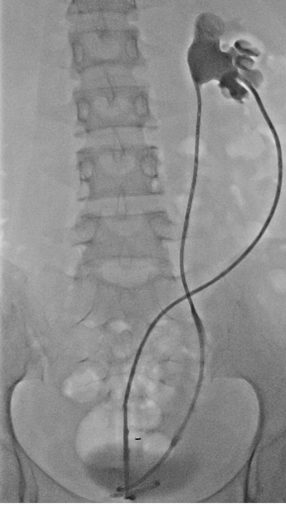
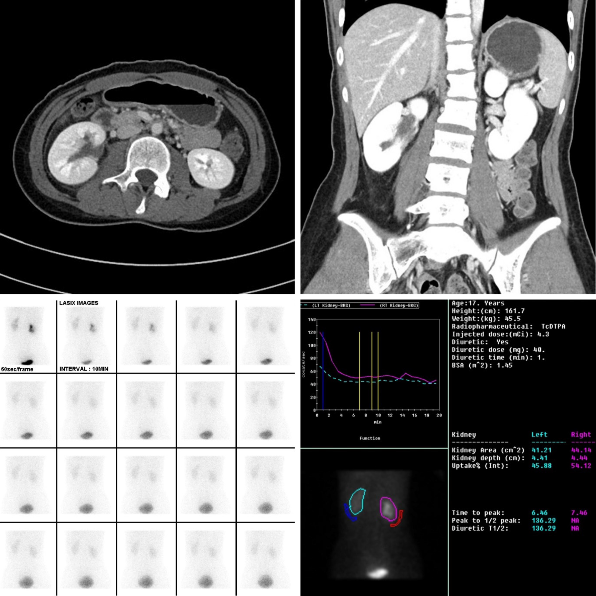
The surgical time was 180 min, the estimated blood loss was 50 mL, and the patient was discharged on postoperative day 10 without any complications. The urethral catheter was removed on postoperative day 2 and the Jackson–Pratt drain was removed on postoperative day 3; the patient could have been discharged at this time, but discharge was delayed for 1 wk at the request of the parents. An antegrade pyelography was performed 3 wk later to confirm the absence of anastomotic leakage (Figure 6). The percutaneous catheter was removed. The ureteric stent was removed 8 wk after the operation. An enhanced abdominal CT scan 3 mo later demonstrated no perinephric fluid collection and mild dilatation of the right renal pelvis. Renal scintigraphy revealed no evidence of obstruction and the furosemide clearance half-time was significantly improved to 6 min (Figure 7). The patient did not complain of any symptoms.
Injury to the genitourinary tract occurs in 10% of abdominal trauma cases[3,4]. The bladder is the structure most often injured, while ureteric trauma is the least common injury type due to the small size, mobility, and protected location of the ureter. Ureteral injuries are often associated with coexisting injuries involving the bowel, vascular structures, and urologic structures. The most common cause of ureteric injury is iatrogenic trauma. Injuries to the ureter are graded using the AAST-OIS and managed according to the extent and type of injury. Retrograde pyelography is a sensitive and specific test for determining the presence, location, and degree of ureteric injury[5]. An intravenous contrast-enhanced abdominal/pelvic CT scan with 10-min delayed images is also a good option to accurately evaluate the ureter. On a CT scan, the presence of contrast extravasation, delayed pyelogram, hydronephrosis, and a lack of contrast in the ureter distal to the site of the suspected injury suggest ureteral injury.
Mid-ureteral and proximal ureteral injuries can be managed with ureteroureterostomy. When a mid- or proximal ureteral injury occurs and the distal ureteral segment is not suitable for anastomosis, Boari bladder flap, transureteroureterostomy, renal autotransplantation, and ureteral substitution are available as alternatives[6]. Pyeloureterostomy is not commonly used for mid- or proximal ureteral injuries. Distal ureteral injuries are usually treated with ureteroneocystostomy.
Pyeloureterostomy was first described by Kummel in 1913, as reported by Diaz-Ball[7]. Laparoscopic and robotic approaches for complex upper-tract reconstruction were first described by Kutikov et al[8] in 2007. Minimally invasive urologic surgery has advanced rapidly over the past two decades, and the range of robot-assisted procedures has expanded[9]. Due to its three-dimensional visualization and more degrees of freedom than traditional laparoscopy, robot-assisted surgery allows surgeons to perform advanced laparoscopic procedures that have traditionally required the open approach[10].
The procedure to restore a damaged ureter is as follows: Remove the necrotic tissue, spatulate the tip of the ureter, create a watertight anastomosis, stent internally, provide an external drain, and separate the repaired ureter. Although open surgery for ureteral reconstruction is still the gold standard, shorter hospital stays without any additional risk of postoperative complications have been reported for laparoscopic (including robot-assisted) surgery[1,11]. Advantages of robot-assisted surgery include the minimal surgeon fatigue and superb visibility.
In this case, even though the ureter was completely separated, a relatively simple procedure was used compared to laparoscopic and open surgery. Although the operative time was 180 min, this included robot docking and positioning. Considering the difficulty of this case, it was a relatively brief procedure and the surgeon did not experience fatigue during the operation. The patient was discharged without complications: The ureter was restored to its original state without damage to other organs or the need for a nephrectomy.
Robot-assisted laparoscopic pyeloureterostomy is a feasible alternative to open and laparoscopic techniques for treating a proximal ureteral injury. The procedure is safe and effective, and reduces the hospital stay and postoperative complications. Additional studies are necessary to confirm the cost effectiveness and clinical efficacy of robot-assisted laparoscopic pyeloureterostomy.
Manuscript source: Unsolicited manuscript
Specialty type: Medicine, research and experimental
Country/Territory of origin: South Korea
Peer-review report’s scientific quality classification
Grade A (Excellent): 0
Grade B (Very good): B
Grade C (Good): C, C
Grade D (Fair): 0
Grade E (Poor): E
P-Reviewer: Desai M, Hofer M, Mrzljak A S-Editor: Gao CC L-Editor: A P-Editor: Wang LL
| 1. | Engel O, Rink M, Fisch M. Management of iatrogenic ureteral injury and techniques for ureteral reconstruction. Curr Opin Urol. 2015;25:331-335. [RCA] [PubMed] [DOI] [Full Text] [Cited by in Crossref: 28] [Cited by in RCA: 50] [Article Influence: 5.6] [Reference Citation Analysis (0)] |
| 2. | Fifer GL, Raynor MC, Selph P, Woods ME, Wallen EM, Viprakasit DP, Nielsen ME, Smith AM, Pruthi RS. Robotic ureteral reconstruction distal to the ureteropelvic junction: a large single institution clinical series with short-term follow up. J Endourol. 2014;28:1424-1428. [RCA] [PubMed] [DOI] [Full Text] [Cited by in Crossref: 23] [Cited by in RCA: 27] [Article Influence: 2.7] [Reference Citation Analysis (0)] |
| 3. | Bryk DJ, Zhao LC. Guideline of guidelines: a review of urological trauma guidelines. BJU Int. 2016;117:226-234. [RCA] [PubMed] [DOI] [Full Text] [Cited by in Crossref: 87] [Cited by in RCA: 124] [Article Influence: 12.4] [Reference Citation Analysis (0)] |
| 4. | Phillips B, Holzmer S, Turco L, Mirzaie M, Mause E, Mause A, Person A, Leslie SW, Cornell DL, Wagner M, Bertellotti R, Asensio JA. Trauma to the bladder and ureter: a review of diagnosis, management, and prognosis. Eur J Trauma Emerg Surg. 2017;43:763-773. [RCA] [PubMed] [DOI] [Full Text] [Cited by in Crossref: 35] [Cited by in RCA: 47] [Article Influence: 5.9] [Reference Citation Analysis (0)] |
| 5. | Brandes S, Coburn M, Armenakas N, McAninch J. Diagnosis and management of ureteric injury: an evidence-based analysis. BJU Int. 2004;94:277-289. [RCA] [PubMed] [DOI] [Full Text] [Cited by in Crossref: 171] [Cited by in RCA: 152] [Article Influence: 7.2] [Reference Citation Analysis (0)] |
| 6. | Burks FN, Santucci RA. Management of iatrogenic ureteral injury. Ther Adv Urol. 2014;6:115-124. [RCA] [PubMed] [DOI] [Full Text] [Cited by in Crossref: 110] [Cited by in RCA: 135] [Article Influence: 12.3] [Reference Citation Analysis (0)] |
| 7. | Diaz-Ball FL, Fink A, Moore CA, Gangai MP. Pyeloureterostomy and ureteroureterostomy: alternative procedures to partial nephrectomy for duplication of the ureter with only one pathological segment. J Urol. 1969;102:621-626. [RCA] [PubMed] [DOI] [Full Text] [Cited by in Crossref: 18] [Cited by in RCA: 20] [Article Influence: 0.4] [Reference Citation Analysis (0)] |
| 8. | Kutikov A, Nguyen M, Guzzo T, Canter D, Casale P. Laparoscopic and robotic complex upper-tract reconstruction in children with a duplex collecting system. J Endourol. 2007;21:621-624. [RCA] [PubMed] [DOI] [Full Text] [Cited by in Crossref: 36] [Cited by in RCA: 28] [Article Influence: 1.6] [Reference Citation Analysis (0)] |
| 9. | Brandao LF, Laydner H, Akca O, Autorino R, Zargar H, De S, Krishnam J, Pallavi P, Monga M, Stein RJ, Magi-Galluzzi C, Andreoni C, Kaouk JH. Robot-assisted ureteral reconstruction using a tubularized peritoneal flap: a novel technique in a chronic porcine model. World J Urol. 2017;35:89-96. [RCA] [PubMed] [DOI] [Full Text] [Cited by in Crossref: 7] [Cited by in RCA: 6] [Article Influence: 0.7] [Reference Citation Analysis (0)] |
| 10. | Korets R, Hyams ES, Shah OD, Stifelman MD. Robotic-assisted laparoscopic ureterocalicostomy. Urology. 2007;70:366-369. [RCA] [PubMed] [DOI] [Full Text] [Cited by in Crossref: 21] [Cited by in RCA: 14] [Article Influence: 0.8] [Reference Citation Analysis (0)] |
| 11. | Mei H, Pu J, Yang C, Zhang H, Zheng L, Tong Q. Laparoscopic vs open pyeloplasty for ureteropelvic junction obstruction in children: a systematic review and meta-analysis. J Endourol. 2011;25:727-736. [RCA] [PubMed] [DOI] [Full Text] [Cited by in Crossref: 116] [Cited by in RCA: 95] [Article Influence: 6.8] [Reference Citation Analysis (0)] |


