Published online Jan 26, 2020. doi: 10.12998/wjcc.v8.i2.318
Peer-review started: October 2, 2019
First decision: December 4, 2019
Revised: December 17, 2019
Accepted: December 22, 2019
Article in press: December 22, 2019
Published online: January 26, 2020
Processing time: 106 Days and 13.2 Hours
Unilateral exophthalmos is often caused by inflammation, neoplasm, infection, metabolic disease, vascular disorder and several other less common conditions. Reflex sympathetic dystrophy related to unilateral exophthalmos has not been reported in the past literature.
We describe a 45-year-old female with unilateral exophthalmos caused by reflex sympathetic dystrophy and its unexpected spontaneous disappearance after a standard anterior cervical discectomy and fixation operation with two PEEK interbody cages and a plate. To our surprise, the patient’s left unilateral exophthalmos improved spontaneously in the morning on postoperative day 2-with no relapse, without any further medication, as of seven years. We have named this condition “cervicogenic exophthalmos.”
We would inform other clinicians that unilateral exophthalmos was caused not only by inflammation, vascular disorder, infection, neoplasm, or metabolic disease, but also by reflex sympathetic dystrophy related with cervicogenic spondylosis. To the best of our knowledge, ours is the first related case report and use of the term “cervicogenic exophthalmos” after reviewing previous literature.
Core tip: “Cervicogenic” headache, vertigo, dizziness, hypertension, or tinnitus had been reported that patients with cervical spondylosis caused by sympathetic symptoms including headache, vertigo, dizziness, hypertension, or tinnitus and treated successfully with operation of anterior cervical discectomy and fixation using PEEK cages and a plate. Here we described a patient with spondylosis of C4/5/6 with unilateral exophthalmos. She suffered from unexpected spontaneous improvement of unilateral exophthalmos after an operation of anterior cervical discectomy and fixation with two PEEK interbody cages and a plate. Therefore, we have named the condition “cervicogenic exophthalmos”. To the best of our knowledge, ours is the first related case report and use of the term “cervicogenic exophthalmos” after reviewing previous literature.
- Citation: Wu CM, Liao HE, Hsu SW, Lan SJ. Cervicogenic exophthalmos: Possible etiology and pathogenesis. World J Clin Cases 2020; 8(2): 318-324
- URL: https://www.wjgnet.com/2307-8960/full/v8/i2/318.htm
- DOI: https://dx.doi.org/10.12998/wjcc.v8.i2.318
Patients who have exophthalmos of one eye often present a diagnostic challenge, perplexing not only to the ophthalmologist, but also to the orthopedist, neurosurgeon, and other clinicians. The etiologies of unilateral exophthalmos could be Graves' disease[1]; infection; trauma; periorbital region hemorrhage and hematoma; dural arteriovenous fistula, increased orbital fat; increased extraocular muscle volume and tension; head or neck surgical complications; or various intra- and extra-orbital tumors. A patient with proptosis seems to have an angry staring eye and a poor appearance. Unilateral exophthalmos not only causes headaches, proptosis, diplopia, and loss of vision, but also endangers a patient’s life.
Reflex sympathetic dystrophy (RSD), a diffuse pain syndrome and unknown pathophysiology[2], is applied for a great variety of quasi unrelated disorders variously described as causalgia, algodystrophy, Sudeck’s atrophy, traumatic angiospasm, peripheral acute trophoneurosis, post-traumatic osteoporosis, shoulder-hand syndrome, minor causalgia, reflex dystrophy[3], and complex regional pain syndrome type I. The clinical characteristics of RSD are diffuse pain, soft tissue swelling, hyperhidrosis, joint stiffness, skin discoloration, vasodilation or vasoconstriction, and osteoporosis in an extremity caused by an unusual and prolonged response of the sympathetic nervous system[4]. To the best of our knowledge, RSD induced unilateral exophthalmos has never been reported in previous literature. Here we report a case of unilateral exophthalmos caused by RSD, which improved suddenly after an anterior cervical discectomy and fixation (ACDF) operation.
A female patient aged 45 years visited our outpatient service on April 24, 2012. She presented with neck pain, both upper limbs numbness, weakness, and pain that had been ongoing for about 4 years.
Patient had been diagnosed with hyperthyroidism with oral methylprednisolone at another medical center for 3 years. She had visited the ophthalmic section at the same medical center to evaluate her unilateral exophthalmos with diplopia over the left eyeball 2 years ago. The impression was thyroid eye disease (Graves’ ophtha-lmopathy). The ophthalmologist suggested the ophthalmic surgery, but she refused and received regular steroid treatment.
She had no special history including smoking or drinking. There was no similar history in the family.
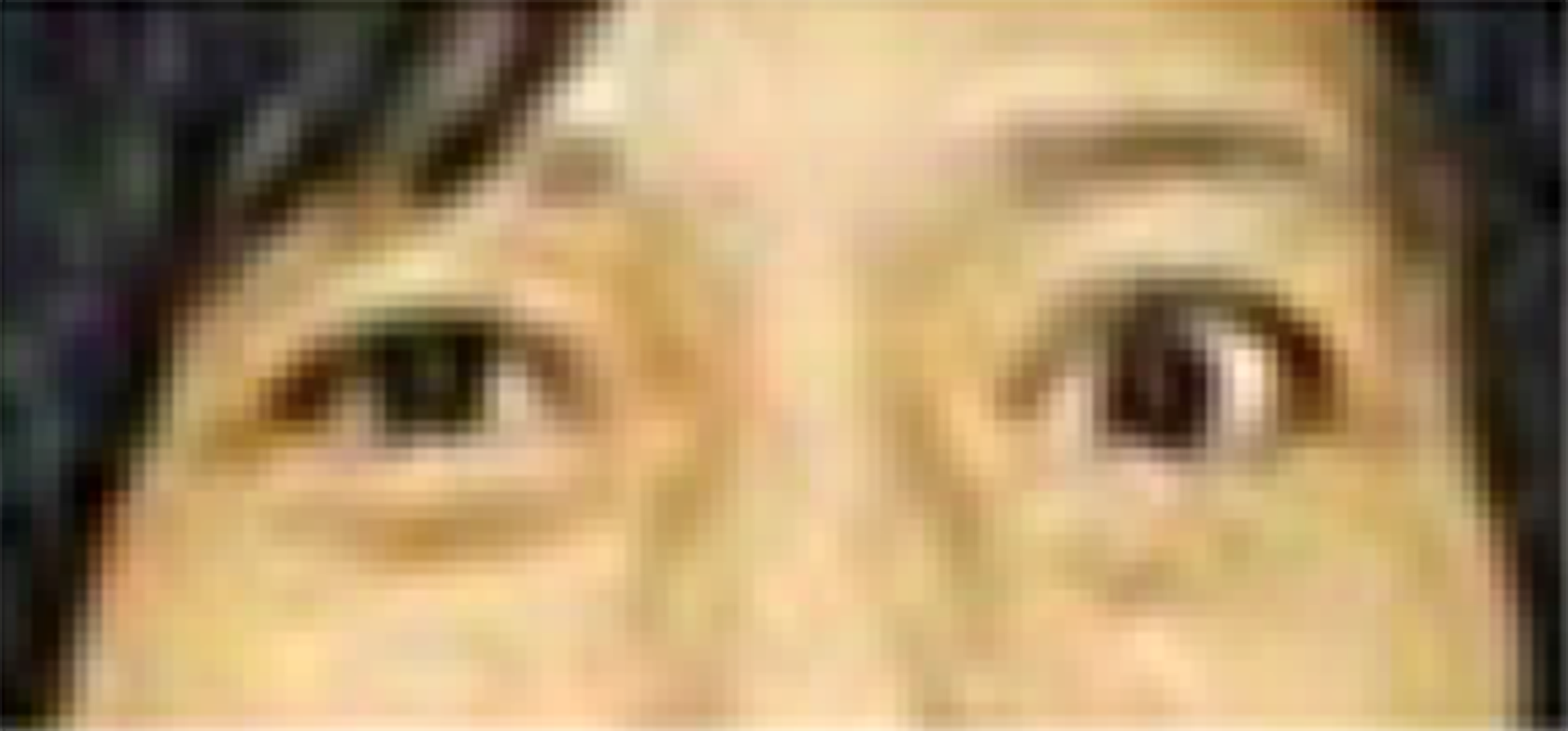
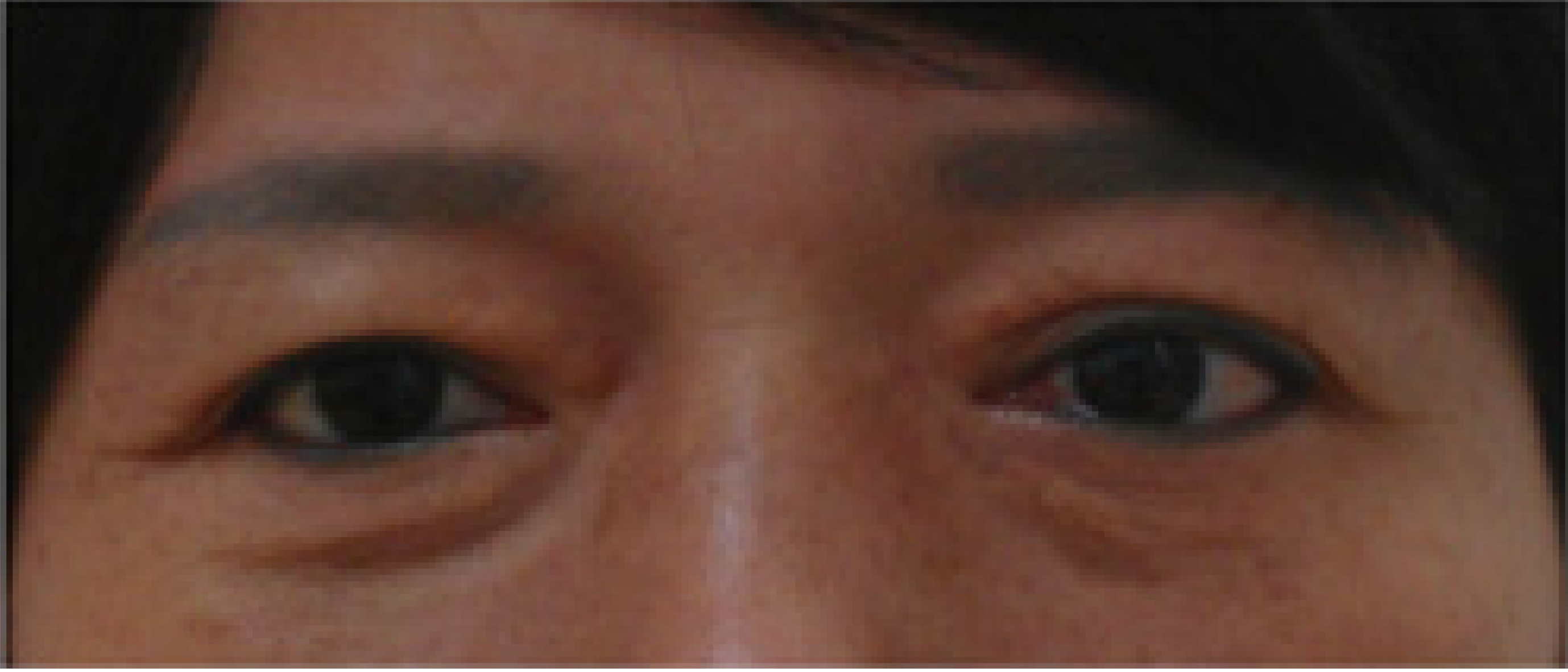
The patient appeared to have a protruding eyeball on the left side (Figure 1). Her left eyelid retraction was 7-8/5 according to the other hospital’s chart (versus 5/5 for the right eye). The distance from upper to lower marginal reflex of left eye was 13 mm (versus 10 mm for the right eye). The neurological examination of muscle strength for both biceps was 3/5 with decreased reflexes. A Hertel exophthalmometer revealed that the distance between the apex of the left cornea and the orbital wall was about 20 mm (versus 18 mm for the right eye).
The thyroid function tests, T3, TSH, free-T4, and other blood chemistry and hematologic parameters were all within normal range.
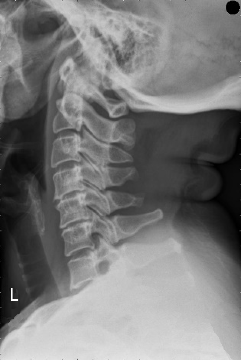
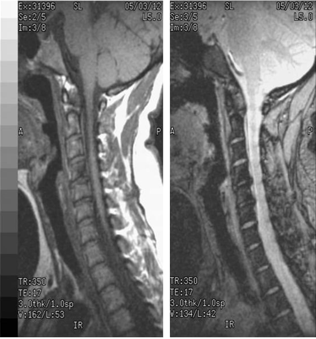
A brain computed tomography report revealed that the left extraconal muscle had increased volume, including left superior and inferior rectus muscle enlargement. An X-ray and magnetic resonance imaging of the cervical spine revealed that marginal osteophytes with posterior disc protrusion and disc space narrowing caused posterior longitudinal ligaments (PLL) and cervical cord compression at the C4/5/6 vertebrae (Figure 2 and Figure 3). Thyroid sonography showed a well-defined hypoechoic lesion about 5 mm × 7 mm × 2 mm over the right lobe, but the size of the left thyroid was normal.
The main impression was spondylosis of C4/5/6 with radiculopathy; the alternative impression was unilateral exophthalmos, possibly caused by Graves’ ophtha-lmopathy.
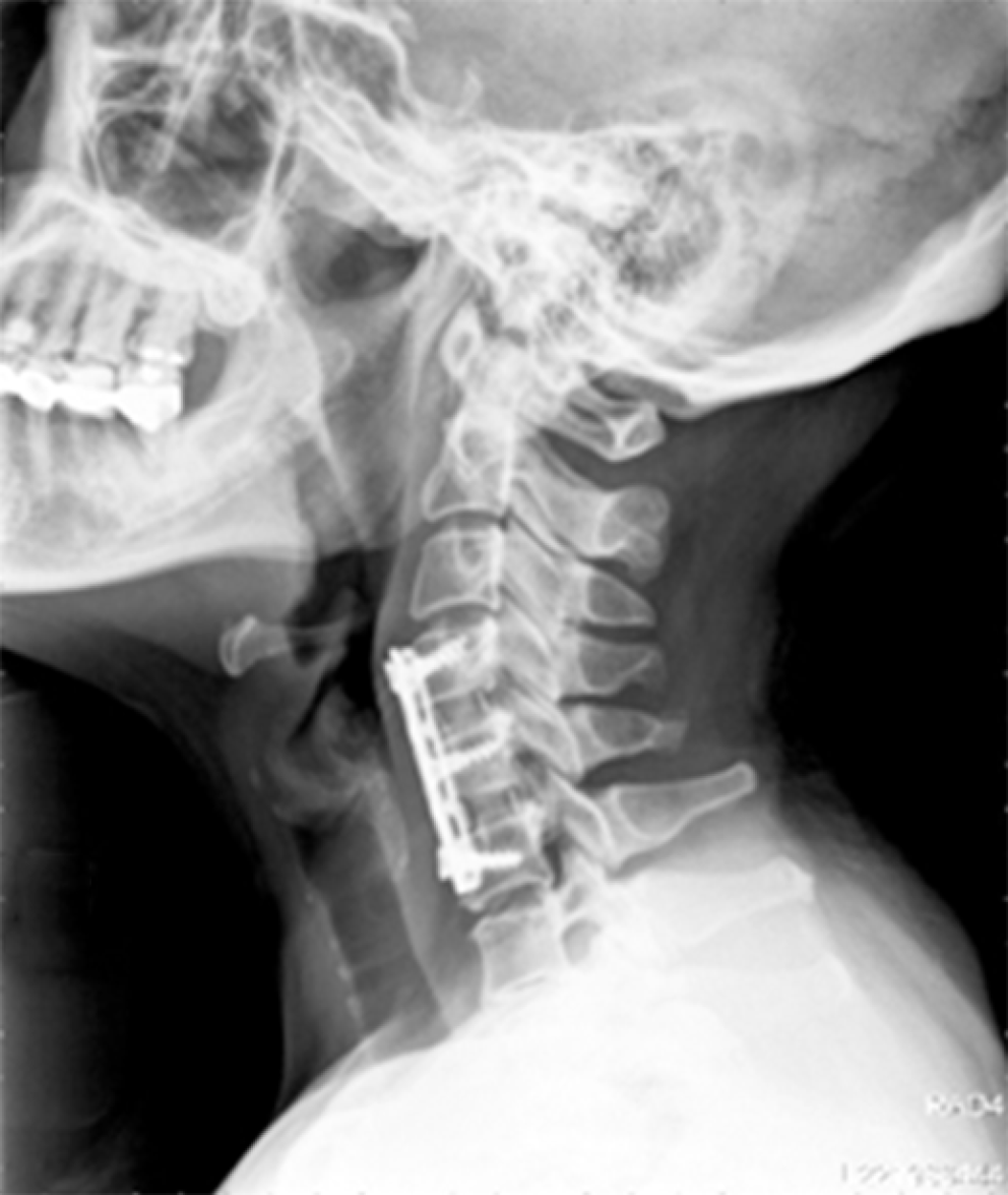
Because her neck pain and numbness in both arms had worsened, the patient received an ACDF operation with two PEEK interbody cages and a plate at C4/5/6 (Figure 4).
The patient was placed supine and her neck was mildly extended to achieve a normal lordotic curve under general anesthesia. After sterilization and draping, a left-side transverse incision was made from midline to lateral edge of the sternocleidomastoid muscle, and then the platysma muscle was opened. The sternocleidomastoid muscle and carotid sheath were retracted laterally, and then the omohyoid and sternohyoid muscles were retracted medially along the trachea and esophagus. The prevertebral space was palpated at the midline. The C4/5/6 disks level were identified with a spinal needle insertion checked by fluoroscopy. The longus coli muscle and anterior longitudinal ligament were dissected and incised, and the cervical disks were exposed. Finally, all disks as well as the cartilaginous materials around the end plates and PLL over C4/5/6 were removed completely with curettes and rongeurs. Two suitably sized PEEK interbody cages were placed into the C4/5 and C5/6 disc spaces. The C4/5/6 spine was fixed with a plate.
After surgery, the patient was given antibiotics of cephalosporin for 1 d to prevent wound infection, and nonsteroidal antiinflammatory drug to reduce her wound pain. The patient’s recovery was good except for mild numbness over her left arm for 1 postoperative day. We administered the drugs methasone (5 mg) in the morning and mepron (40 mg) in the afternoon, by way of intravenous injection, in an attempt to improve her left arm numbness. To our surprise, the patient’s left unilateral exophthalmos improved spontaneously in the morning on postoperative day 2 (Figure 5)-with no relapse, without any further medication, as of 7 years.
Most unilateral exophthalmos is caused by Graves' disease, periorbital trauma, hemorrhage, hematoma, cellulitis and infection, head or neck surgical complications, increased orbital fat, increased extraocular muscle volume and tension, or various intra- and extraorbital tumors.
Exophthalmos can also be caused by other less common conditions. Friedgood[5] mentioned that exophthalmosis often associated with hyperactivity of the sympathetic nervous system. Muller described the possibility that the contraction of the ocular unstriped muscle bands, which are controlled by the sympathetic nerves, may induce widening of the palpebral fissure. It has been demonstrated that changes occur in the sympathetic ganglia over the cervical region in exophthalmic goiter[6]. Several authorities have reported that the thyrotropic hormone fails to produce exophthalmos if the cervical sympathetic is divided simultaneously, whereas others maintain that it acts directly on the unstriped muscle fibers or the sympathetic nerve endings around the orbit[7].
RSD is a chronic condition characterized by diffuse burning pain; soft tissue swelling; trophic changes; and abnormalities in the sensory, motor, and autonomic nervous systems. Presently, there is some agreement that RSD results from an abnormal sympathetic nervous reflex[3]. The sympathetic nervous system controls all body structures, including muscles, synovium, tendons, ligaments, dura, disks, bones, and even the internal organs. If sympathetic nerves are damaged, as in RSD, the injured neural segments and abnormal feedback influence the internuncial centers of the spinal cord. The resulting error message would cause an abnormal sympathetic reflex with increased sympathetic activity.
Some literature describes that the cervical discs, like the lumbar discs, may indeed have a nerve supply. Li et al[8] found that a large number of sympathetic postganglionic fibers exist in the cervical PLL of every vertebra in rabbits. The distribution of sympathetic postganglionic fibers is also assumed in human specimens. Nathan et al[9] thought that there are sympathetic fibers distributed in the cervical uncovertebral joint capsule, PLL, rear of the annulus, and dura sac in humans[8]. Hovelacque reported that the vertebral nerve and its branch of the sinuvertebral nerve, a kind of sympathetic plexus accompanying the vertebral artery, enters into cervical intervertebral discs. Wang et al[10] hypothesized that (1) there are sympathetic nerve postganglionic fibers distributed in the PLL or discs; (2) pathological changes secondary to degeneration of the intervertebral disc may irritate sympathetic nerve fibers in the PLL or discs, leading to sympathetic symptoms via certain pathways; and (3) the removal of the PLL, or stabilization of the segment that decreases the irritation of the PLL, can help to eliminate the sympathetic symptoms[8,10].
Our case was spondylosis of C4/5/6 with radiculopathy. After performing the ACDF operation, the unilateral exophthalmos spontaneously and unexpectedly improved. The possible mechanism was that a degenerative and protruded disc compressed the PLL with sympathetic nerve fiber at C4/5/6 vertebrae, inducing an abnormal sympathetic reflex with increased sympathetic activity over the disc and PLL. This is called RSD, and it caused the enlargement of the left superior and inferior rectus muscles with intraoribtal space narrowing, ultimately leading to left unilateral exophthalmos. Thus, the sympathetic ganglion was no longer compressed after the complete removal of the cervical disk and PLL, such that the patient’s unilateral exophthalmos improved spontaneously.
Hong et al[11] described that cervical spondylosis patients with sympathetic symptoms including headache, vertigo, dizziness, hypertension, nausea, vomiting, or tinnitus may be treated successfully with ACDF surgery using PEEK cages[11-13]. They named these conditions “cervicogenic” headache[14], vertigo[8], dizziness[12], or hypertension[13]. Therefore, we have named the condition we encountered in this case “cervicogenic exophthalmos.”
The methods of treatment for Graves’ ophthalmopathy was different depending on its severity of clinical signs and symptoms. In the mild stage of Graves’ ophthalmopathy, we could encourage patient to change his life style, such as smoking cessation, using cold compression, lubricants, ointments, artificial tears, sunglasses, and prisms for symptomatic relief [15]. If the disease progressed to moderate or severity, drugs of glucocorticoid agents with local, oral or intravenous pathway, somatostatin analogues or orbital irradiation might be suggested[16]. Surgery should be performed for exophthalmos if high-dose glucocorticoids didn’t ameliorate this condition. The surgical methods had orbial decompressive surgery and thyroid surgery. Thyroid surgery for Graves’ ophthalmopathy commonly included three categories: (1) Total thyroidectomy; (2) Bilateral subtotal thyroidectomy; and (3) Unilateral total and contralateral subtotal thyroidectomy, or the Dunhill procedure[17].
Generally, a high dose of corticosteroid administered over a long term could treat an ocular disease, yet in this case, doses of less than 20 mg prednisolone or 100 mg hydrocortisone had been ineffective for reducing the exophthalmos[18]. Intravenous glucocorticoid could be used to decelerate thyroid eye disease activity, but the course of treatment would involve weekly treatments over a 10–12 wk period; a commonly used regimen consists of 12 weekly infusions of methylprednisolone (50 mg weekly for 6 wk, then 250 mg weekly for 6 wk) with a cumulative dose of 4.5 g[16]. Our case only used intravenous methasone 5 mg and mepron 40 mg, but the corticosteroid could not be the reason that the unilateral exophthalmos subsided because the dose was too low and the usage period too short.
On the other hand, patients with Graves’ disease associated exophthalmos received the surgery of thyroidectomy. There were 73% of cases would regressed with significant symptomatic relief at 1 year[19]. Our case received the ACDF operation with two PEEK interbody cages and a plate at C4/5/6. Her left unilateral exophthalmos improved spontaneously less than 48 h after cervical surgery-with no relapse, without any further medication, as of 7 years. We thought that the patient’s left unilateral exophthalmos didn’t caused by Graves’ ophthalmopathy.
Unilateral exophthalmos is often caused by inflammation, vascular disorder, infection, neoplasm, or metabolic disease[20]. If we were unaware of this etiology, we might think several other less common conditions, such as RSD, had been caused by the cervicogenic spondylosis. We have reported this case to inform other clinicians that unilateral exophthalmos maybe produced by cervicogenic spondylosis. To the best of our knowledge, ours is the first use of the term “cervicogenic exophthalmos”, and the first relative case report after reviewing past literature.
Manuscript source: Unsolicited Manuscript
Specialty type: Medicine, research and experimental
Country of origin: Taiwan
Peer-review report classification
Grade A (Excellent): 0
Grade B (Very good): B
Grade C (Good): C
Grade D (Fair): 0
Grade E (Poor): 0
P-Reviewer: Vaudo G, Bloomfield DA S-Editor: Wang YQ L-Editor: A E-Editor: Liu MY
| 1. | Furth ED, Becker DV, Ray BS, Kane JW. Appearance of unilateral infiltrative exophthalmos of Graves' disease after the successful treatment of the same process in the contralateral eye by apparently total surgical hypophysectomy. J Clin Endocrinol Metab. 1962;22:518-524. [RCA] [PubMed] [DOI] [Full Text] [Cited by in Crossref: 39] [Cited by in RCA: 42] [Article Influence: 0.7] [Reference Citation Analysis (0)] |
| 2. | Kemler MA, Barendse GA, van Kleef M, de Vet HC, Rijks CP, Furnée CA, van den Wildenberg FA. Spinal cord stimulation in patients with chronic reflex sympathetic dystrophy. N Engl J Med. 2000;343:618-624. [RCA] [PubMed] [DOI] [Full Text] [Cited by in Crossref: 642] [Cited by in RCA: 545] [Article Influence: 21.8] [Reference Citation Analysis (0)] |
| 3. | Veldman PH, Reynen HM, Arntz IE, Goris RJ. Signs and symptoms of reflex sympathetic dystrophy: prospective study of 829 patients. Lancet. 1993;342:1012-1016. [RCA] [PubMed] [DOI] [Full Text] [Cited by in Crossref: 825] [Cited by in RCA: 694] [Article Influence: 21.7] [Reference Citation Analysis (0)] |
| 4. | Jobe MT, Martinez SF. Peripheral Nerve Injuries, In: Campbell’s Operative Orthopaedics. 10th ed. 2003;3221-3283. |
| 5. | Friedgood HB. The Ocular Manifestations of Sympathetic Nervous System Hyperactivity in Conditions Other Than Exophthalmic Goiter and Especially in Essential Hypertension. Am J Med Sci. 1930;180:836. [RCA] [DOI] [Full Text] [Cited by in Crossref: 4] [Cited by in RCA: 4] [Article Influence: 0.0] [Reference Citation Analysis (0)] |
| 6. | Tilley JH. Exophthalmos: The Mechanism of its production in exophthalmic goitre. Ann Surg. 1926;84:647-650. [PubMed] |
| 7. | Stallard HB. A Case of "Exophthalmic Ophthalmoplegia with Thyrotoxicosis". Br J Ophthalmol. 1936;20:612-619. [RCA] [PubMed] [DOI] [Full Text] [Cited by in Crossref: 12] [Cited by in RCA: 12] [Article Influence: 0.7] [Reference Citation Analysis (0)] |
| 8. | Li Y, Peng B. Pathogenesis, Diagnosis, and Treatment of Cervical Vertigo. Pain Physician. 2015;18:E583-E595. [PubMed] |
| 9. | Nathan PW, Smith MC. The location of descending fibres to sympathetic preganglionic vasomotor and sudomotor neurons in man. J Neurol Neurosurg Psychiatry. 1987;50:1253-1262. [RCA] [PubMed] [DOI] [Full Text] [Cited by in Crossref: 55] [Cited by in RCA: 45] [Article Influence: 1.2] [Reference Citation Analysis (0)] |
| 10. | Wang Z, Wang X, Yuan W, Jiang D. Degenerative pathological irritations to cervical PLL may play a role in presenting sympathetic symptoms. Med Hypotheses. 2011;77:921-923. [RCA] [PubMed] [DOI] [Full Text] [Cited by in Crossref: 23] [Cited by in RCA: 26] [Article Influence: 1.9] [Reference Citation Analysis (0)] |
| 11. | Hong L, Kawaguchi Y. Anterior cervical discectomy and fusion to treat cervical spondylosis with sympathetic symptoms. J Spinal Disord Tech. 2011;24:11-14. [RCA] [PubMed] [DOI] [Full Text] [Cited by in Crossref: 32] [Cited by in RCA: 30] [Article Influence: 2.1] [Reference Citation Analysis (0)] |
| 12. | Wrisley DM, Sparto PJ, Whitney SL, Furman JM. Cervicogenic dizziness: a review of diagnosis and treatment. J Orthop Sports Phys Ther. 2000;30:755-766. [RCA] [PubMed] [DOI] [Full Text] [Cited by in Crossref: 154] [Cited by in RCA: 124] [Article Influence: 5.0] [Reference Citation Analysis (0)] |
| 13. | Liu H, Ploumis A. Cervicogenic hypertension: A possible etiology and pathogenesis of essential hypertension. Hypothesis. 2012;10:e4. [RCA] [DOI] [Full Text] [Cited by in Crossref: 4] [Cited by in RCA: 4] [Article Influence: 0.3] [Reference Citation Analysis (0)] |
| 14. | Blume HG. Cervicogenic headaches: radiofrequency neurotomy and the cervical disc and fusion. Clin Exp Rheumatol. 2000;18:S53-S58. [PubMed] |
| 15. | Bartalena L, Tanda ML. Clinical practice. Graves' ophthalmopathy. N Engl J Med. 2009;360:994-1001. [RCA] [PubMed] [DOI] [Full Text] [Cited by in Crossref: 214] [Cited by in RCA: 208] [Article Influence: 13.0] [Reference Citation Analysis (0)] |
| 16. | Yang DD, Gonzalez MO, Durairaj VD. Medical management of thyroid eye disease. Saudi J Ophthalmol. 2011;25:3-13. [RCA] [PubMed] [DOI] [Full Text] [Cited by in Crossref: 14] [Cited by in RCA: 15] [Article Influence: 1.0] [Reference Citation Analysis (0)] |
| 17. | Liu ZW, Masterson L, Fish B, Jani P, Chatterjee K. Thyroid surgery for Graves’ disease and Graves’ ophthalmopathy. Cochrane Database of Systematic Reviews. 2015;11:Art. No.: CD010576. [RCA] [DOI] [Full Text] [Cited by in Crossref: 28] [Cited by in RCA: 22] [Article Influence: 2.2] [Reference Citation Analysis (0)] |
| 18. | CORTISONE in exophthalmos: report on a therapeutic trial of cortisone and corticotrophin (A.C.T.H.) in exophthalmos and exophthalmic ophthalmoplegia by a panel appointed by the Medical Research Council. Lancet. 1955;268:6-9. [PubMed] |
| 19. | Bhargav PRK, Sabaretnam M, Kumar SC, Zwalitha S, Devi NV. Regression of Ophthalmopathic Exophthalmos in Graves' Disease After Total Thyroidectomy: a Prospective Study of a Surgical Series. Indian J Surg. 2017;79:521-526. [RCA] [PubMed] [DOI] [Full Text] [Cited by in Crossref: 6] [Cited by in RCA: 3] [Article Influence: 0.4] [Reference Citation Analysis (0)] |
| 20. | Tsai MH, Chao SC, Lin HY. Exophthalmos Related to Paranasal Sinusitis with Subperiosteal Abscess-a Case Report. Taiwan J Ophthalmos. 2006;45:103-108. [DOI] [Full Text] |


