Published online Aug 6, 2020. doi: 10.12998/wjcc.v8.i15.3249
Peer-review started: April 9, 2020
First decision: April 29, 2020
Revised: May 11, 2020
Accepted: July 14, 2020
Article in press: July 14, 2020
Published online: August 6, 2020
Processing time: 119 Days and 0.1 Hours
Glaucoma is an irreversible optic neuropathy with the loss of visual field and decrease of vision. The variable clinical manifestations may result in differential diagnostic difficulties. The early screening and diagnosis of glaucoma are currently experiencing a demand for anterior segment analysis tools that can gather more information with one short measurement. Therefore, we analyzed the agreement, difference, and correlation of chamber angle parameters such as angel opening distance at 500 μm (AOD500) and trabeculo-iris space area at 500 μm2 (TISA500) measured by anterior segment optical coherence tomography (AS-OCT) and ultrasound biomicroscopy (UBM).
To compare the differences, correlation, and agreement in measuring AOD500 and TISA500 by AS-OCT and UBM.
Both AS-OCT and UBM were performed to measure AOD500 and TISA500 in 45 subjects (72 eyes). All subjects without glaucoma were collected from October 2015 to August 2016 at the Ophthalmology Department of the Fourth Affiliated Hospital of China Medical University. Data of the two groups (AOD500 and TISA500) were compared by nonparametric tests. Pearson correlative analysis and Bland–Altman analysis were used to compare the correlation and agreement.
There were no significant differences between AS-OCT and UBM in measuring AOD500 (P1 = 0.110, P2 = 0.633, P3 = 0.078, and P4 = 0.474) and TISA500 (P1 = 0.584, P2 = 0.889, P3 = 0.297, and P4 = 0.550) of the four quadrants of the anterior chamber angle. There was a high correlation in measuring AOD500 (r1 = 0.562, r2 = 0.671, r3 = 0.635, and r4 = 0.720; P < 0.001) and TISA500 (r1 = 0.584, r2 = 0.889, r3 = 0.297, and r4 = 0.550; P < 0.001). There was a good agreement in measuring AOD500 and TISA500 by the two modalities.
There is a high correlation and agreement between AOD500 and TISA500 measurements by AS-OCT and UBM. They are interchangeable under some circumstances. AS-OCT proves to be a better early screening tool for glaucoma.
Core tips: Glaucoma is an irreversible optic neuropathy with the loss of visual field and decrease of vision. The early screening and diagnosis of glaucoma are currently experiencing a demand for anterior segment analysis tools that can gather more information with one short measurement. In this study, we compared the agreement, difference, and correlation of anterior chamber angle parameters such as angel opening distance at 500 μm and trabeculo-iris space area at 500 μm2 measured by anterior segment optical coherence tomography and ultrasound biomicroscopy.
- Citation: Yu ZY, Huang T, Lu L, Qu B. Comparison of measurements of anterior chamber angle via anterior segment optical coherence tomography and ultrasound biomicroscopy. World J Clin Cases 2020; 8(15): 3249-3258
- URL: https://www.wjgnet.com/2307-8960/full/v8/i15/3249.htm
- DOI: https://dx.doi.org/10.12998/wjcc.v8.i15.3249
Glaucoma is an irreversible optic neuropathy with the loss of visual field and decrease of vision. There are no obvious clinical manifestations in the early stage of glaucoma; typical visual field defects or progressive loss of visual acuity and other symptoms may appear in the moderate or severe phase of glaucoma[1,2]. Approximately 80% of aqueous humor flows out through the anterior chamber angle, when aqueous humor outflow obstruction is caused by abnormality of anterior chamber angle structure or function, and compensatory imbalance of aqueous humor leads to increased intraocular pressure.
At present, gonioscopy, anterior segment optical coherence tomography (AS-OCT), and ultrasound biomicroscopy (UBM) are commonly used to evaluate anterior chamber angle[3-5]. AS-OCT and other non-contact imaging modalities provide a quick, user-friendly, and objective non-invasive method of assessing the anterior segment parameters and anterior chamber angle, are well tolerated by the patient, and correlate well with the information provided by gonioscopy[6,7].
In this study, we evaluated the agreement, difference, and correlation of chamber angle parameters such as angel opening distance at 500 μm (AOD500) and trabeculo-iris space area at 500 μm2 (TISA500) measured by AS-OCT and UBM.
Forty-five subjects (15 males and 30 females; age range, 50–80 years) without glaucoma who visited the Fourth Affiliated Hospital of China Medical University from October 2015 to August 2016 were selected. All subjects underwent routine ophthalmological examinations (including visual acuity, intraocular pressure, integrated optometry, slit lamp examination, and fundus examination) and AS-OCT and UBM. None of the subjects had an intraocular surgery or laser procedure, and none of the subjects had evidence of glaucoma, dry eye disease, ocular inflammatory activity, or other ocular diseases.
This study was approved by the Fourth Affiliated Hospital of China Medical University Committee on Human Research. The registry’s URL is www.medresman.org and the registration number is ChiCTR1900021378. All study procedures adhered to the Declaration of Helsinki for research involving human participants. All participants provided informed consent after explanation of possible consequences of the study to them.
The patients were examined in the order of AS-OCT (sitting position) and UBM (supine position). The interval of examinations was about 5 min; all the examinations with the same instrument were performed three times in the same brightness environment and by the same operator. The average values of AOD500 and TISA500 of four quadrants for each eye were recorded.
All descriptive analyses, difference analyses, and correlation analyses were performed with SPSS version 19.0, and agreement analysis was performed with MedCalc version 15.0. All descriptive results are expressed as the mean ± SD. The differences in the two instruments were analyzed by paired t-test if the data were in accordance with the normal distribution and the variance was homogeneous; otherwise, nonparametric tests were used. Correlations and agreements between the instruments were performed by Pearson correlation analysis and Bland–Altman analysis. P < 0.05 was considered statistically significant.
A total of 15 male subjects (24 eyes) and 30 female subjects (48 eyes) were examined by UBM and AS-OCT for anterior chamber angles parameters (AOD500 and TISA500), with a mean age of 59.47 ± 14.481 years.
AOD500 at 90° (12 o’clock position) was 0.2453 ± 0.2566 mm by AS-OCT and 0.1642 ± 0.1502 mm by UBM. AOD500 at 0° (3 o’clock position) was 0.2493 ± 0.2065 mm by AS-OCT and 0.2090 ± 0.1344 mm by UBM. AOD500 at 270° (6 o’clock position) was 0.2828 ± 0.2587 mm by AS-OCT and 0.1914 ± 0.1410 mm by UBM. AOD500 at 180° (9 o’clock position) was 0.2661 ± 0.2300 mm by AS-OCT and 0.2194 ± 0.1523 mm by UBM.
TISA500 at 90° (12 o’clock position) was 0.0912 ± 0.0884 mm by AS-OCT and 0.0749 ± 0.0630 mm by UBM. TISA500 at 0° (3 o’clock position) was 0.0997 ± 0.0716 mm by AS-OCT and 0.0982 ± 0.0676 mm by UBM. TISA500 at 270° (6 o’clock position) was 0.1066 ± 0.0941 mm by AS-OCT and 0.0842 ± 0.0625 mm by UBM. TISA500 at 180° (9 o’clock position) was 0.1071 ± 0.0773 mm by AS-OCT and 0.1004 ± 0.0765 mm by UBM.
For AOD500 and TISA500 of the four quadrants of the chamber angles, measurements by AS-OCT were greater than those by UBM. AOD500 measured by AS-OCT from wide to narrow were 6 o’clock, 9 o’clock, 3 o’clock, and 12 o’clock positions. AOD500 measured by UBM from big to small were 9 o’clock, 3 o’clock, 6 o’clock, and 12 o’clock positions. TISA500 measurements by AS-OCT from wide to narrow were 9 o’clock, 6 o’clock, 3 o’clock, and 12 o’clock positions. TISA500 measurements by UBM from big to small were 9 o’clock, 3 o’clock, 6 o’clock, and 12 o’clock positions.
The measurements of AOD500 and TISA500 using the two methods were not normally distributed, and the homogeneity of variance (Mann–Whitney test) and the differences in them were compared using nonparametric tests. There was no significant differences between the two methods in measuring AOD500 of the four quadrants of the chamber angles (P1 = 0.110, P2 = 0.633, P3 = 0.078, and P4 = 0.474). There was no significant differences between the two methods in measuring TISA500 of the four quadrants of the chamber angles (P1 = 0.584, P2 = 0.889, P3 = 0.297, and P4 = 0.550). Comparing the AOD500 and TISA500 measured by AS-OCT and UBM, the results were not significantly different (P > 0.05).
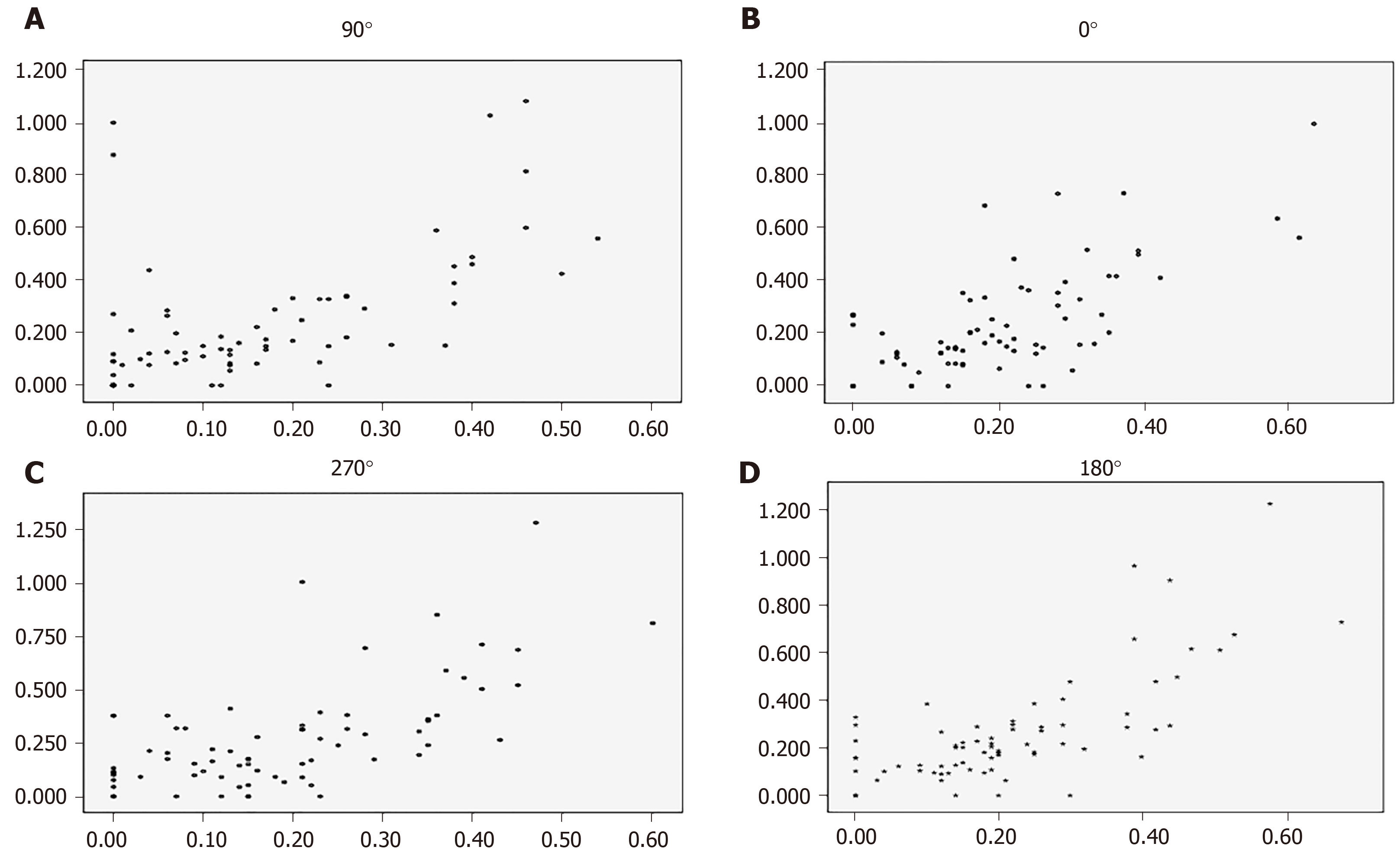
The AOD500 measurements by AS-OCT and UBM at 90° (r = 0.562, P < 0.001), 0° (r = 0.671, P < 0.001), 270° (r = 0.635, P < 0.001), and 180° (r = 0.720, P < 0.001) were highly correlated (Figure 1).
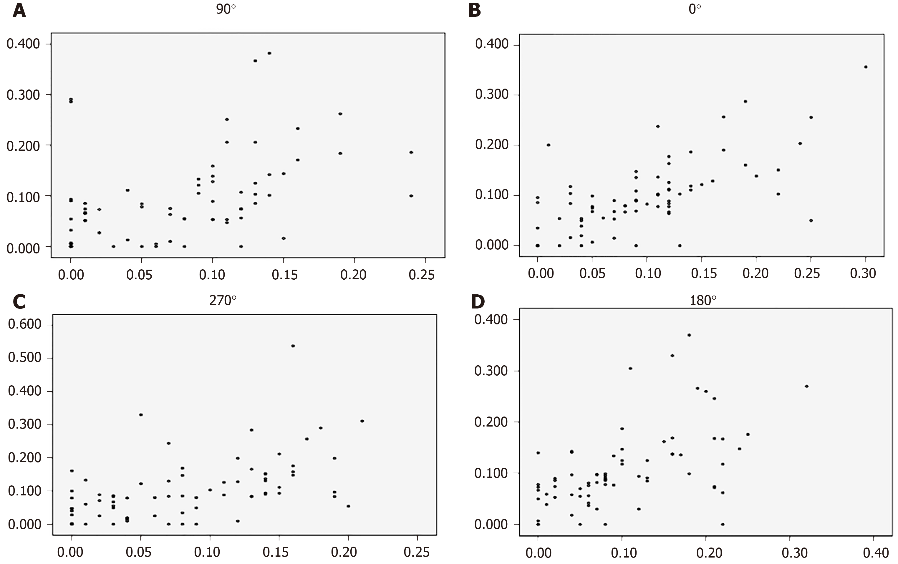
The TISA500 measurements by AS-OCT and UBM at 90° (r = 0.466, P < 0.001), 0° (r = 0.652, P < 0.001), 270° (r = 0.507, P < 0.001), and 180° (r = 0.545, P < 0.001) were highly correlated (Figure 2).
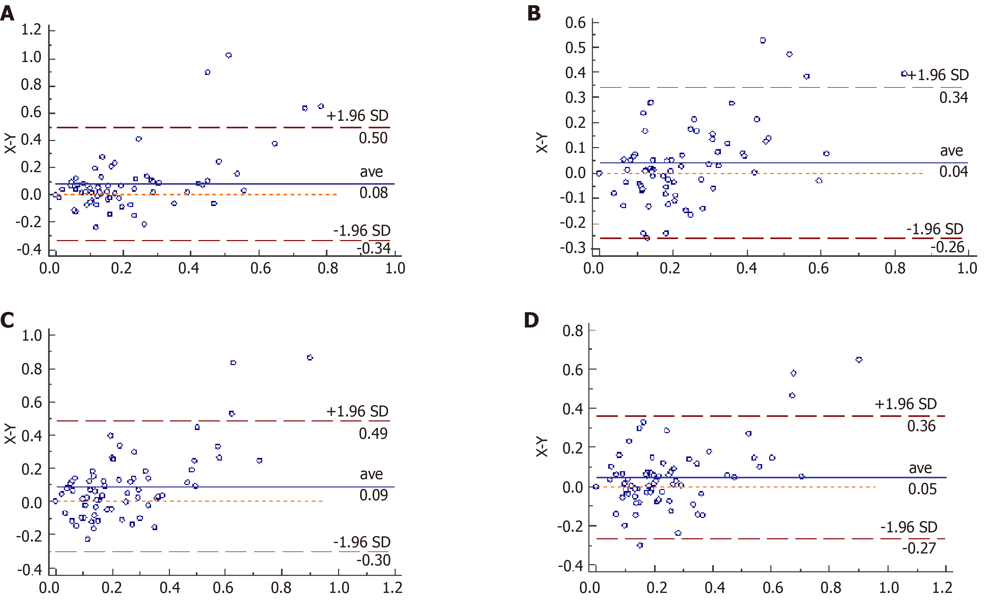
Agreement of the AOD500 measurements by the two methods was analyzed by Bland–Altman diagram at the four quadrants of the anterior chamber angles (Figure 3). At 90°, four (2.78%) of 144 points were outside the confidence interval. At 0°, four of 144 points were outside the confidence interval. At 270°, three (2.08%) of 144 points were outside the confidence interval. At 180 °, four of 144 points were outside the confidence interval. The Bland–Altman diagram of AOD500 measurements at any quadrants of the chamber angles showed that > 95% of the data points were in the 95% agreement interval and the confidence interval was relatively narrow, with no significant difference, and could be clinically accepted. Bland–Altman diagram analysis showed that the results of AOD500 measurements at four quadrants of the chamber angles measured by the two methods were in good agreement.
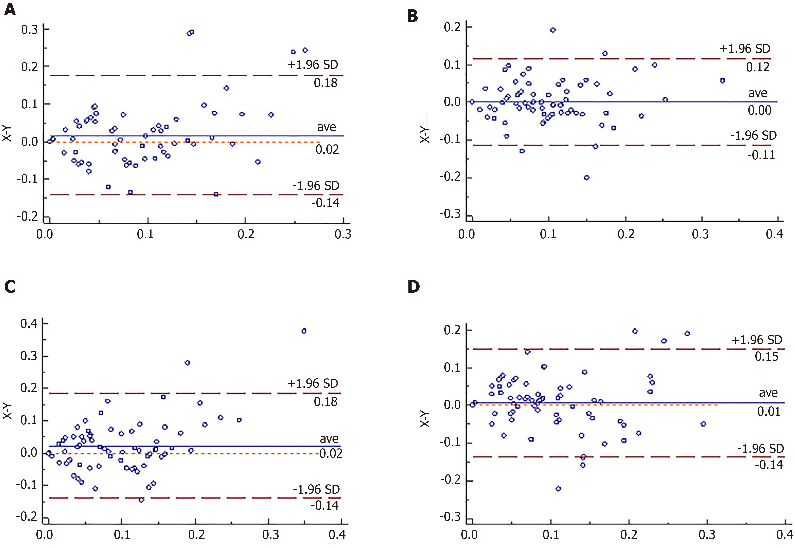
Agreements of the TISA500 measurements by the two methods were analyzed by Bland–Altman diagram at the four quadrants of the anterior chamber angles (Figure 4). At 90°, four (2.78%) of 144 points were outside the confidence interval. At 0°, five (3.47%) of 144 points were outside the confidence interval. At 270°, three (2.08%) of 144 points were outside the confidence interval. At 180°, five (3.47%) of 144 points were outside the confidence interval. The Bland–Altman diagram of TISA500 measurements at any quadrants of the chamber angles showed that > 95% of the data points were in the 95% agreement interval and the confidence interval was relatively narrow, with no significant difference, and could be clinically accepted. Bland–Altman diagram analysis showed that the results of TISA500 measurements at four quadrants of the chamber angles measured by the two methods were in good agreement.
Microstructures of the anterior chamber angle (such as iris root, scleral protrusion, and trabecular meshwork) can be observed to estimate the degree of opening of the anterior chamber angle, and its structure and function can be estimated by gonioscopy[8,9]. Observation of a scleral spur suggests complete exposure of the functional trabecular meshwork, and this is a marker in imaging examination and the first step in observation[10]. AOD500 and TISA500 measurements are affected by body position, eye position, whether there is contact or pressure on the eye during measurement, and whether front and rear boundary measurements are accurate[11,12]. Under the influence of gravity, the cornea can be compressed by the upper eyelid, and the normal anterior chamber angle shows that the top position is narrowest and the bottom position is widest, followed by nasal and temporal position[13].
At present, objective methods of visualizing anterior chamber angle are UBM and AS-OCT. AOD500 and TISA500 measurements by AS-OCT were, respectively, 0.2453 ± 0.2566 mm and 0.0912 ± 0.0884 mm at 90°, 0.2828 ± 0.2587 mm and 0.1066 ± 0.0941 mm at 270°, 0.2493 ± 0.2065 mm and 0.2661 ± 0.2300 mm at 0°, and 0.0997 ± 0.0716 mm and 0.1071 ± 0.0773 mm at 180°. AS-OCT is a non-contact examination with the subject in the sitting position, and it is not affected by body and eye positions[14]. In AOD500 and TISA500 measurements, the top position angle is narrowest and coincides with the physiological state. In AOD500 measurements, the bottom position angle is widest and coincides with the physiological state. In TISA500 measurements, related to the measurement error, the 270° angle is widest but not much different from the bottom position angle. There are many reasons for the measurement error. The eyeball may be completely exposed while measuring, which can exert pressure on the eyeball, thus affecting the accuracy of measurement. The measurements are automatically identified by computer, resulting in inaccuracy of the positioning boundaries.
AOD500 and TISA500 measurements by UBM were, respectively, 0.1642 ± 0.1502 mm and 0.0749 ± 0.0630 mm at 90° position, 0.1914 ± 0.1410 mm and 0.0842 ± 0.0625 mm at 270°, 0.2090 ± 0.1344 mm and 0.2194 ± 0.1523 mm at 0°, and 0.0982 ± 0.0676 mm and 0.1004 ± 0.0765 mm at 180°. In AOD500 and TISA500 measurements, the top position angle is narrowest, the 180° position angle is widest, and the 0° position takes second place. Considering that UBM is performed with the patient in the supine position, change of barycenter leads to redistribution of aqueous humor; the width of the horizontal anterior chamber angle increases, and the width of the lower anterior chamber angle decreases. When measuring in one direction, the patient’s eyeball rotates and eye position changes in that direction, which can cause measurement error. It is a contact examination and has a greater possibility of eye compression, both of which can affect the measurement results. Moreover, UBM resolution is lower, and manual measurement can introduce human error.
Comparing the degree of openness of the anterior chamber angle can determine opening and closing and degree of stenosis of the anterior chamber angle. Moreover, it can be used to compare preoperative and postoperative chamber angle changes[15-17]. By comparing the AOD500 and TISA500 measurements via UBM and AS-OCT, we conclude that the agreement and correlation are excellent, and there is no significant difference between them, proving that they are interchangeable. Previous studies also support this viewpoint[1,18-21]. Compared with UBM, AS-OCT has better repeatability, noninvasiveness, rapidity, high resolution, and automatic analysis. These features can avoid measurement errors caused by eye compression, eyeball rotation, and body position change, making anterior chamber angle parameters more coincident with those under physiological conditions. AS-OCT can also avoid human measurement error, making the data in good repeatability. As a non-contact device, with short measure time, fast and clear imaging, and easily follow-up, AS-OCT can be widely used in glaucoma screening, open eye injury, and other diseases. However, UBM can be used to diagnose ciliary body detachment, ciliary body tumor, peripheral retinal detachment, and other diseases, while AS-OCT cannot.
The effect of intragroup differences on the results was considered. Age, sex, and laterality of the eye were not distinguished. The measurements were not classified based on the width of anterior chamber angle and status of diseases. There were only 72 eyes, and the data did not satisfy a normal distribution. The influence of the above factors on the measurement results should be investigated further using a larger study sample size.
In conclusion, there is a good correlation and agreement between AOD500 and TISA500 measurements by AS-OCT and UBM. The two methods are interchangable for measurement of the anterior segment angle and cannot be completely replaced by each other. AS-OCT can be used as the first choice for early glaucoma screening, and can be used for follow-up of glaucoma surgery.
Glaucoma is an irreversible optic neuropathy with the loss of visual field and decrease of vision. A quick, user-friendly, and non-invasive method of assessing the anterior segment parameters is essential for early screening and diagnosis of glaucoma.
The main topic is to compare the difference between anterior chamber angle parameters measured by different methods.
To investigate the agreement, difference, and correlation of chamber angle parameters such as angel opening distance at 500 μm (AOD500) and trabeculo-iris space area at 500 μm2 (TISA500) measured by anterior segment optical coherence tomography (AS-OCT) and ultrasound biomicroscopy (UBM) in normal subjects.
Both AS-OCT and UBM were performed to measure AOD500 and TISA500 in 45 subjects (72 eyes). All subjects without glaucoma were collected from October 2015 to August 2016 at the Ophthalmology Department of the Fourth Affiliated Hospital of China Medical University. Data of the two groups (AOD500 and TISA500) were compared by nonparametric tests. Pearson correlative analysis and Bland–Altman analysis were used to compare the correlation and agreement.
There were no significant differences between AS-OCT and UBM in measuring AOD500 and TISA500 of the four quadrants of the anterior chamber angle. There was a high correlation in measuring AOD500 and TISA500. There was a good agreement in measuring AOD500 and TISA500.
There is a high correlation and agreement between AOD500 and TISA500 measurements by AS-OCT and UBM. They are interchangeable under some circumstances. AS-OCT proves to be a better early screening tool for glaucoma.
AS-OCT and UBM are interchangable for AOD500 and TISA500 measurements and cannot be completely replaced by each other. AS-OCT can be used as the first choice for early glaucoma screening, especially for primary open-angle glaucoma patients.
Manuscript source: Unsolicited manuscript
Specialty type: Medicine, research and experimental
Country/Territory of origin: China
Peer-review report’s scientific quality classification
Grade A (Excellent): 0
Grade B (Very good): B, B
Grade C (Good): C
Grade D (Fair): 0
Grade E (Poor): 0
P-Reviewer: Apisarnthanarax S, Snowdon VK, Voigt M S-Editor: Wang JL L-Editor: Wang TQ E-Editor: Xing YX
| 1. | Kuerten D, Plange N, Becker J, Walter P, Fuest M. Evaluation of Long-term Anatomic Changes Following Canaloplasty With Anterior Segment Spectral-domain Optical Coherence Tomography and Ultrasound Biomicroscopy. J Glaucoma. 2018;27:87-93. [RCA] [PubMed] [DOI] [Full Text] [Cited by in Crossref: 8] [Cited by in RCA: 10] [Article Influence: 1.7] [Reference Citation Analysis (0)] |
| 2. | Tham YC, Li X, Wong TY, Quigley HA, Aung T, Cheng CY. Global prevalence of glaucoma and projections of glaucoma burden through 2040: a systematic review and meta-analysis. Ophthalmology. 2014;121:2081-2090. [RCA] [PubMed] [DOI] [Full Text] [Cited by in Crossref: 5390] [Cited by in RCA: 4475] [Article Influence: 406.8] [Reference Citation Analysis (4)] |
| 3. | Lata S, Venkatesh P, Temkar S, Selvan H, Gupta V, Dada T, Upadhyay AD, Sihota R. Comparative Evaluation of Anterior Segment Optical Coherence Tomography, Ultrasound Biomicroscopy, and Intraocular Pressure Changes After Panretinal Photocoagulation by Pascal and Conventional Laser. Retina. 2020;40:537-545. [RCA] [PubMed] [DOI] [Full Text] [Cited by in Crossref: 2] [Cited by in RCA: 2] [Article Influence: 0.5] [Reference Citation Analysis (0)] |
| 4. | Xu BY, Burkemper B, Lewinger JP, Jiang X, Pardeshi AA, Richter G, Torres M, McKean-Cowdin R, Varma R. Correlation between Intraocular Pressure and Angle Configuration Measured by OCT: The Chinese American Eye Study. Ophthalmol Glaucoma. 2018;1:158-166. [RCA] [PubMed] [DOI] [Full Text] [Cited by in Crossref: 32] [Cited by in RCA: 35] [Article Influence: 5.0] [Reference Citation Analysis (0)] |
| 5. | Sedaghat MR, Mohammad Zadeh V, Fadakar K, Kadivar S, Abrishami M. Normative values and contralateral comparison of anterior chamber parameters measured by Pentacam and its correlation with corneal biomechanical factors. Saudi J Ophthalmol. 2017;31:7-10. [RCA] [PubMed] [DOI] [Full Text] [Full Text (PDF)] [Cited by in Crossref: 6] [Cited by in RCA: 6] [Article Influence: 0.7] [Reference Citation Analysis (0)] |
| 6. | Porporato N, Baskaran M, Tun TA, Sultana R, Tan M, Quah JH, Allen JC, Perera S, Friedman DS, Cheng CY, Aung T. Understanding diagnostic disagreement in angle closure assessment between anterior segment optical coherence tomography and gonioscopy. Br J Ophthalmol. 2020;104:795-799. [RCA] [PubMed] [DOI] [Full Text] [Cited by in Crossref: 15] [Cited by in RCA: 27] [Article Influence: 4.5] [Reference Citation Analysis (0)] |
| 7. | Phu J, Wang H, Khou V, Zhang S, Kalloniatis M. Remote Grading of the Anterior Chamber Angle Using Goniophotographs and Optical Coherence Tomography: Implications for Telemedicine or Virtual Clinics. Transl Vis Sci Technol. 2019;8:16. [RCA] [PubMed] [DOI] [Full Text] [Full Text (PDF)] [Cited by in Crossref: 13] [Cited by in RCA: 12] [Article Influence: 2.0] [Reference Citation Analysis (0)] |
| 8. | Phu J, Wang H, Khuu SK, Zangerl B, Hennessy MP, Masselos K, Kalloniatis M. Anterior Chamber Angle Evaluation Using Gonioscopy: Consistency and Agreement between Optometrists and Ophthalmologists. Optom Vis Sci. 2019;96:751-760. [RCA] [PubMed] [DOI] [Full Text] [Cited by in Crossref: 17] [Cited by in RCA: 21] [Article Influence: 4.2] [Reference Citation Analysis (0)] |
| 9. | Rahmatnejad K, Pruzan NL, Amanullah S, Shaukat BA, Resende AF, Waisbourd M, Zhan T, Moster MR. Surgical Outcomes of Gonioscopy-assisted Transluminal Trabeculotomy (GATT) in Patients With Open-angle Glaucoma. J Glaucoma. 2017;26:1137-1143. [RCA] [PubMed] [DOI] [Full Text] [Cited by in Crossref: 62] [Cited by in RCA: 96] [Article Influence: 12.0] [Reference Citation Analysis (0)] |
| 10. | Swain DL, Ho J, Lai J, Gong H. Shorter scleral spur in eyes with primary open-angle glaucoma. Invest Ophthalmol Vis Sci. 2015;56:1638-1648. [RCA] [PubMed] [DOI] [Full Text] [Cited by in Crossref: 20] [Cited by in RCA: 20] [Article Influence: 2.0] [Reference Citation Analysis (0)] |
| 11. | Salim S. The role of anterior segment optical coherence tomography in glaucoma. J Ophthalmol. 2012;2012:476801. [RCA] [PubMed] [DOI] [Full Text] [Full Text (PDF)] [Cited by in Crossref: 16] [Cited by in RCA: 23] [Article Influence: 1.8] [Reference Citation Analysis (0)] |
| 12. | Leung CK, Weinreb RN. Anterior chamber angle imaging with optical coherence tomography. Eye (Lond). 2011;25:261-267. [RCA] [PubMed] [DOI] [Full Text] [Cited by in Crossref: 70] [Cited by in RCA: 75] [Article Influence: 5.4] [Reference Citation Analysis (0)] |
| 13. | Liu S, Yu M, Ye C, Lam DS, Leung CK. Anterior chamber angle imaging with swept-source optical coherence tomography: an investigation on variability of angle measurement. Invest Ophthalmol Vis Sci. 2011;52:8598-8603. [RCA] [PubMed] [DOI] [Full Text] [Cited by in Crossref: 75] [Cited by in RCA: 83] [Article Influence: 5.9] [Reference Citation Analysis (0)] |
| 14. | Fu H, Xu Y, Lin S, Zhang X, Wong DWK, Liu J, Frangi AF, Baskaran M, Aung T. Segmentation and Quantification for Angle-Closure Glaucoma Assessment in Anterior Segment OCT. IEEE Trans Med Imaging. 2017;36:1930-1938. [RCA] [PubMed] [DOI] [Full Text] [Cited by in Crossref: 63] [Cited by in RCA: 45] [Article Influence: 5.6] [Reference Citation Analysis (0)] |
| 15. | Moghimi S, Chen R, Johari M, Bijani F, Mohammadi M, Khodabandeh A, He M, Lin SC. Changes in Anterior Segment Morphology After Laser Peripheral Iridotomy in Acute Primary Angle Closure. Am J Ophthalmol. 2016;166:133-140. [RCA] [PubMed] [DOI] [Full Text] [Cited by in Crossref: 25] [Cited by in RCA: 29] [Article Influence: 3.2] [Reference Citation Analysis (0)] |
| 16. | Ramakrishnan R, Mitra A, Abdul Kader M, Das S. To Study the Efficacy of Laser Peripheral Iridoplasty in the Treatment of Eyes With Primary Angle Closure and Plateau Iris Syndrome, Unresponsive to Laser Peripheral Iridotomy, Using Anterior-Segment OCT as a Tool. J Glaucoma. 2016;25:440-446. [RCA] [PubMed] [DOI] [Full Text] [Cited by in Crossref: 25] [Cited by in RCA: 26] [Article Influence: 3.3] [Reference Citation Analysis (0)] |
| 17. | Kokubun T, Kunikata H, Tsuda S, Himori N, Maruyama K, Nakazawa T. Quantification of the filtering bleb's structure with anterior segment optical coherence tomography. Clin Exp Ophthalmol. 2016;44:446-454. [RCA] [PubMed] [DOI] [Full Text] [Cited by in Crossref: 13] [Cited by in RCA: 13] [Article Influence: 1.4] [Reference Citation Analysis (0)] |
| 18. | Mansouri K, Sommerhalder J, Shaarawy T. Prospective comparison of ultrasound biomicroscopy and anterior segment optical coherence tomography for evaluation of anterior chamber dimensions in European eyes with primary angle closure. Eye (Lond). 2010;24:233-239. [RCA] [PubMed] [DOI] [Full Text] [Cited by in Crossref: 25] [Cited by in RCA: 27] [Article Influence: 1.7] [Reference Citation Analysis (0)] |
| 19. | Kwon J, Sung KR, Han S, Moon YJ, Shin JW. Subclassification of Primary Angle Closure Using Anterior Segment Optical Coherence Tomography and Ultrasound Biomicroscopic Parameters. Ophthalmology. 2017;124:1039-1047. [RCA] [PubMed] [DOI] [Full Text] [Cited by in Crossref: 25] [Cited by in RCA: 28] [Article Influence: 3.5] [Reference Citation Analysis (0)] |
| 20. | Wan T, Yin H, Yang Y, Wu F, Wu Z, Yang Y. Comparative study of anterior segment measurements using 3 different instruments in myopic patients after ICL implantation. BMC Ophthalmol. 2019;19:182. [RCA] [PubMed] [DOI] [Full Text] [Full Text (PDF)] [Cited by in Crossref: 10] [Cited by in RCA: 21] [Article Influence: 3.5] [Reference Citation Analysis (0)] |
| 21. | Loya-Garcia D, Hernandez-Camarena JC, Valdez-Garcia JE, Rodríguez-Garcia A. Cogan-Reese syndrome: image analysis with specular microscopy, optical coherence tomography, and ultrasound biomicroscopy. Digit J Ophthalmol. 2019;25:26-29. [RCA] [PubMed] [DOI] [Full Text] [Cited by in Crossref: 5] [Cited by in RCA: 5] [Article Influence: 0.8] [Reference Citation Analysis (0)] |


