Published online Jun 26, 2020. doi: 10.12998/wjcc.v8.i12.2634
Peer-review started: March 7, 2020
First decision: April 22, 2020
Revised: May 14, 2020
Accepted: May 26, 2020
Article in press: May 26, 2020
Published online: June 26, 2020
Processing time: 108 Days and 7.7 Hours
Acetabular anterior wall fracture with preservation of the pelvic brim is extremely rare. It is different from anterior wall fracture classified by Judet and Letournel. Few studies have reported cases treated by open reduction and internal fixation via the Smith-Petersen or iliofemoral approach.
We report a 48-year-old Chinese woman who had difficulty moving her right hip from abduction and external rotation after falling from 3 m. Pelvic radiograph and three-dimensional reconstruction of computed tomography revealed acetabular anterior wall fractures combined with fractures of the anterior inferior iliac spine and the iliac wing but not involving the pelvic brim. First, the patient underwent interim management by closed reduction of the hip dislocation and skin traction for 6 d. Then, we used a modified pararectus approach for treatment to fix the acetabular fractures with a reconstruction plate and nonlocking T-shape plate. At the 9-mo follow-up, the patient could walk painlessly without necrosis of the femoral head or heterotopic ossification, and the X-rays and computed tomography scan reconstructions showed good bone union.
The modified pararectus approach described here can facilitate exposure, reduction, and osteosynthesis for atypical acetabular fracture with less invasiveness.
Core tip: Acetabular anterior wall fracture without involvement of the pelvic brim is extremely rare. We present a case about the surgical outcome of a patient with atypical acetabular anterior wall fracture. The patient underwent open reduction and internal fixation via a modified pararectus approach that has not been reported. Follow-up X-ray and computed tomography scans showed satisfactory bone union. The function of the hip was excellent at the 9-mo follow-up after surgery.
- Citation: Wang JJ, Ni JD, Song DY, Ding ML, Huang J, He GX, Li WZ. Modified pararectus approach for treatment of atypical acetabular anterior wall fracture: A case report. World J Clin Cases 2020; 8(12): 2634-2640
- URL: https://www.wjgnet.com/2307-8960/full/v8/i12/2634.htm
- DOI: https://dx.doi.org/10.12998/wjcc.v8.i12.2634
Before Judet et al[1] and Letournel et al[2] proposed and developed the classification and management of acetabular fracture in the late 20th century, acetabular fractures were treated inappropriately. According to the Judet-Letournel classification, the anterior wall fracture is a separation of the anterior wall of the articular surface, together with the corresponding segment of the iliopectineal line[2]. However, lesions of the anterior wall with preservation of the pelvic brim were not included in the Judet-Letournel classification, but have been described by others[3,4]. The morphology of the isolated anterior wall fracture is similar to that of the posterior wall fracture. But the anterior wall fracture without involvement of the pelvic brim is rare, so it is named atypical anterior wall fracture in some literature[4]. In this type of acetabular fracture, there is always an extraarticular component of different sizes accompanying with the articular component, including the anterior inferior iliac spine (AIIS) and the interspinous region, and even the anterior superior iliac spine (ASIS) or iliac wing[3,4].
Indications for surgical treatment of acetabular fracture are incongruent or unstable fractures with verified dislocation greater than 5 mm[5]. The modified Smith-Peterson approach described by Judet et al[1] is used for the treatment of anterior wall fracture[3,4,5]. Several modifications of the Smith-Peterson approach have been introduced, with different osteotomies or muscle striping for atypical anterior wall fracture[4,6,7].
This report describes a rare acetabular anterior wall fracture with preservation of the pelvic brim, but consisting of the AIIS and iliac wing. A modified pararectus approach was used for therapeutic management and was successful in achieving a good outcome. The advantages accruing from this approach include minimal tissue damage and sufficient exposure of the osseous lesions.
A 48-year-old Chinese woman presented with difficulty moving her right hip from abduction and external rotation after falling from 3 m that occurred 3 d ago.
Due to right hip pain, the patient underwent an X-ray examination in the outpatient department and was diagnosed with “right acetabular fracture,” after which she was admitted to the hospital.
The patient exhibited pain and external rotation of the right hip. Tenderness was present near the hip joint and movements were painful and limited. The neurovascular status was normal.
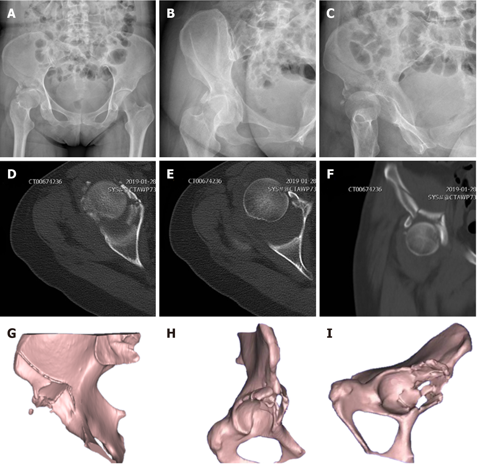
Standard radiographs at admission revealed fractures of the right acetabular anterior wall in association with fractures of the AIIS and the iliac wing, with preservation of the pelvic brim (Figure 1). Computed tomography showed fractures of the acetabular anterior wall, the AIIS, and the iliac wing with retained integrity of the pelvic brim (Figure 1).
The patient underwent interim management by closed reduction of the hip dislocation and skin traction for 6 d.
For the treatment of right atypical acetabular anterior wall fracture and iliac fracture, open reduction and rigid internal fixation were performed via a modified pararectus approach under general anesthesia.
The surgery was performed on the 7th day following the traumatic injury. Under general anesthesia, the patient was placed supine on a radiolucent operating table and the ipsilateral leg was prepared and draped freely, facilitating reduction maneuvers. To reduce the tension of the iliopsoas muscle and protect the femoral nerve, hip joints were slightly flexed by putting a bump underneath both knees. In order to monitor potential iatrogenic bladder injury during surgery, a Foley catheter was placed in the bladder before surgery.
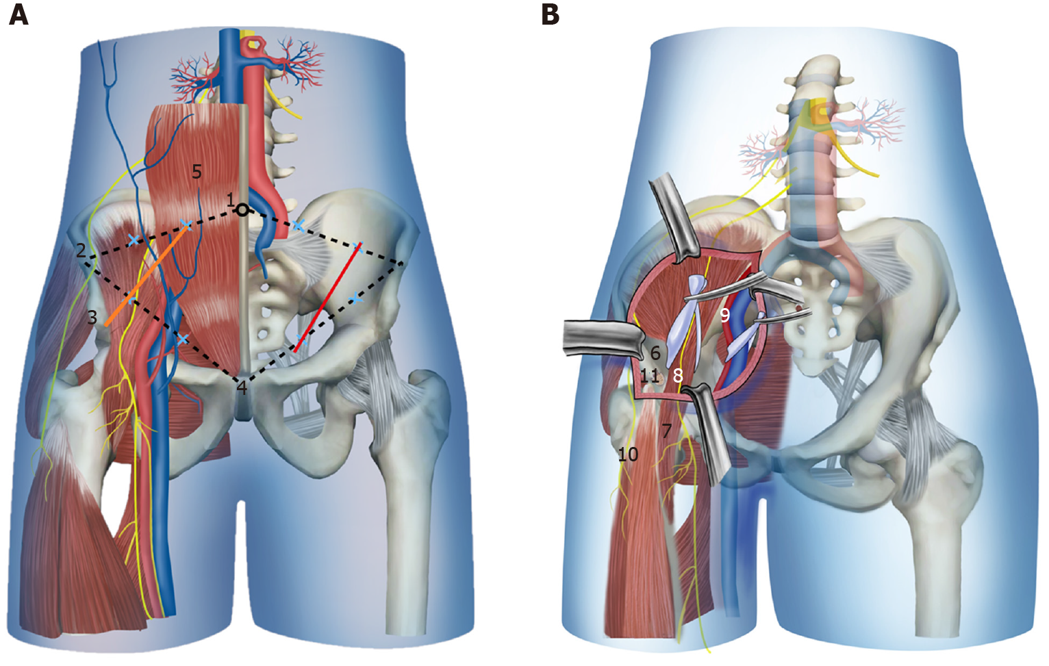
A modified pararectus approach was used, in which the skin incision was started from the junction of the medial and middle thirds of the line connecting the ASIS with the umbilicus to the junction of the middle and lateral thirds of the line connecting the symphysis with the ASIS, and extended distally-laterally to the AIIS (Figure 2A).
To facilitate exposure of the anterior layer of rectus sheath, the subcutaneous fatty tissue, fascia of Camper, fascia of Scarpa, and external oblique muscle aponeurosis were dissected. The incision of the anterior fascial layer of rectus sheath and the transversalis fascia was made to expose the extraperitoneal space in the true pelvis, with care taken to identify the round ligament and the inferior epigastric vessels. The expanded first window of pararectus approach[8] was developed between the iliac crest and iliopsoas muscle by dissecting the iliacus muscle along the inner surface of the iliac crest and dragging it medially. By retracting the iliopsoas muscle laterally and the external iliac vessels medially, the second window[8] was created. Through the expanded first and second windows, both the anterior wall and anterior column could be seen (Figure 2B).
Once the approach was complete, the fracture fragments were identified, namely, two main fragments of the anterior wall with superior extension of fracture above the iliopectineal line into iliac wing, and a fragment of AIIS that was attached by the direct head of the rectus femoris tendon. The reflected head of the rectus femoris tendon was released to allow mobilization of the fragment. Manual distraction was performed by placing a subtrochanteric Schanz screw parallel to the femoral neck, which was then used to visualize the joint after incision through the hip capsule, and the articular impaction was addressed.
The fracture fragments were anatomically reduced under direct visualization, and the cancellous bone void left beneath the elevated impacted fragments was filled with allograft. Interfragmentary lag screws were used to fix the AIIS and iliac wing. The fixation of anterior wall was achieved by using a nonlocking, precontoured, low-profile reconstruction plate and a nonlocking T-shape plate. The T-shape plate was situated near the border of the acetabulum, and was slightly under-bent to enable compressing and buttressing of the fragments. Intraoperative fluoroscopy was used to evaluate the reduction and position of the implants. The hip capsule and rectus release were repaired, and the anterior layer of rectus sheath, subcutaneous tissue, and skin were sutured sequentially. A suction drain was placed for 24 h.
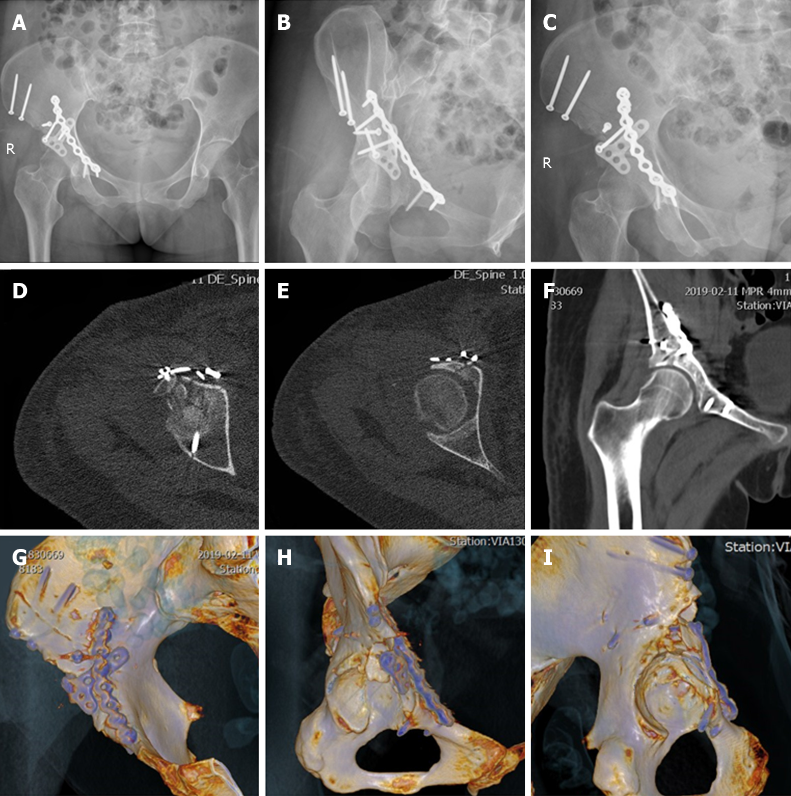
Prophylactic intravenous antibiotic was used from 30 min before surgery to 24 h postoperatively. Rivaroxaban was employed for 5 wk as prophylaxis against deep venous thromboembolism. The patient was started on muscle strengthening as soon as medically possible but with restricted mobilization to toe-touch weight-bearing for the first 3 mo. The postoperative X-rays and computed tomography scans revealed good reduction of the fracture (Figure 3).
Right acetabular anterior wall fracture with preservation of the pelvic brim, associated with fracture of the AIIS and the iliac wing.
The patient was treated by open reduction and internal fixation with interfragmentary lag screws, reconstruction plate and T-shape plate via a modified pararectus approach.
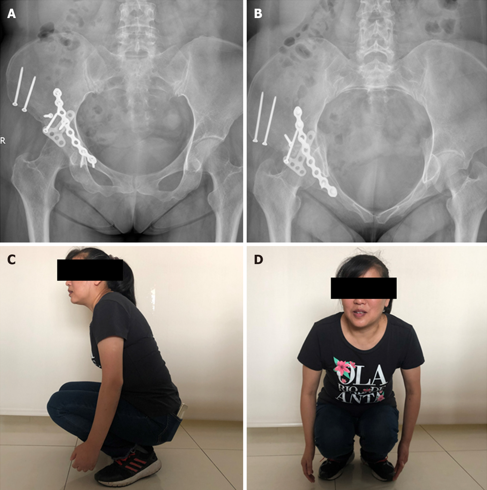
The patient was followed monthly. At 9 mo postoperatively, an X-ray showed that the fracture lines were blurred. The patient was satisfied with the function of the injured hip (Figure 4).
Acetabular anterior wall fractures with preservation of the pelvic brim are very uncommon. As far as we know, there are only ten cases reported previously in the literature[3,4,9]. Mirovsky et al[9] reported an acetabular anterior wall fracture with anterior hip dislocation treated by hip arthroplasty in a 67-year-old woman. Badelon et al[10] and Piriou et al[3] reported three similar cases, but they were treated by open reduction and internal fixation. Lenarz et al[4] reported a series of anterior acetabula fracture without involvement of the pelvic brim, and defined the special form of acetabular anterior wall fracture as atypical anterior wall fracture of the acetabulum.
Previously reported cases and the present case have common features. The lesions, in all cases, included the anterior wall of the acetabulum with preservation of the pelvic brim, unlike the anterior wall fracture described in the Judet-Letournel classification. The anteriorsuperior part of the acetabulum, rather than the entire roof, was fractured, differing from the anterior wall fracture described in the Judet-Letournel classification[2]. Furthermore, the fractures extended to the AIIS in all cases and even the interspinous region in some cases. The indications for surgical treatment of acetabular fracture are incongruent or unstable fractures with a dislocation verified to be greater than 5 mm[5], so that no alternative treatment to surgery was considered in our case.
The operative approach should provide access to the anterior column, anterior wall, AIIS, and hip joint for direct reduction and osteosynthesis. According to Tannast’s study about survivorship of the hip in 810 patients with operatively treated acetabular fractures, anterior wall fractures had the worst survivorship, which might be related to marginal impaction and nonanatomical articular reduction associated with limited exposure[11]. Because the Smith-Petersen approach could provide exposure of the anterior column and wall extending from the inner table to the outer table, and a complete intra-articular view to judge reduction and address articular impaction[6], it was employed for open reduction and rigid internal fixation in all of the nine cases previously reported. The ilioinguinal approach, which is often used as a recommended treatment for anterior lesions, is absolutely incapable of treating such fractures because of limited potential for intraarticular manipulation through fracture line. Despite the excellent exposure of the Smith-Petersen approach for anterior acetabular fractures, it requires detachment of iliacus muscle, release of rectus femoris muscle, and even release of the attachment of the gluteus medius muscle or osteotomy of the ASIS for more extensile exposure.
The modified pararectus approach described here combines advantages of the pararectus approach and Smith-Petersen approach. It provides access to the anterior column and wall as with the pararectus approach but with less invasive tissue dissection[8]. Furthermore, by changing the orientation of the skin incision towards the AIIS, the anterior impacted articular lesion could be manipulated under direct vision, after retracting fractured AIIS. Although the modified pararectus approach provides a less exposed area of intra-articular surface and outer table of iliac wing than the Smith-Petersen approach, it offered adequate view and operation space in our case and prevented further damage to the iliac wing from osteotomy.
The major advantage of the modified pararectus approach compared with the original pararectus approach is the enlarged first and second windows, so that fractures of the low anterior column with marginal impaction can be well operated. On the other hand, more dissection of subcutaneous tissue is needed, and a smaller range of the superior ramus of the pubis is exposed.
We report a rare acetabular anterior wall fracture with preservation of the pelvic brim, but consisting of the AIIS and iliac wing in a 48-year-old woman. This atypical acetabular anterior wall fracture is different from the Judet-Letournel classification in terms of morphology and surgical procedure. A modified pararectus approach was used in our case, which could facilitate exposure, reduction, and osteosynthesis for atypical acetabular fracture with less invasiveness.
Manuscript source: Unsolicited manuscript
Specialty type: Medicine, research and experimental
Country/Territory of origin: China
Peer-review report’s scientific quality classification
Grade A (Excellent): 0
Grade B (Very good): B
Grade C (Good): 0
Grade D (Fair): 0
Grade E (Poor): 0
P-Reviewer: Aprato A S-Editor: Zhang L L-Editor: Wang TQ E-Editor: Ma YJ
| 1. | Judet R, Judet J, Letournel E. Fractures of the acetabulum: Classification and surgical approaches for open reduction. J Bone Joint Surg Am. 1964;46:1615-1646. [RCA] [PubMed] [DOI] [Full Text] [Cited by in Crossref: 800] [Cited by in RCA: 730] [Article Influence: 25.2] [Reference Citation Analysis (0)] |
| 2. | Letournel E. Acetabulum fractures: classification and management. Clin Orthop Relat Res. 1980;151:81-106. [PubMed] |
| 3. | Piriou P, Siguier T, De Loynes B, Charnley G, Judet T. Anterior wall acetabular fractures: report of two cases and new strategies in operative management. J Trauma. 2002;53:553-557. [RCA] [PubMed] [DOI] [Full Text] [Cited by in Crossref: 9] [Cited by in RCA: 8] [Article Influence: 0.3] [Reference Citation Analysis (0)] |
| 4. | Lenarz CJ, Moed BR. Atypical anterior wall fracture of the acetabulum: case series of anterior acetabular rim fracture without involvement of the pelvic brim. J Orthop Trauma. 2007;21:515-522. [RCA] [PubMed] [DOI] [Full Text] [Cited by in Crossref: 11] [Cited by in RCA: 12] [Article Influence: 0.7] [Reference Citation Analysis (0)] |
| 5. | Grubor P, Krupic F, Biscevic M, Grubor M. Controversies in treatment of acetabular fracture. Med Arch. 2015;69:16-20. [RCA] [PubMed] [DOI] [Full Text] [Full Text (PDF)] [Cited by in Crossref: 14] [Cited by in RCA: 22] [Article Influence: 2.2] [Reference Citation Analysis (0)] |
| 6. | Lefaivre KA, Starr AJ, Reinert CM. A modified anterior exposure to the acetabulum for treatment of difficult anterior acetabular fractures. J Orthop Trauma. 2009;23:370-378. [RCA] [PubMed] [DOI] [Full Text] [Cited by in Crossref: 11] [Cited by in RCA: 10] [Article Influence: 0.6] [Reference Citation Analysis (0)] |
| 7. | Mears DC, Gordon RG. Internal fixation of acetabular fractures. Techniques in Orthopaedics. 1990;4:36-51. [RCA] [DOI] [Full Text] [Cited by in Crossref: 6] [Cited by in RCA: 6] [Article Influence: 0.2] [Reference Citation Analysis (0)] |
| 8. | Keel MJB, Siebenrock KA, Tannast M, Bastian JD. The Pararectus Approach: A New Concept. JBJS Essent Surg Tech. 2018;8:e21. [RCA] [PubMed] [DOI] [Full Text] [Full Text (PDF)] [Cited by in Crossref: 23] [Cited by in RCA: 42] [Article Influence: 6.0] [Reference Citation Analysis (0)] |
| 9. | Mirovsky Y, Fischer S, Hendel D, Halperin N. Traumatic anterior dislocation of the hip joint with fracture of the acetabulum: a case report. J Trauma. 1988;28:1597-1599. [RCA] [PubMed] [DOI] [Full Text] [Cited by in Crossref: 6] [Cited by in RCA: 9] [Article Influence: 0.2] [Reference Citation Analysis (0)] |
| 10. | Badelon O, Leroux D, Huten D, Duparc J. [Anterior luxation of the hip associated with a fracture of the anterior aspect of the acetabulum. Apropos of a case]. Ann Chir. 1986;40:38-40. [PubMed] |
| 11. | Tannast M, Najibi S, Matta JM. Two to twenty-year survivorship of the hip in 810 patients with operatively treated acetabular fractures. J Bone Joint Surg Am. 2012;94:1559-1567. [RCA] [PubMed] [DOI] [Full Text] [Cited by in Crossref: 235] [Cited by in RCA: 285] [Article Influence: 21.9] [Reference Citation Analysis (0)] |


