Published online Mar 26, 2019. doi: 10.12998/wjcc.v7.i6.773
Peer-review started: January 10, 2019
First decision: January 19, 2019
Revised: January 31, 2019
Accepted: February 18, 2019
Article in press: February 18, 2019
Published online: March 26, 2019
Processing time: 75 Days and 20.9 Hours
Crossed fused renal ectopia is a rare congenital anomaly of the ascent of the kidney. This anomaly may be observed as a solitary kidney during initial evaluation. A solitary kidney must be evaluated for associated anomalies such as duplication, horseshoe kidney, or crossed renal ectopia.
An anomaly was observed in a 9-mo-old male child who was subsequently diagnosed with crossed fused renal ectopia and vesicoureteral reflux (VUR). In this condition, recurrent febrile urinary tract infection can be a serious problem, and can easily cause renal damage due to relatively short ureters and high pressure in the kidney.
To prevent urosepsis and preserve renal function, early diagnosis and proper management including surgical correction should be considered for the management of renal ectopia with VUR.
Core tip: Crossed fused renal ectopia is a rare congenital anomaly in pediatric urology. The majority of the cases shows a favorable prognosis with conservative management, while early surgical intervention should be considered in selective cases. Up to now, the guideline for the disease has not established, and this case may be beneficial to determine therapeutic plan, especially for the necessity of surgery. Furthermore, the helpful diagnostic and therapeutic methods are mentioned based on our experience.
- Citation: Choi T, Yoo KH, Song R, Lee DG. Lump type crossed fused renal ectopia with bilateral vesicoureteral reflux: A case report. World J Clin Cases 2019; 7(6): 773-777
- URL: https://www.wjgnet.com/2307-8960/full/v7/i6/773.htm
- DOI: https://dx.doi.org/10.12998/wjcc.v7.i6.773
Crossed fused renal ectopia is a rare anomaly characterized by the ectopic kidney crossing the midline with the ureter at the ureterovesical junction (UVJ) in the orthotopic position. Vesicoureteral reflux (VUR) is a relatively common urologic disorder, and is traditionally categorized as primary or secondary according to its etiology. VUR is no longer thought to be a disease in itself; it is now considered a marker of heterogeneous conditions of the entire urinary tract, including congenital renal hypoplasia and dysplasia, primary reflux caused by an incompetent UVJ, altered lower urinary tract function, and inherent predisposition to urinary tract infection (UTI). It poses many challenges to the urologist for diagnosis and treatment.
Generally, renal fusion anomalies are classified into two groups: horseshoe kidney and crossed fused ectopia. Crossed fused ectopic kidney is a rare anomaly of the ascent of the kidney, which can be associated with VUR, but has not been reported widely.
We report our experience with a male neonate who suffered from recurrent UTI due to crossed fused renal ectopia, accompanied by bilateral VUR.
A 1-mo-old male child was referred to the department of urology for evaluation and management of abnormal prenatal ultrasonography (US) findings.
Prenatal US had shown an empty renal fossa, and a pelvic kidney suggestive of renal ectopia with agenesis had been observed.
The patient’s gestational period was 38 + 2 wk, with birth weight 2520 g, and perinatal abnormalities such as meconium staining or premature rupture of membranes were not present. There was not any significant risk factor or family history.
Physical examination was unremarkable.
Urinalysis demonstrated significant pyuria.
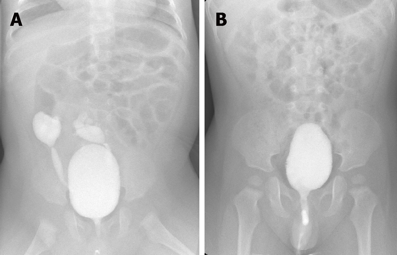
Technetium-99m dimercaptosuccinic acid (DMSA) scan demonstrated sufficient uptake indicating a single ectopic right kidney with normal function, with left renal agenesis, which was consistent with the US findings. Voiding cystourethrogram (VCUG) was performed for possible associated VUR, and showed 2 dilated ureters with VUR (right: grade IV, left: grade IV) leading out from a fused kidney situated at the right side of the lower abdomen.
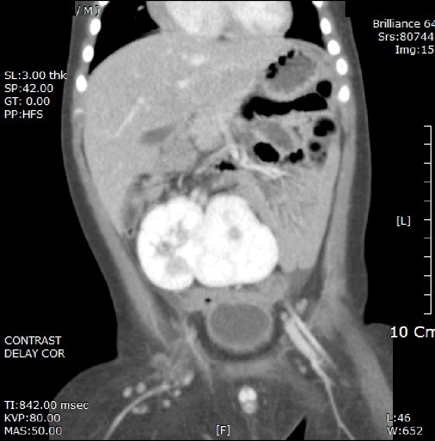
A second breakthrough infection occurred at 9 mo of age and abdominal contrast computed tomography (CT) was performed for further evaluation. It showed the left kidney crossing the midline to fuse to the right kidney situated in the right abdomen, from the aortic bifurcation to the iliac crest. Both renal hila faced to the front, indicative of an associated rotational anomaly. The right kidney was supplied by the right renal artery and a branch of the left common iliac artery, while the left kidney was fed by only the left renal artery. Pyelonephritis that had not been observed previously was noted (Figures 1 and 2).
We diagnosed crossed fused renal ectopia (“lump” type) with associated bilateral VUR.
Initially, continuous low-dose antibiotic prophylaxis was started. Despite antibiotic prophylaxis, he was readmitted through the emergency room 3 months later because of a febrile UTI.
In order to prevent these repeated events of infection, we decided to perform a bilateral ureteroneocystostomy to manage VUR. During the operation, we observed both ureters to be very short, particularly on the left side, as expected preoperatively. The length of the ureters was insufficient, and therefore the Politano-Leadbetter technique was carried out instead of the Cohen procedure.
There was no significant postoperative complication and the patient was discharged after a week. No abnormal findings were reported on VCUG three months after surgery, and the patient had no febrile UTI or other complications during the follow-up period of six years. As a biennial routine follow-up, serum creatinine was 0.78 mg/dL and US showed no evidence of hydronephrosis.
Crossed fused renal ectopia is a rare congenital abnormality of the urinary tract. It is the second most common fusion anomaly, with an incidence placed at 1 in 2000 autopsies, with male predominance (3:2)[1]. About 2% of the anomalies are complete crossed fused ectopia, with either lump or disc kidneys. The ectopic kidney crosses the midline to lie on the opposite side from its ureteral insertion into the bladder. Most cases of crossed renal ectopia are discovered incidentally, often by antenatal sonography when two kidneys are not identified. US evaluation of the newborn infant may underestimate the hydronephrosis and dilated ureter, due to physiologic dehydration[2]. The typical US findings in crossed fused renal ectopia include an anterior/posterior notch with distinguishable orientation of the collecting systems in the kidneys. In addition, US can provide important information on the presence of abnormal vasculature, hydronephrosis, or urolithiasis. CT is also useful to demonstrate the precise anatomy and provide functional information about such anomalies. A DMSA radionuclide scan is used in evaluating renal morphology and structure.
Patients may remain asymptomatic throughout life, but symptoms may occur due to minor trauma associated with the abnormal location. They may have abdominal or flank pain, a palpable mass, or dysuria. When symptoms do occur, they are often related to infection, obstruction, or urolithiasis. One previous study reported that about one-third of patients had a history of pyelonephritis and one-quarter had hydronephrosis[3]. The life-long clinical course in the aspect of renal function is still remained unclear.
There are six different varieties of crossed ectopia with fusion. The most common form is unilateral fused type with inferior ectopia, in which the upper pole of the crossed kidney is fused to the lower pole of the normally positioned kidney. The second most common type is the sigmoid, or S-shaped, kidney. The crossed kidney is inferior, but the two renal pelvises face in opposite directions. The lump, or “cake,” kidney and the “disc” kidney both involve extensive fusion of the two renal masses. With an L-shaped kidney, the crossed kidney assumes a transverse position. With a superior ectopic kidney, the least common type is the crossed ectopic kidney, which lies superior to the normal kidney. Among these, only the unilateral lump kidney and disc kidney are completely fused. In our case, the fusion corresponded to the lump type crossed fused ectopic kidney. Pannorlus first described this condition as an extreme variant of horseshoe kidney in 1654[4].
It has been suggested that there is a significant correlation between genitourinary abnormalities and malformations such as musculoskeletal, gastrointestinal, and cardiovascular anomalies[5]. For this reason, early and complete evaluation is needed for the patient with crossed renal ectopia.
One of the most common associated abnormalities is VUR, which is frequently noted in the ectopic kidney. The rate of spontaneous resolution was about 35%-40% in one-year-old infants regardless of the VUR grades[6]. Surgical correction can be considered in certain circumstances that patient has a history of febrile UTI, worsening hydronephrosis, abnormal kidney function on renal scan, or preference for surgical treatment. The predictive factors of surgery included older age at initial diagnosis, the presence of antenatal hydronephrosis, bilateral and high grade VUR in a large-scaled cohort study[7]. Despite hopeful chance of spontaneous VUR resolution based on the previous studies, we predicted little chance of spontaneous resolution because of the complicated VUR with crossed fused kidney. And early surgical repair was considered and performed. Previous study demonstrated that VUR occurred in 20% of crossed renal ectopy, 30% of simple renal ectopy, and 70% of bilateral simple renal ectopy cases[8]. Less common problems include ureteropelvic junction obstruction, renal dysplasia, and renal tumors.
In our case, the Politano-Leadbetter technique was used to correct VUR instead of the Cohen procedure, due to the short ureters. In the Politano-Leadbetter technique, we create a long tunnel and perform retrograde catheterization via an intact ureteral orifice. However, making a new cephalad hiatus is challenging for the operating surgeon. The Paquin technique is another common option for the management of similar cases. It has an advantage over the Politano-Leadbetter technique because it is performed under direct vision, thus reducing the risk of peritoneal injury.
Crossed fused renal ectopia is often misdiagnosed as a solitary kidney. As suggested above, urologists should be aware of such anomalies. The length of the ureter of the ectopic kidney is usually shorter than normal, and that kidney appears to sustain more pressure with VUR compared with the normal state; this causes renal damage more easily. Early diagnosis and proper management of renal ectopia with VUR are necessary. During surgery, the Politano-Leadbetter or Paquin technique may be preferred if there is insufficient length of the ureters.
Manuscript source: Unsolicited manuscript
Specialty type: Medicine, research and experimental
Country of origin: South Korea
Peer-review report classification
Grade A (Excellent): 0
Grade B (Very good): B
Grade C (Good): C, C
Grade D (Fair): D
Grade E (Poor): 0
P-Reviewer: Markic D, Stavroulopoulos A, Tanaka H, Yorioka N S-Editor: Ji FF L-Editor: A E-Editor: Wu YXJ
| 1. | Patel TV, Singh AK. Crossed fused ectopia of the kidneys. Kidney Int. 2008;73:662. [RCA] [PubMed] [DOI] [Full Text] [Cited by in Crossref: 22] [Cited by in RCA: 27] [Article Influence: 1.6] [Reference Citation Analysis (0)] |
| 2. | Hains DS, Bates CM, Ingraham S, Schwaderer AL. Management and etiology of the unilateral multicystic dysplastic kidney: a review. Pediatr Nephrol. 2009;24:233-241. [RCA] [PubMed] [DOI] [Full Text] [Cited by in Crossref: 108] [Cited by in RCA: 82] [Article Influence: 5.1] [Reference Citation Analysis (0)] |
| 3. | Abeshouse BS, Bhisitkul I. Crossed renal ectopia with and without fusion. Urol Int. 1959;9:63-91. [RCA] [PubMed] [DOI] [Full Text] [Cited by in Crossref: 97] [Cited by in RCA: 76] [Article Influence: 2.8] [Reference Citation Analysis (0)] |
| 4. | Kaufman MH, Findlater GS. An unusual case of complete renal fusion giving rise to a 'cake' or 'lump' kidney. J Anat. 2001;198:501-504. [RCA] [PubMed] [DOI] [Full Text] [Cited by in Crossref: 20] [Cited by in RCA: 22] [Article Influence: 0.9] [Reference Citation Analysis (0)] |
| 5. | Rai AS, Taylor TK, Smith GH, Cumming RG, Plunkett-Cole M. Congenital abnormalities of the urogenital tract in association with congenital vertebral malformations. J Bone Joint Surg Br. 2002;84:891-895. [RCA] [PubMed] [DOI] [Full Text] [Cited by in RCA: 9] [Reference Citation Analysis (0)] |
| 6. | Wildbrett P, Schwebs M, Abel JR, Lode H, Barthlen W. Spontaneous vesicoureteral reflux resolution in children: A ten-year single-centre experience. Afr J Paediatr Surg. 2013;10:9-12. [RCA] [PubMed] [DOI] [Full Text] [Cited by in Crossref: 6] [Cited by in RCA: 9] [Article Influence: 0.8] [Reference Citation Analysis (0)] |
| 7. | Szymanski KM, Oliveira LM, Silva A, Retik AB, Nguyen HT. Analysis of indications for ureteral reimplantation in 3738 children with vesicoureteral reflux: a single institutional cohort. J Pediatr Urol. 2011;7:601-610. [RCA] [PubMed] [DOI] [Full Text] [Cited by in Crossref: 11] [Cited by in RCA: 11] [Article Influence: 0.8] [Reference Citation Analysis (0)] |
| 8. | Guarino N, Tadini B, Camardi P, Silvestro L, Lace R, Bianchi M. The incidence of associated urological abnormalities in children with renal ectopia. J Urol. 2004;172:1757-9; discussion 1759. [RCA] [PubMed] [DOI] [Full Text] [Cited by in Crossref: 83] [Cited by in RCA: 69] [Article Influence: 3.3] [Reference Citation Analysis (0)] |


