Copyright
©The Author(s) 2019.
World J Clin Cases. Oct 6, 2019; 7(19): 3126-3131
Published online Oct 6, 2019. doi: 10.12998/wjcc.v7.i19.3126
Published online Oct 6, 2019. doi: 10.12998/wjcc.v7.i19.3126
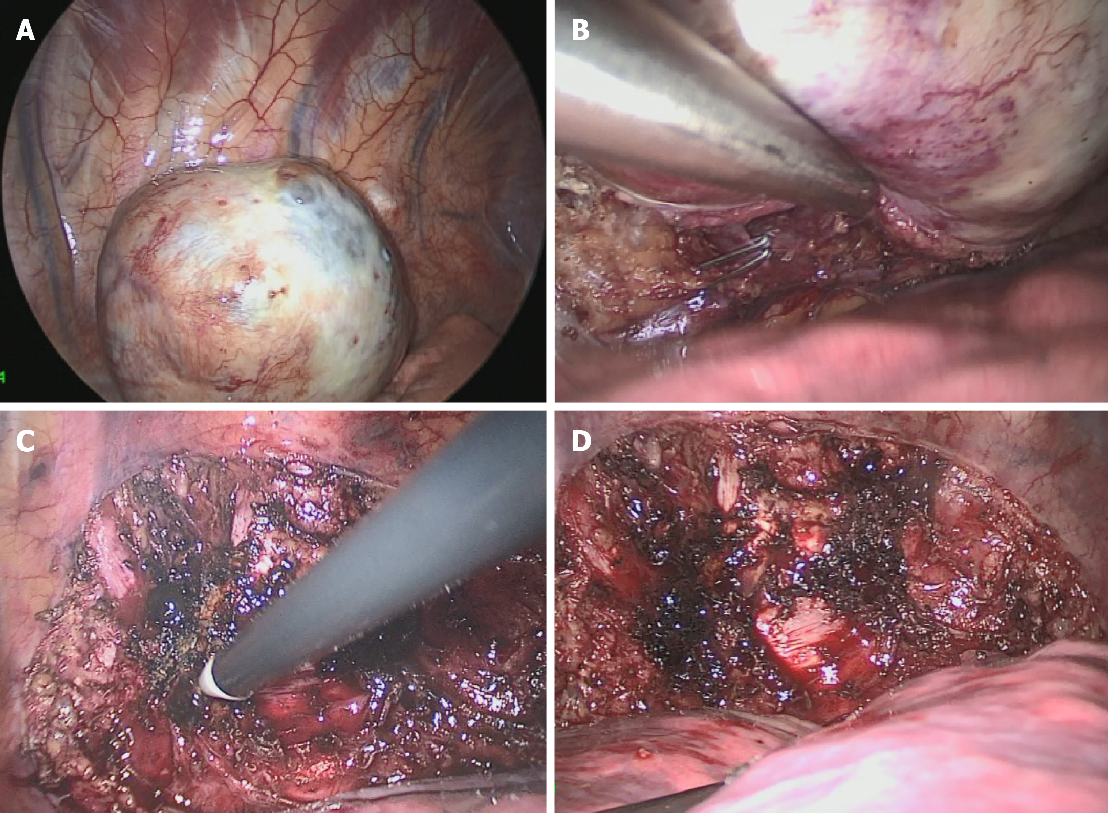
Figure 2 A video-assisted thoracoscopic surgery was performed to remove the mass.
A: The tumor located on the posterior chest wall nearby the 9th thoracic vertebra; B: The drain vein to the hemiazygos vein was ligated by hemoclips; C: The tumor was dissected carefully by the electrocautery and Harmonic in turns; D: The base plane on the chest wall was deal with the electrocoagulation.
- Citation: Chen YT, Yang Z, Li H, Ni CH. Clear cell sarcoma of soft tissue in pleural cavity: A case report. World J Clin Cases 2019; 7(19): 3126-3131
- URL: https://www.wjgnet.com/2307-8960/full/v7/i19/3126.htm
- DOI: https://dx.doi.org/10.12998/wjcc.v7.i19.3126


