Copyright
©The Author(s) 2019.
World J Clin Cases. May 26, 2019; 7(10): 1133-1141
Published online May 26, 2019. doi: 10.12998/wjcc.v7.i10.1133
Published online May 26, 2019. doi: 10.12998/wjcc.v7.i10.1133
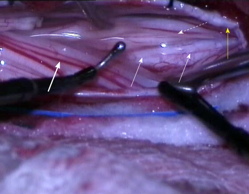
Figure 3 The intraoperative separation of motor and sensory roots and the identification of the groove that divides the roots anatomically.
The electrophysiological probe can be seen in place (right) as well as the groove on conus medullaris, separating the both groups of roots (thin arrows). The thick arrow indicates sensory roots and the dotted arrow indicates motor roots. The yellow arrow indicates the dural rim, held in place by tack-up sutures.
- Citation: Velnar T, Spazzapan P, Rodi Z, Kos N, Bosnjak R. Selective dorsal rhizotomy in cerebral palsy spasticity - a newly established operative technique in Slovenia: A case report and review of literature. World J Clin Cases 2019; 7(10): 1133-1141
- URL: https://www.wjgnet.com/2307-8960/full/v7/i10/1133.htm
- DOI: https://dx.doi.org/10.12998/wjcc.v7.i10.1133


