Published online Oct 26, 2018. doi: 10.12998/wjcc.v6.i12.538
Peer-review started: July 4, 2018
First decision: August 20, 2018
Revised: September 13, 2018
Accepted: October 10, 2018
Article in press: October 10, 2018
Published online: October 26, 2018
Processing time: 114 Days and 23.8 Hours
Colovesical fistulas (CVFs) are rare complications of very advanced cancers of the abdominal or pelvic cavity and often cause diagnostic troubles. CVFs are found more often in males, whereas females usually suffer from rectovaginal or vesicovaginal fistulas. This article presents a case of a female patient who was admitted to the hospital because of acute diarrhea, presumably of infectious origin, and with only subtle abnormalities in blood tests and urinalysis. Owing to the ineffectiveness of the performed treatment and progressive intensification of symptoms, diagnostics were extended to include a computed tomography scan, sigmoidoscopy and cystography. The imaging results revealed a large heterogeneous conglomerate of solid and fluid structures in the pelvis, which involved reproductive organs, the bladder and sigmoid colon. The excrement leaking from the digestive tract was urine, and CVF was the first manifestation of colon cancer. Shortly after the final diagnosis, the patient deteriorated and eventually died after an urgent colostomy was performed because of a bowel obstruction.
Core tip: Colovesical fistulas are a rare complication of very advanced cancers of the abdominal or pelvic cavity. This article presents a case of a female patient whose first manifestation of colon cancer was a misleading watery diarrhea, which was later found to be urination through a pathologic junction between the bladder and distal colon.
- Citation: Skierucha M, Barud W, Baraniak J, Krupski W. Colovesical fistula as the initial manifestation of advanced colon cancer: A case report and review of literature. World J Clin Cases 2018; 6(12): 538-541
- URL: https://www.wjgnet.com/2307-8960/full/v6/i12/538.htm
- DOI: https://dx.doi.org/10.12998/wjcc.v6.i12.538
Colovesical fistulas (CVFs) are rare medical findings and mostly represent complications of diverticulitis, cancer, or Crohn’s disease. Advanced tumors, which grow abundantly in the abdominal or pelvic cavity, are responsible for approximately 20%-30% of CVFs[1]. Most frequently, CVFs originate from colon adenocarcinomas but may also occur as a consequence of neoplasms in other pelvic organs[2,3]. In any case, CVFs are observed at very advanced stages of neoplastic diseases[4,5].
This article presents a case of a patient with late manifestations and a rapid course of an advanced colon cancer. The first symptom of the disease was urination through a pathologic junction between the bladder and distal colon, which was misinterpreted as diarrhea.
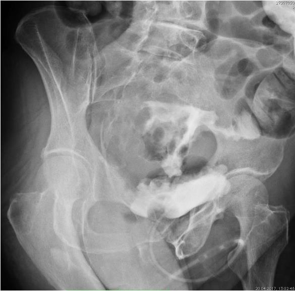
A female aged 81, previously healthy, physically fit and active was admitted to the hospital because of diarrhea, which had intensified within a few preceding days, despite maintaining a complete diet. On admission, the patient presented with a mild but recurring fever, abdominal cramps, nausea and a lack of appetite. The abnormalities in laboratory blood tests, performed upon the admission, were as follows: low potassium (2.68 mmol/L) and albumin (2.1 g/dL) levels, leukocytosis with a predominance of granulocytes (18.52 × 10⁹/L) and an increased level of C-reactive protein (191.8 mg/L; norm: < 5 mg/L). Urinalysis revealed mild bacteriuria and leukocyturia. An abdominal ultrasound showed mild hepatic steatosis with multiple solid, hyperechoic lesions in the entire liver, which were described as simple cysts (the largest measuring 20 mm). Initially, the patient was treated for diarrhea and urinary tract infection. After a week of ineffective antibiotic therapy, ruling out the initial diagnosis of infectious diarrhea, the patient was constantly deteriorating, and the symptoms of abdominal pain, massive watery diarrhea and nausea were progressively intensifying. The patient had no control urinalysis because of defecating to incontinence pads and a high possibility of contamination. She refused to be catheterized. An abdominal computed tomography scan revealed a 112 mm × 78 mm × 10 mm heterogeneous conglomerate of solid and fluid structures, which presumably originated from some gynecologic or colon cancer and involved reproductive organs, the bladder and sigmoid colon (Figure 1). Locoregional lymph nodes were irregularly enlarged to approximately 20 mm and formed large masses, suggestive of peritoneum involvement. Moreover, there were numerous hypodense solid lesions in the liver and both adrenal glands, as well as one in the right kidney cortex, all suggestive of metastases. By sigmoidoscopy, 10 cm from the anal verge, an irregular infiltrating mass and leakage of a colorless fluid were found, which were suggestive of CVF. The diagnosis was confirmed by cystography (Figure 2). While undergoing to all the diagnostic examinations, the patient collapsed, and her condition severely worsened. She started vomiting feces, and after consultation with a surgeon, she was qualified for an urgent colostomy because of a bowel obstruction. The patient died within few hours after the surgery. The postmortem pathology diagnosis was a G3 poorly differentiated colorectal adenocarcinoma.
From the literature, it seems that fistulas between the bladder and digestive tract are uncommon and often cause diagnostic troubles. The patients are mostly characterized by recurrent urinary tract infection, dysuria, fecaluria, and pneumaturia and less often by intestinal abnormalities such as diarrhea, melena and hematemesis[6]. Electrolyte and acid-base disturbances depend on the localization of the pathologic junction. Enterovesical fistulas cause pathological reuptake of electrolytes[4,7]. In cases of CVFs, cells of the colon epithelium exchange bicarbonates for ammonium and chloride ions[8]. CVFs tend to cause severe physical complications, and in most cases, patients end up with a close death[4,9-11].
Wei et al[6] have described a case of a patient with CVF, whose symptoms were comparable to those described in our case, i.e., chronic diarrhea with only mild urinary tract symptoms. However, their patient was a male, and the outstanding feature of our case is that the patient was a female. CVFs are more likely to occur in males, whereas females usually suffer from rectovaginal or vesicovaginal fistulas.
The presented patient experienced an unexpectedly late manifestation of a very advanced cancer. Supposedly, she must have had some alarming symptoms before the admission to the hospital, but they were not troublesome enough to make her seek any help. The bothering problems started from the perforation of the tumor and the formation of a fistula that caused a misleading urine passage through the digestive tract, which appeared as diarrhea. If control urinalysis was performed, the diagnosis would have been made a few days earlier. In this particular case, an earlier diagnosis would not have probably changed the course of events. Nevertheless, the most basic test, a urinalysis, might have been as informative as were the complicated, time-consuming and expensive radiological examinations. The concluding remark from the case is that the excrement from the digestive tract may not always be only feces, and diarrhea may be a misleading symptom of CVF.
Misinterpretation of diarrhea in a female patient with a colovesical fistula (CVF) due to an advanced colon cancer.
Acute diarrhea, recurring fever, abdominal cramps, nausea and a lack of appetite.
Infectious diarrhea.
Hypokalemia, hypoalbuminemia, leukocytosis with a predominance of granulocytes and elevated C-reactive protein. Bacteriuria and leukocyturia in urinalysis.
A computed tomography scan revealed a heterogeneous tumor, enlarged lymph nodes in the pelvis and metastases in the liver and peritoneum. Cystography showed a CVF.
G3 poorly differentiated colorectal adenocarcinoma.
An urgent colostomy conducted because of a bowel obstruction.
Wei et al have described a case of a male patient with CVF, whose symptoms were comparable to those described in our case. However, to our limited knowledge, this is the first case report describing CVF in a female.
CVF is a pathologic junction between the bladder and colon. Approximately 20%-30% of CVFs develop as complications of advanced tumors of the abdominal or pelvic cavity.
Diarrhea may be a misleading symptom of CVF. A basic test such as urinalysis should never be neglected. In our case, repeated urinalysis may have been very informative. Patients with diarrhea of unknown reason should be catheterized to control the renal loss of fluids and the quality of urine.
CARE Checklist (2013) statement: The authors have read the CARE Checklist (2013), and the manuscript was prepared and revised according to the CARE Checklist (2013).
Manuscript source: Invited manuscript
Specialty type: Medicine, research and experimental
Country of origin: Poland
Peer-review report classification
Grade A (Excellent): 0
Grade B (Very good): B
Grade C (Good): C
Grade D (Fair): 0
Grade E (Poor): 0
P- Reviewer: Yu S, To KKW S- Editor: Dou Y L- Editor: A E- Editor: Tan WW
| 1. | Golabek T, Szymanska A, Szopinski T, Bukowczan J, Furmanek M, Powroznik J, Chlosta P. Enterovesical fistulae: aetiology, imaging, and management. Gastroenterol Res Pract. 2013;2013:617967. [RCA] [PubMed] [DOI] [Full Text] [Full Text (PDF)] [Cited by in Crossref: 78] [Cited by in RCA: 93] [Article Influence: 7.8] [Reference Citation Analysis (0)] |
| 3. | McCaffrey JC, Foo FJ, Dalal N, Siddiqui KH. Benign multicystic peritoneal mesothelioma associated with hydronephrosis and colovesical fistula formation: report of a case. Tumori. 2009;95:808-810. [PubMed] |
| 4. | Bolmers MD, Linthorst GE, Soeters MR, Nio YC, van Lieshout JJ. Green urine, but no infection. Lancet. 2009;374:1566. [RCA] [PubMed] [DOI] [Full Text] [Cited by in Crossref: 7] [Cited by in RCA: 6] [Article Influence: 0.4] [Reference Citation Analysis (0)] |
| 5. | Yahagi N, Kobayashi Y, Ohara T, Suzuki N, Kiguchi K, Ishizuka B. Ovarian carcinoma complicated by sigmoid colon fistula formation: a case report and review of the literature. J Obstet Gynaecol Res. 2011;37:250-253. [RCA] [PubMed] [DOI] [Full Text] [Cited by in Crossref: 6] [Cited by in RCA: 7] [Article Influence: 0.5] [Reference Citation Analysis (0)] |
| 6. | Wei XQ, Zou Y, Wu ZE, Abassa KK, Mao W, Tao J, Kang Z, Wen ZF, Wu B. Acute diarrhea and metabolic acidosis caused by tuberculous vesico-rectal fistula. World J Gastroenterol. 2014;20:15462-15466. [RCA] [PubMed] [DOI] [Full Text] [Full Text (PDF)] [Cited by in CrossRef: 2] [Cited by in RCA: 1] [Article Influence: 0.1] [Reference Citation Analysis (0)] |
| 7. | Murakami K, Tomita M, Kawamura N, Hasegawa M, Nabeshima K, Hiki Y, Sugiyama S. Severe metabolic acidosis and hypokalemia in a patient with enterovesical fistula. Clin Exp Nephrol. 2007;11:225-229. [RCA] [PubMed] [DOI] [Full Text] [Cited by in Crossref: 9] [Cited by in RCA: 9] [Article Influence: 0.5] [Reference Citation Analysis (0)] |
| 8. | Pillinger T, Abdelrahman M, Jones G, D’Souza F. Intractable metabolic acidosis in a patient with colovesical fistula. N Z Med J. 2012;125:74-76. [PubMed] |
| 9. | Law WL, Chu SM. An unusual enterovesical fistula. Am J Surg. 2008;195:814-815. [RCA] [PubMed] [DOI] [Full Text] [Cited by in Crossref: 5] [Cited by in RCA: 5] [Article Influence: 0.3] [Reference Citation Analysis (0)] |
| 10. | Kim SJ, Kimoto Y, Nakamura H, Taguchi T, Tanji Y, Izukura M, Shiba E, Takai S. Ovarian carcinoma with fistula formation to the sigmoid colon and ileum: report of a case. Surg Today. 1999;29:449-452. [PubMed] |
| 11. | Bats AS, Rockall AG, Singh N, Reznek RH, Jeyarajah A. Perforation of a malignant ovarian tumor into the recto-sigmoid colon. Acta Obstet Gynecol Scand. 2010;89:1362-1363. [RCA] [PubMed] [DOI] [Full Text] [Cited by in Crossref: 6] [Cited by in RCA: 7] [Article Influence: 0.5] [Reference Citation Analysis (0)] |


