Published online Jul 16, 2014. doi: 10.12998/wjcc.v2.i7.293
Revised: April 21, 2014
Accepted: May 15, 2014
Published online: July 16, 2014
Processing time: 208 Days and 1.9 Hours
Focal epithelial hyperplasia (FEH), or Heck’s disease, is a rare disease of the oral mucosa; it is mostly found in children or young adults who are immunosuppressed and who live in regions with low socioeconomic status. It is characterized by asymptomatic papules on the oral mucosa, gingiva, tongue, and lips. Healing can be spontaneous, and treatment is indicated if there are aesthetic or functional complications. Human papillomavirus, especially genotypes 13 and 32, has been associated with FEH and is detected in the majority of lesions. Histopathologically, FEH is characterized by parakeratosis, epithelial hyperplasia, focal acanthosis, and fusion and horizontal outgrowth of epithelial ridges. A 37-year-old male patient was referred to the Department of Oral and Maxillofacial Sciences at the Sapienza University of Rome, complaining of numerous exophytic lesions in his mouth. He stated that the lesions were not painful but he had experienced occasional bleeding after incidental masticatory trauma. He had received no previous treatment for the oral lesions. His medical history revealed that he was human immuno-deficiency virus positive and was a smoker with numerous, asymptomatic oral papules clinically and histologically corresponding to FEH. The labial and buccal mucosa were especially affected by lesions. Surgical treatment was performed using a 532-nm potassium titanyl phosphate laser (SmartLite, Deka, Florence, Italy) in continuous mode with a 300 μm fiber and power of 1.4 W (power density 1980.22 W/cm2). After anesthesia without vasoconstrictors, the lesions were tractioned with sutures or an Allis clamp and then completely excised. The lesions were preserved in 10% formalin for histological examination, which confirmed the clinical diagnosis of FEH. In this case, the laser allowed excellent control of bleeding, without postoperative sutures, and optimal wound healing.
Core tip: Focal epithelial hyperplasia (FEH), or Heck’s disease, is a rare disease of the oral mucosa, characterized by asymptomatic papules in the oral cavity. Human papillomaviruses have been associated with FEH and have been detected in the majority of lesions. Histopathologically, FEH is characterized by parakeratosis, epithelial hyperplasia, and acanthosis. Here, the case of a 37-year-old male patient, human immuno-deficiency virus-positive, smoker, with numerous asymptomatic oral papules clinically and histologically corresponding to FEH is described. Surgical treatment was performed using a 532-nm potassium-titanyl-phosphate laser. In this case, the laser allowed excellent control of bleeding without postoperative sutures and optimal wound healing.
- Citation: Galanakis A, Palaia G, Tenore G, Vecchio AD, Romeo U. Focal epithelial hyperplasia in a human immuno-deficiency virus patient treated with laser surgery. World J Clin Cases 2014; 2(7): 293-296
- URL: https://www.wjgnet.com/2307-8960/full/v2/i7/293.htm
- DOI: https://dx.doi.org/10.12998/wjcc.v2.i7.293
Focal epithelial hyperplasia (FEH), or Heck’s disease, is an uncommon, benign disease of the oral mucosa; it is mostly found in children and young adults. It has also been described in some Native American communities in North and South America, as well as in Eskimos of Greenland[1,2]. FEH is characterized by numerous nodules or papules, usually painless, with sizes that vary from 1 mm to 1 cm, that are mainly found on the lips, buccal mucosa, tongue and palate[3]. Human papillomavirus (HPV) has been detected in the lesions with both electron microscopy and DNA testing[4,5]. The most frequently involved viruses are HPV 13 and HPV 32[5]. The case of a young HIV-positive adult affected by FEH is reported below.
An African male patient, 37-year-old, was referred to the Department of Oral and Maxillofacial Sciences at the Sapienza University of Rome, complaining about numerous exophytic lesions in his mouth. He stated that the lesions were not painful but that he had experienced occasional bleeding after incidental masticatory trauma. He had not received previous treatment for the oral lesions. His medical history revealed that he was human immuno-deficiency virus (HIV)-positive. The diagnosis of HIV was made 2 years previously. He was, at the time of the visit, in treatment with lopinavir and ritonavir (Kaletra, Abbott Italy, Campoverde di Aprilia, Italy), emtricitabine and tenofovir (Truvada, Gilead, Foster City, CA, United States), and in prophylaxis with sulfamethoxazole and trimetoprim (Bactrim, Roche, Milan, Italy). He also smoked 10 cigarettes per day, but he did not make use of alcoholic drinks.
According to the classification of the Centers for Disease Control, he was classified as stage A3.
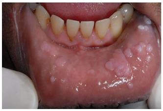
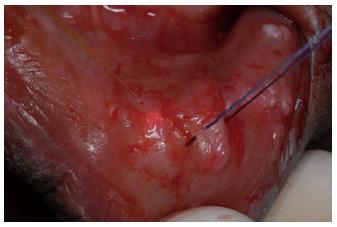
Extraoral examination did not reveal any signs of other diseases. Intraoral examination showed the presence of 17 sessile, soft, normochromic lesions in the oral cavity. The lesions were localized on the lower lip and the buccal mucosa, on both sides (Figure 1). After the examination, a diagnostic hypothesis of FEH was assumed. Excisional biopsy was performed on one of the lesions to confirm the diagnostic hypothesis. The biopsy was performed using a potassium-titanyl-phosphate (KTP) laser with a wavelength of 532 nm (SmartLite, Deka, Florence, Italy) in continuous mode with a 300 μm fiber, power 1.4 W (power density 1980.22 W/cm2). After anesthesia without vasoconstrictors (Mepivacaina Pierrel 30 mg/mL, Pierrel, Milan, Italy) the lesions were tractioned with sutures or an Allis clamp and then completely excised (Figure 2). The specimens were preserved in 10% buffered formalin for histological examination, which confirmed the clinical diagnosis of FEH.
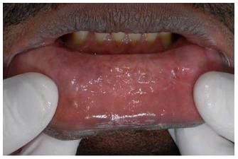
In this case, the laser allowed excellent control of bleeding, without postoperative sutures, and optimal wound healing. After the first excisional biopsy, all of the lesions were surgically removed, in several steps, using the same operative approach. The patient was monitored with follow-up visits for one year, during which no recurrence of the pathology was observed (Figure 3), and he was asked to return if any lesions reappeared in the future.
FEH is a rare condition in Italy and Europe. Here, we describe the case of an immunosuppressed African man with FEH. This pathology has been extensively described among native South and North American populations[1,6-8]. It seems that there could be a genetic predilection for FEH, as cases seem to be limited to specific ethnic groups in certain geographic regions[1,6]. Ledesma-Montes et al[7] suggested a series of factors that could contribute to the development of FEH: poverty, genetic predisposition (ethnic factors), and a deficient hygienic lifestyle. FEH lesions are most frequently localized on the buccal mucosa, lip, tongue, and commissures; the retromolar area, palate, and mouth floor are rarer localizations[7]. FEH must be included in differential diagnosis with several pathological conditions that can be observed in the oral cavity[8], namely, condylomata acuminata, verrucous carcinoma, inflammatory fibrous hyperplasia, inflammatory papillary hyperplasia, and verruciform xanthoma[3]. Condyloma may appear similarly because it is caused by the same virus, but FEH lesions are more numerous and flatter, with typical localizations (buccal mucosa, lip and tongue)[9]. Verrucous carcinoma is a malign neoplasm that usually occurs in a different age group, usually in the sixth decade of life, with epidemiological characteristics that are similar to other oral carcinomas[10].
The last three pathologies are reactive lesions that usually occur with an irritating stimulus[9]. The diagnosis of FEH can be performed on the basis of clinical observation and can be confirmed by histological examination[11-13].
The histological features of this disease are parakeratosis, acanthosis, elongation of rete ridges, some of which may be anastomosed (the so-called “bronze age battle-axe” appearance[14], and usually koilocytes, as well as other cellular modifications that can indicate viral infection[5,14]. Cells with nuclear degeneration, called mitosoid cells, can also appear[15]. FEH can be associated with HIV infection, although the relationship between these conditions has not yet been completely clarified[12]. Suppression of the immune system leaves the patient vulnerable to opportunistic infections, including HPV infections[12].
There is no agreement in the literature on the potential malignant transformation of FEH lesions in immunocompromised patients. Moerman et al[12] stated that FEH lesions may have a high risk of malignant transformation in immunocompromised patients. Durso et al[13] tended to consider FEH a benign condition and stated that to date, no research has demonstrated the potential for malignant transformation of FEH lesions with HPV 13 and 32 subtypes. Only one case of malignant transformation of FEH caused by HPV type 24 has been reported[16]. Further studies are required to clarify this point.
Several therapeutic approaches have been proposed throughout the years. Some authors advise against removing the lesions because spontaneous regression can be observed[17], especially in children. Steinhoff et al[18] successfully treated FEH with topical applications of interferon beta. Other described methods include scalpel surgery, electrocoagulation, electrodessication, cryosurgery, and laser surgery[19,20]. In this case, laser surgery allowed excellent control of bleeding, without postoperative sutures, and optimal wound healing. Moreover, histological analysis is always possible with laser surgery when the proper parameters and correct surgical technique are used[21].
A 37-year-old male with a history of human immuno-deficiency virus (HIV) infection presented with numerous asymptomatic lesions in the oral cavity.
Seventeen sessile, soft, normochromic lesions on the lower lip and the buccal mucosa, on both sides.
Condylomata acuminata, verrucous carcinoma, inflammatory fibrous hyperplasia, inflammatory papillary hyperplasia, verruciform xanthoma.
Cluster of differentiation 4 receptors (CD4) 129/μL; HIV RNA < 37 copies/mL; metabolic panel and coagulation within normal limits.
Histological examination revealed parakeratosis, acanthosis, presence of koilocytes and mitosoid cells.
Excisional biopsy with a 532-nm potassium titanyl phosphate (KTP) laser.
Focal epithelial hyperplasia (FEH) can be associated with HIV infection, although the relationship between these two conditions has not yet been completely clarified; most likely, suppression of the immune system leaves the patient vulnerable to opportunistic infections, including HPV infections.
KTP lasers are powerful tools for oral surgery and oral pathology, as are other types of lasers.
Oral lesions can be a manifestation of more complex systemic diseases; advanced surgical techniques can be useful tools in the management of multiple oral viral lesions.
This paper is worthy of publication as an interesting case report of uncommon oral mucosa disease in HIV infected patient. Particularly, an information of efficiency of KTP laser surgery in treatment of Heck’s disease will be useful for clinicians.
P- Reviewers: Bugaj AM, Kaliyadan F S- Editor: Ji FF L- Editor: A E- Editor: Wu HL
| 1. | Archard HO, heck JW, Stanley HR. Focal epithelial hyperplasia: an unusual oral mucosal lesion found in indian children. oral surg oral med oral pathol. 1965;20:201-212. [RCA] [PubMed] [DOI] [Full Text] [Cited by in Crossref: 152] [Cited by in RCA: 134] [Article Influence: 4.6] [Reference Citation Analysis (0)] |
| 2. | Clausen FP, Mogeltoft M, Roed-Petersen B, Pindborg JJ. Focal epithelial hyperplasia of the oral mucosa in a south-west Greenlandic population. Scand J Dent Res. 1970;78:287-294. [PubMed] |
| 3. | Terezhalmy GT, Riley CK, Moore WS. Focal epithelial hyperplasia (Heck’s disease). Quintessence Int. 2001;32:664-665. [PubMed] |
| 4. | Hanks CT, Fishman SL, Guzman MN: Focal epithelial hyperplasia. A light and electron microscopic study. Oral Surg Oral Med Oral Pathol. 1972;33:934-943. [RCA] [PubMed] [DOI] [Full Text] [Cited by in Crossref: 30] [Cited by in RCA: 30] [Article Influence: 0.6] [Reference Citation Analysis (0)] |
| 5. | Falaki F, Amir Chaghmaghi M, Pakfetrat A, Delavarian Z, Mozaffari PM, Pazooki N. Detection of human papilloma virus DNA in seven cases of focal epithelial hyperplasia in Iran. J Oral Pathol Med. 2009;38:773-776. [RCA] [PubMed] [DOI] [Full Text] [Cited by in Crossref: 14] [Cited by in RCA: 16] [Article Influence: 1.0] [Reference Citation Analysis (0)] |
| 6. | Witkop CJ, Niswander JD. Focal epithelial hyperplasia in central and south american indians and ladinos. Oral Surg Oral Med Oral Pathol. 1965;20:213-217. [RCA] [PubMed] [DOI] [Full Text] [Cited by in Crossref: 59] [Cited by in RCA: 62] [Article Influence: 2.1] [Reference Citation Analysis (0)] |
| 7. | Ledesma-Montes C, Garcés-Ortíz M, Hernández-Guerrero JC. Clinicopathological and immunocytochemical study of multifocal epithelial hyperplasia. J Oral Maxillofac Surg. 2007;65:2211-2217. [RCA] [PubMed] [DOI] [Full Text] [Cited by in Crossref: 15] [Cited by in RCA: 12] [Article Influence: 0.7] [Reference Citation Analysis (0)] |
| 8. | Cubie HA. Diseases associated with human papillomavirus infection. Virology. 2013;445:21-34. [RCA] [PubMed] [DOI] [Full Text] [Cited by in Crossref: 179] [Cited by in RCA: 201] [Article Influence: 16.8] [Reference Citation Analysis (0)] |
| 9. | Borborema-Santos CM, Castro MM, Santos PJ, Talhari S, Astolfi-Filho S. Oral focal epithelial hyperplasia: report of five cases. Braz Dent J. 2006;17:79-82. [RCA] [PubMed] [DOI] [Full Text] [Cited by in Crossref: 29] [Cited by in RCA: 22] [Article Influence: 1.2] [Reference Citation Analysis (0)] |
| 10. | Zhu LK, Ding YW, Liu W, Zhou YM, Shi LJ, Zhou ZT. A clinicopathological study on verrucous hyperplasia and verrucous carcinoma of the oral mucosa. J Oral Pathol Med. 2012;41:131-135. [RCA] [PubMed] [DOI] [Full Text] [Cited by in Crossref: 15] [Cited by in RCA: 18] [Article Influence: 1.3] [Reference Citation Analysis (0)] |
| 11. | Bassioukas K, Danielides V, Georgiou I, Photos E, Zagorianakou P, Skevas A. Oral focal epithelial hyperplasia. Eur J Dermatol. 2000;10:395-397. [PubMed] |
| 12. | Moerman M, Danielides VG, Nousia CS, Van Wanzeele F, Forsyth R, Vermeersch H. Recurrent focal epithelial hyperplasia due to HPV13 in an HIV-positive patient. Dermatology. 2001;203:339-341. [RCA] [PubMed] [DOI] [Full Text] [Cited by in Crossref: 21] [Cited by in RCA: 19] [Article Influence: 0.8] [Reference Citation Analysis (0)] |
| 13. | Durso BC, Pinto JM, Jorge J, de Almeida OP. Extensive focal epithelial hyperplasia: case report. J Can Dent Assoc. 2005;71:769-771. [PubMed] |
| 14. | Marvan E, Firth N. Focal epithelial hyperplasia in an HIV positive man. An illustrated case and review of the literature. Aust Dent J. 1998;43:305-310. [RCA] [PubMed] [DOI] [Full Text] [Cited by in Crossref: 18] [Cited by in RCA: 18] [Article Influence: 0.7] [Reference Citation Analysis (0)] |
| 15. | Segura-Saint-Gerons R, Toro-Rojas M, Ceballos-Salobreña A, Aparicio-Soria JL, Fuentes-Vaamonde H. Focal epithelial hyperplasia. A rare disease in our area. Med Oral Patol Oral Cir Bucal. 2005;10:128-131. [PubMed] |
| 16. | Niebrügge B, Villiers E, Gerlach K, Franke I, Gollnick H. Demonstration of HPV 24 in long-standing Heck’s disease with malignant transformation. Eur J Dermatol. 1999;9:477-479. [PubMed] |
| 17. | Pfister H, Hettich I, Runne U, Gissmann L, Chilf GN. Characterization of human papillomavirus type 13 from focal epithelial hyperplasia Heck lesions. J Virol. 1983;47:363-366. [PubMed] |
| 18. | Steinhoff M, Metze D, Stockfleth E, Luger TA. Successful topical treatment of focal epithelial hyperplasia (Heck’s disease) with interferon-beta. Br J Dermatol. 2001;144:1067-1069. [RCA] [PubMed] [DOI] [Full Text] [Cited by in Crossref: 33] [Cited by in RCA: 33] [Article Influence: 1.4] [Reference Citation Analysis (0)] |
| 19. | Akyol A, Anadolu R, Anadolu Y, Ekmekci P, Gürgey E, Akay N. Multifocal papillomavirus epithelial hyperplasia: successful treatment with CO2 laser therapy combined with interferon alpha-2b. Int J Dermatol. 2003;42:733-735. [RCA] [PubMed] [DOI] [Full Text] [Cited by in Crossref: 23] [Cited by in RCA: 22] [Article Influence: 1.0] [Reference Citation Analysis (0)] |
| 20. | Said AK, Leao JC, Fedele S, Porter SR. Focal epithelial hyperplasia - an update. J Oral Pathol Med. 2013;42:435-442. [RCA] [PubMed] [DOI] [Full Text] [Cited by in Crossref: 49] [Cited by in RCA: 48] [Article Influence: 3.7] [Reference Citation Analysis (0)] |
| 21. | Romeo U, Palaia G, Del Vecchio A, Tenore G, Gambarini G, Gutknecht N, De Luca M. Effects of KTP laser on oral soft tissues. An in vitro study. Lasers Med Sci. 2010;25:539-543. [RCA] [PubMed] [DOI] [Full Text] [Cited by in Crossref: 21] [Cited by in RCA: 25] [Article Influence: 1.7] [Reference Citation Analysis (0)] |


