Published online Aug 26, 2025. doi: 10.12998/wjcc.v13.i24.107384
Revised: April 17, 2025
Accepted: May 13, 2025
Published online: August 26, 2025
Processing time: 86 Days and 13.4 Hours
Slipping rib syndrome (SRS) is caused by abnormal subluxation of floating ribs, resulting in chronic pain and possible tissue damage. Its prevalence is often overlooked, as it shares symptoms with other musculoskeletal conditions, and is more common in young females and athletes. Symptoms include pain along the lower rib margin, aggravated by trunk movements, deep breathing and coughing. Treatment usually starts conservatively with physiotherapy and analgesics. In severe cases, extrapleural rib resection may be required.
In April 2023, a 24-year-old woman presented with persistent right hemithorax pain in the eleventh rib for one year. Instrumental examinations, including ultra
Minimally invasive rib-preserving surgery effectively reduces pain and hospitalization time, offering a valid alternative to conventional rib resection for refractory SRS.
Core Tip: Slipping rib syndrome (SRS) remains underdiagnosed due to its nonspecific presentation and lack of definitive diagnostic tools. Traditional surgical treatment involves complete rib resection, but minimally invasive approaches with rib preservation are emerging as effective alternatives. This case report presents a novel hybrid technique that combines cartilaginous resection and rib stabilization, resulting in significant pain reduction and functional improvement. Our findings highlight the importance of tailored surgical strategies, minimizing postoperative complications and optimizing patient outcomes, thus offering a viable alternative for refractory SRS cases.
- Citation: Spinelli F, Petrella F, Cara A, Cassina E, Libretti L, Pirondini E, De Simone M, Cioffi U, Tuoro A, Cioffi G, Raveglia F. Minimally invasive surgical approach for slipping rib syndrome: A case report. World J Clin Cases 2025; 13(24): 107384
- URL: https://www.wjgnet.com/2307-8960/full/v13/i24/107384.htm
- DOI: https://dx.doi.org/10.12998/wjcc.v13.i24.107384
Slipping rib syndrome (SRS) is a clinical condition defined by the atypical subluxation of the floating ribs, which can lead to persistent thoracic pain and potential injury to adjacent soft tissues. The underlying mechanism of SRS is believed to be related to the detachment of interchondral cartilaginous junctions, resulting in a structural discontinuity of the costal margin[1]. Given their high sensitivity, intercostal nerves are particularly vulnerable to injury and may generate chronic pain when irritated. Repeated and subtle movements of the displaced rib, pressing against the adjacent fixed rib, may provoke mechanical irritation of the intercostal nerve[2].
The incidence of SRS is frequently underestimated due to its symptomatologic overlap with other musculoskeletal conditions and the absence of a definitive diagnostic test. Owing to underdiagnosis, reliable epidemiological data are lacking; however, the syndrome appears to be more prevalent among young female individuals and athletes, often exerting a significant impact on both professional and socioeconomic functioning[3].
Diagnosis is primarily clinical, based on anamnesis and targeted physical examination. Specific tests, such as the “hooking maneuver”, may reproduce the characteristic pain and aid in confirmation. Clinical presentation is variable, but typically includes pain localized along the lower costal margin, which tends to exacerbate with movements of the trunk, forced inspiration, or coughing, thereby compromising routine daily functions[4].
First-line management is conservative, with a primary focus on symptom control via physiotherapeutic interventions, administration of analgesic drugs, and corticosteroid therapy[5]. Nonetheless, according to current literature, in cases unresponsive to conservative treatments or presenting with severe symptoms, extrapleural surgical resection of the involved costal segment may become necessary. The absence of a standardized diagnostic method, however, raises legi
This case report presents the use of a minimally invasive, rib-sparing surgical technique in the management of a patient with slipping rib syndrome refractory to conventional medical therapies.
In April 2023, a 24-year-old woman presented with persistent right hemithorax pain at the level of the eleventh rib for one year.
The patient described the pain as debilitating, exacerbated by twisting movements of the chest, respiratory excursions, and coughing. It initially presented as episodic but progressively worsened into a constant symptom that significantly limited her daily activities.
She had no history of previous surgeries or chronic illnesses.
No special notes.
No clinically significant findings were observed on physical examination. Or asymmetries of the chest wall; on palpation, the patient tested positive for the hooking maneuver. This maneuver, first described by Heinz and Zavala, reproduces subluxation and intercostal nerve compression, causing pain in patients affected by SRS.
No special notes.
Instrumental examinations, including abdominal ultrasonography and magnetic resonance imaging (MRI), revealed no abdominal or osteochondral abnormalities. Therefore, osteochondral pathologies were ruled out. The intensity of the pain was quantified using the numeric rating scale (NRS), with the patient reporting a score of 8/10 at her first outpatient visit.
The diagnosis of SRS was primarily based on a thorough medical history and a targeted physical examination, with particular emphasis on the Hooking maneuver, which reproduced the characteristic pain associated with the mobilization of the floating ribs. However, to confirm the diagnosis, it was necessary to rule out other conditions with overlapping symptoms through imaging techniques and a rigorous clinical evaluation.
In this case, ultrasound and MRI were used to exclude osteochondral or visceral pathologies; the diagnostic investigations performed proved to be decisive in the differential diagnosis. The use of computed tomography was considered but ultimately deemed unnecessary in agreement with our radiologist. Therefore, the absence of abnormal findings further supported the diagnostic hypothesis of SRS. The main differential diagnoses considered included costochondritis, which is characterized by localized pain at the costochondral junction without evidence of subluxation or abnormal rib mobility, and nerve entrapment syndromes, such as intercostal nerve neuropathy or post-surgical neuropathies. In these cases, neuropathic pain is generally elicited by direct palpation of the nerve rather than by the Hooking maneuver.
The patient had previously undergone medical treatment with analgesic therapy, consisting of 400 mg of ibuprofen taken twice a day, followed by as-needed administration of paracetamol/codeine 500 mg/30 mg every 8 hours. However, she experienced no significant symptom relief. After a thorough literature review and informing the patient of the possibility of an unconventional hybrid surgical approach, we proceed to surgery.
During the surgical planning, we carefully considered and evaluated potential approaches in the event that the procedure would not be resolutive. The patient was informed that, should the intervention fail to alleviate the pain symptoms, the next planned approach would be a more extensive resection of the affected rib segment.
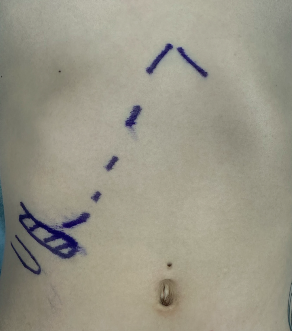
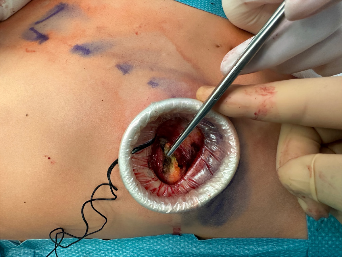
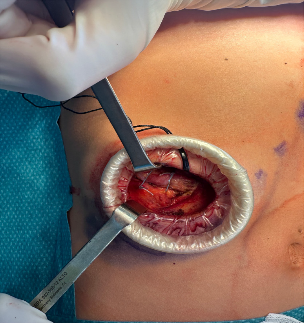
Under general anesthesia and single-lumen endotracheal intubation, the procedure was conducted with the patient in the supine position. We considered the use of a single-lumen tube sufficient due to the low risk of pleural breach during surgical planning. In the event of an accidental pleural opening at the level of the tenth rib during the procedure, the risks associated with placing a chest drain and inducing temporary apnea were deemed lower than those related to the use of a double-lumen endotracheal tube. A 4-cm incision was made along the anterior third of the eleventh right rib (Figure 1). Upon reaching the costal plane through the external oblique muscle using a muscle-sparing technique, an Alexis S tissue retractor was positioned (Figure 2). Once the costal plane was exposed, a non-physiological floating movement was observed, leading to overlapping with the overlying rib. The terminal cartilaginous segment was isolated and resected for approximately 2.5 cm. Subsequently, the eleventh rib was anchored to the overlying rib using transfixing stitches with non-absorbable sutures (Figure 3). No pleural drainage placement was required. The postoperative chest X-ray showed normal surgical outcomes.
No postoperative complications occurred. The patient was discharged on the first postoperative day. She underwent follow-up through outpatient visits at one week, one month, and three months postoperatively. At one week postoperatively, the patient reported using analgesic therapy, taking 1000 mg of paracetamol twice a day and oxycodone/naloxone 10/5 mg twice a day for six days, as stated in the discharge letter. She reported a pain level of 6/10 on the NRS before taking the analgesic medication. After one month, she had discontinued analgesic use, with a pain level reduced to 1/10 on the NRS, and the chest X-ray showed normal postoperative findings. At three months postoperatively, the patient reported being very satisfied with the surgical outcome and described fluctuating pain with a score of 2/10 on the NRS, occasionally requiring analgesics such as 1000 mg of paracetamol, but without interfering with daily activities. The histological examination of the resected specimen showed no cartilaginous abnormalities. During the outpatient follow-up visits, the patient was satisfied with the result and stated that she would undergo the surgical procedure again if needed. The patient provided written informed consent for the publication of this case report and the associated images.
SRS is frequently challenging to diagnose due to its nonspecific clinical manifestations and the absence of definitive radiological findings. Management strategies range from conservative therapies to surgical intervention. The traditional surgical approach often involves complete rib resection, which may be associated with prolonged postoperative recovery and a non-negligible risk of complications; therefore, surgical treatment is generally approached with caution. Nonethe
Madeka et al[4] describe a technique involving isolated cartilaginous rib excision (CRE), performed through a limited incision along the inferior costal margin. In this approach, the hypermobile cartilaginous segment is identified and resected up to the costochondral junction. The osseous portion of the rib is preserved, the pleural cavity remains intact, and particular attention is given to preserving the neurovascular bundle. Recurrence may occur due to continued motion of the remaining floating rib segment, which can reproduce symptoms through intermittent compression of the overlying intercostal nerve.
A single-institution descriptive study by MacGregor et al[6] analyzed 13 pediatric patients with SRS who underwent CRE between 2012 and 2020. Among them, 91% reported postoperative pain improvement, with a median follow-up period of 3.5 months. However, the median interval between symptom onset and surgical treatment was approximately two years, a delay consistent with existing literature.
Laparoscopic CRE has also been investigated but has not demonstrated significant advantages over the open tech
Vertical rib plating has emerged as a strategy to reduce recurrence. In a retrospective review of 85 patients, McMahon et al[5] compared outcomes of SRS patients treated with CRE alone versus those treated with CRE in combination with bioabsorbable vertical rib plating. Plating was employed intraoperatively when the surgeon could manually reproduce rib subluxation relative to adjacent structures. Compared to metallic hardware, bioabsorbable plates exhibit a more favorable safety profile and have demonstrated acceptable tolerability in both adult and pediatric populations. While early outcomes appear promising, long-term data are needed to assess the durability of symptom relief once the plates have fully resorbed—typically by the second postoperative year[5]. Additionally, this technique may require more extensive dissection for adequate exposure and incurs greater costs due to the use of prosthetic materials, factors that contribute to longer operative times and larger incisions.
In a separate retrospective analysis, Hansen et al[3] proposed a minimally invasive method without cartilage excision, performed in 29 patients. The authors stabilized the tenth rib at its costal insertion using two figure-of-eight sutures with orthopedic tape (Tiger Tape), securing the structure superiorly and inferiorly while carefully avoiding neural entrapment. Functional outcomes postoperatively showed significant improvement as early as one week after surgery, with continued benefit noted at one month and six months. Pain and disability scores demonstrated statistically significant reductions (P < 0.001)[3].
In the case presented, based on the available scientific literature and despite the limited number of reported cases, we opted for a hybrid approach combining cartilaginous resection with stabilization of the subluxated rib. In our view, this technique offers complementary benefits by limiting rib mobility while maintaining a minimal skin incision, thereby balancing efficacy and invasiveness.
This case report underscores the complexity of chronic pain management in slipping rib syndrome and illustrates the successful application of a minimally invasive surgical technique. The implementation of an individualized treatment strategy led to effective pain control, minimized postoperative complications and adverse effects, and obviated the need for additional analgesic therapy, all within the context of a 24-hour hospital stay. We share our experience, based on a careful literature review and daily activity in a high-volume chest wall center, with the intention of offering a practical and effective therapeutic option to healthcare professionals encountering similar clinical scenarios.
We thank Dr. Gerardo Cioffi, native speaker, for reviewing the English language.
| 1. | Van Tassel D, McMahon LE, Riemann M, Wong K, Barnes CE. Dynamic ultrasound in the evaluation of patients with suspected slipping rib syndrome. Skeletal Radiol. 2019;48:741-751. [RCA] [PubMed] [DOI] [Full Text] [Cited by in Crossref: 20] [Cited by in RCA: 24] [Article Influence: 4.0] [Reference Citation Analysis (0)] |
| 2. | Gould JL, Rentea RM, Poola AS, Aguayo P, St Peter SD. The effectiveness of costal cartilage excision in children for slipping rib syndrome. J Pediatr Surg. 2016;51:2030-2032. [RCA] [PubMed] [DOI] [Full Text] [Cited by in Crossref: 15] [Cited by in RCA: 19] [Article Influence: 2.1] [Reference Citation Analysis (0)] |
| 3. | Hansen AJ, Toker A, Hayanga J, Buenaventura P, Spear C, Abbas G. Minimally Invasive Repair of Adult Slipped Rib Syndrome Without Costal Cartilage Excision. Ann Thorac Surg. 2020;110:1030-1035. [RCA] [PubMed] [DOI] [Full Text] [Cited by in Crossref: 5] [Cited by in RCA: 12] [Article Influence: 2.4] [Reference Citation Analysis (0)] |
| 4. | Madeka I, Alaparthi S, Moreta M, Peterson S, Mojica JJ, Roedl J, Okusanya O. A Review of Slipping Rib Syndrome: Diagnostic and Treatment Updates to a Rare and Challenging Problem. J Clin Med. 2023;12:7671. [RCA] [PubMed] [DOI] [Full Text] [Cited by in RCA: 2] [Reference Citation Analysis (0)] |
| 5. | McMahon LE. Recurrent Slipping Rib Syndrome: Initial Experience with Vertical Rib Stabilization Using Bioabsorbable Plating. J Laparoendosc Adv Surg Tech A. 2020;30:334-337. [RCA] [PubMed] [DOI] [Full Text] [Cited by in Crossref: 6] [Cited by in RCA: 9] [Article Influence: 1.8] [Reference Citation Analysis (0)] |
| 6. | MacGregor RM, Schulte LJ, Merritt TC, Keller MS, Aubuchon JD, Abarbanell AM. Slipping Rib Syndrome in Children: Natural History and Outcomes Following Costal Cartilage Excision. J Surg Res. 2022;280:204-208. [RCA] [PubMed] [DOI] [Full Text] [Cited by in RCA: 5] [Reference Citation Analysis (0)] |


