Published online Aug 6, 2025. doi: 10.12998/wjcc.v13.i22.104352
Revised: March 11, 2025
Accepted: April 9, 2025
Published online: August 6, 2025
Processing time: 147 Days and 14.5 Hours
Intussusception is the invagination of a segment of the bowel into an adjacent segment. It is the most common cause of intestinal obstruction in children, but in adults, it is rare, accounting for 1% of all intestinal obstructions and 5% of all intussusceptions, with malignancy being the most common cause. In the past, it was typically diagnosed intraoperatively. However, with the availability of computed tomography for abdominal imaging, recognizing the condition's signs has become crucial. Surgical intervention is essential for managing neoplastic cases and their complications.
A 45-year-old female presented with severe abdominal pain encompassing her entire abdomen, abdominal distension, vomiting, and persistent constipation. Over the past two months, she has also experienced considerable weight loss. After an initial history review, examination, and imaging investigations, the patient was diagnosed with ileo cecal intussusception resulting from a colo rectal mass located in the cecum and ascending colon. This condition was surgically managed through an extended right hemi colectomy.
Intussusception is uncommon in adults, but it should be considered in patients with intestinal obstruction. Surgical intervention is essential.
Core Tip: Intussusception, a common cause of intestinal obstruction in children, is rare in adults and often linked to malignancy. Early diagnosis through computed tomography imaging is crucial to prevent complications. This case highlights a 45-year-old female with ileocecal intussusception due to a colorectal mass, successfully treated with extended right hemicolectomy. Recognizing intussusception as a differential diagnosis in adult intestinal obstruction is essential, as timely surgical intervention plays a critical role in patient outcomes.
- Citation: Abdishakur AE, Ahmed MAA. Adult ileo cecal intussusception as a manifestation of colon carcinoma: A case report. World J Clin Cases 2025; 13(22): 104352
- URL: https://www.wjgnet.com/2307-8960/full/v13/i22/104352.htm
- DOI: https://dx.doi.org/10.12998/wjcc.v13.i22.104352
Intussusception is a type of bowel obstruction that occurs when one segment of the intestine (intussusceptum) telescopes into the adjacent distal segment (intussuscipiens), along with its mesenteric fold. This telescoping can compromise blood flow, leading to ischemia or perforation if not addressed.
While intussusception is relatively common in children, it is much less frequent in adults, approximately 6% of all intussusception cases[1].
In adults, intussusception constitutes approximately 1% of all cases of intestinal obstruction and is most often secondary to an underlying pathological condition. Conversely, in children, it is usually idiopathic[2,3]. Intussusception is categorized based on its location (enteroenteric, ileocolic, or Colo colic), underlying cause (idiopathic, benign, or malignant), and the presence of a lead point. Among these classifications, Colo colic intussusception is the least common in adults.
Accounting for only 11% of cases, whereas ileocecal intussusception is the most frequent, followed by enteroenteric intussusception. Studies indicate that intussusception often originates from a lead point, which acts as a focal site of traction, pulling the proximal bowel into the distal segment[3]. In adults, a demonstrable etiology is identified in 70%-90% of cases.
We present a case of intussusception caused by colon cancer in a female patient and provide a review of the literature.
Imaging is essential for determining the cause of intussusception and identifying complex cases. Detecting impaired mesenteric circulation and signs of parietal ischemia is particularly crucial, as these conditions are linked to an increased risk of perforation and peritonitis.
Bowel perforation is an absolute contraindication for enema use in all age groups, requiring mandatory surgical intervention.
Abdominal computed tomography (CT) is the imaging modality of choice for diagnosing intussusception in adults. Characteristic CT findings include a "target" or "sausage-shaped" mass, which aids in identifying the condition. where the outer layer corresponds to the intussuscipiens and the inner layer represents the intussusceptum[4].
In the majority of adult cases, intussusception is caused by an underlying bowel malignancy, making surgical resection the preferred treatment option. Additional possible causes include trauma, Meckel's diverticulum, postoperative adhesions, lipomas, and adenomatous polyps.
The clinical presentation of intussusception in adults is variable. Unlike the classic pediatric triad of vomiting, abdominal pain, and rectal bleeding, adult cases often present with non-specific symptoms such as intermittent abdominal pain and vomiting, making preoperative diagnosis more challenging[5].
A 45-year-old female patient presented to the emergency department with severe abdominal pain, abdominal distention, and constipation.
The abdominal pain started two days before the examination, characterized by severe, excruciating discomfort, which was later accompanied by abdominal distention and constipation. The patient also reported considerable weight loss and generalized fatigue over the past two months.
There was no history of a similar condition or previous surgical operations.
The patient's medical, surgical, and family history were non-contributory.
the general examination revealed that the patient was cachectic, severely dehydrated, and pale. Vital signs included a blood pressure of 100/70 mmHg, a pulse rate of 89 beats per minute, a respiratory rate of 16 breaths per minute, and a temperature of 37.5 °C. Abdominal assessment showed significant distension and generalized tenderness, with pronounced discomfort on the right side.
Following initial fluid resuscitation and pain management, routine investigations were requested: White blood cell count: 17 × 109/L. Hemoglobin: 8.5 g/DL. Renal and liver function tests: Unremarkable. Coagulation profile: Unremarkable.
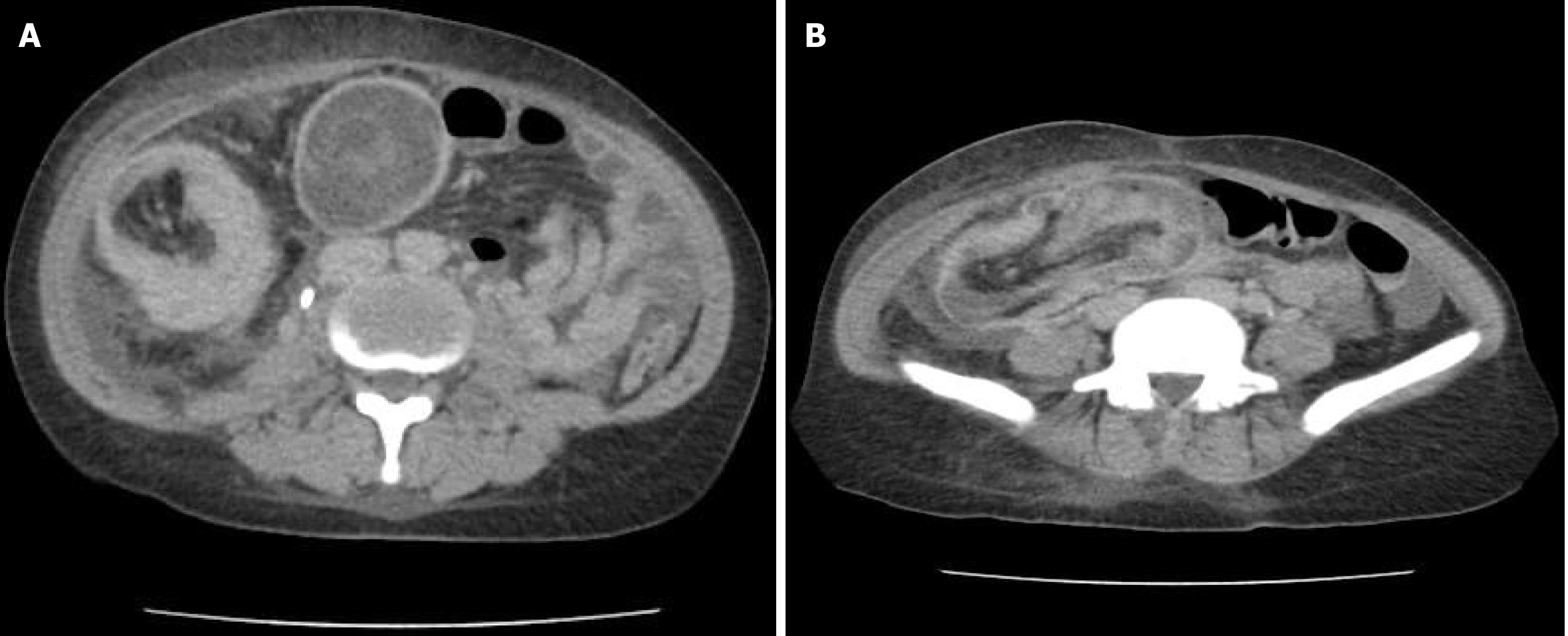
The patient had undergone a colonoscopy three weeks prior, which identified two large masses in the cecum and ascending colon. A contrast-enhanced CT scan of the abdomen revealed ileocolic intussusception along with an associated mass in the cecum and ascending colon (Figure 1A).
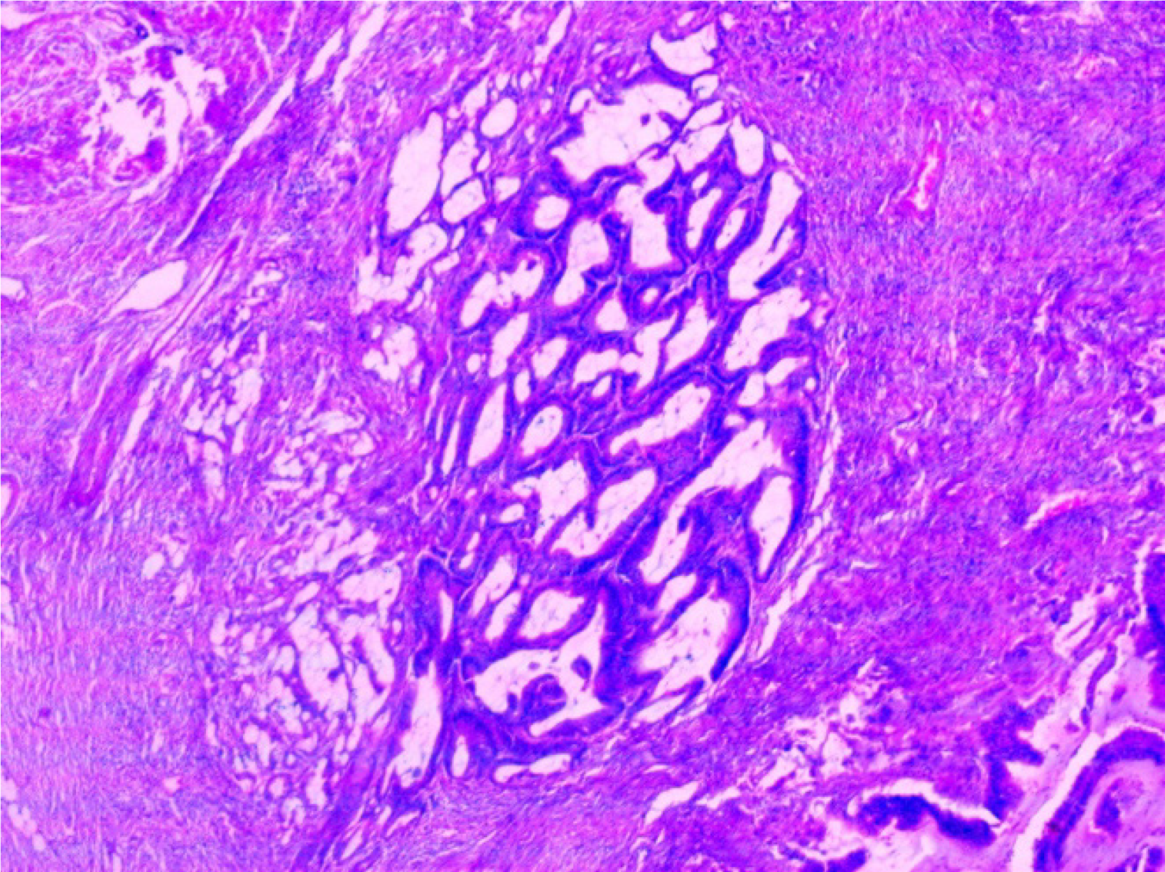
Despite the patient's emergency intestinal obstruction, senior consultants were consulted and recommended emergency exploration. Following surgical resection and histopathological confirmation of adenocarcinoma (Figure 2), the patient was referred to an oncologist abroad for further management owing to the lack of an oncology center in Somalia.
The patient was diagnosed with ileocecal intussusception associated with a cecal and colonic mass (Figure 1B).
After adequate preoperative preparation, including blood transfusion, enema, nasogastric tube placement, and intravenous antibiotics, the patient underwent exploratory laparotomy under general anesthesia.
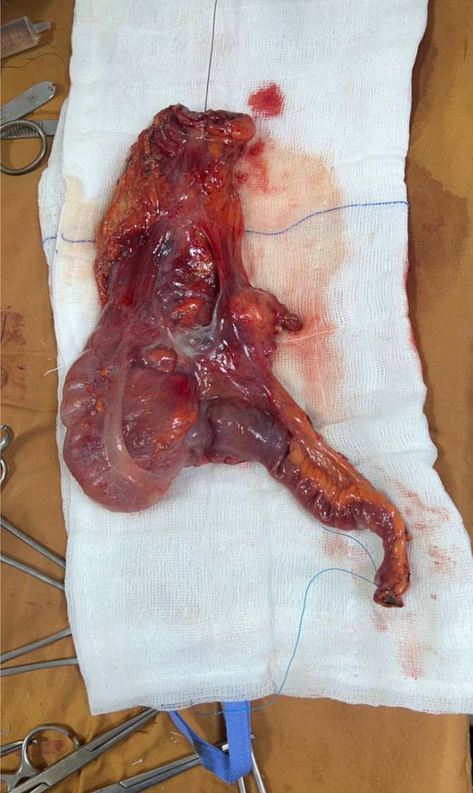
Intraoperatively, ileocolic intussusception was identified and manually reduced. An extended right hemicolectomy (Figure 3) was then performed.
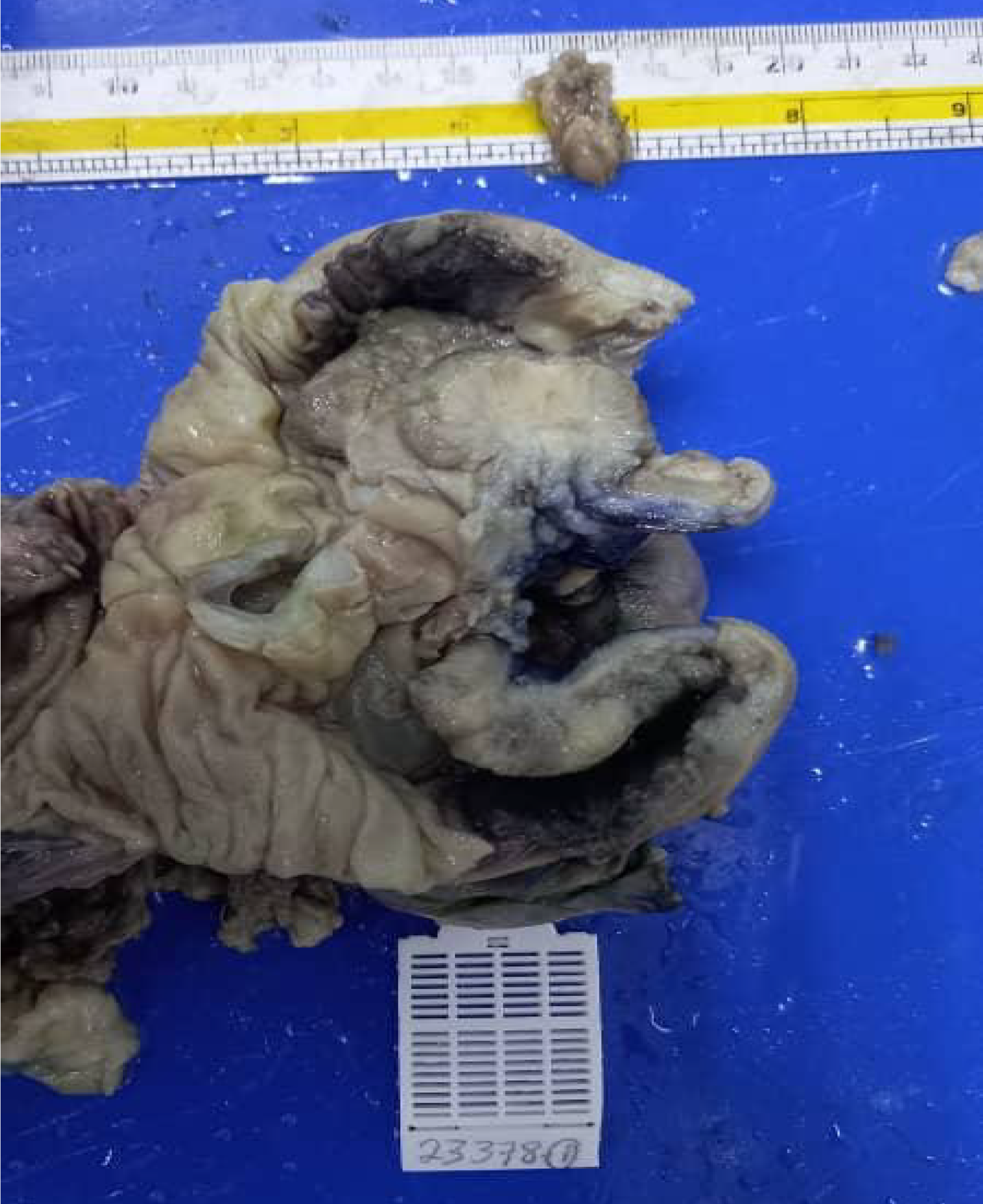
The surgical specimen was sent for histopathological examination, Figure 4 which confirmed the diagnosis of adenocarcinoma of the cecum and ascending colon.
At the three-month post-operative review, the patient was noted to have made a satisfactory recovery. She was free of abdominal pain and had gradually regained weight. No obvious post-operative complications were observed. The patient was advised and referred to an oncologist for ongoing follow-up care.
Adult intussusception is a rare condition, accounting for only 1% of intestinal obstruction and less than 5% of all cases of intussusception[6]. Unlike children, who typically present with characteristic symptoms such as abdominal pain, abdominal mass, and bloody stools, adults may have a wide range of symptoms that can be acute, intermittent, or chronic. Most affected adults experience episodes of intermittent abdominal pain and vomiting prior to diagnosis[7]. In the case presented, the patient's symptoms and signs were non-specific, making it challenging to diagnose and potentially leading to a delay in treatment.
In the case presented, adenocarcinoma of the colon led to ileocecal intussusception, manifesting as severe abdominal pain, distension, and constipation. These symptoms can be non-specific and overlap with other conditions, posing a diagnostic challenge in adult intussusception. This challenge is further compounded by the absence of classic pediatric symptoms, such as vomiting or "red currant jelly" stool. As a result, adult cases often present with chronic, vague abdominal symptoms and are diagnosed incidentally through imaging or during surgical exploration.
Imaging, particularly CT, plays a pivotal role in diagnosing adult intussusception. CT provides a clear visualization of the characteristic "target" or "sausage-shaped" mass, which is pathognomonic for intussusception. In the case presented, CT imaging definitively established the diagnosis, underscoring its significance as a diagnostic tool in adult cases where symptoms may not be immediately suggestive of intussusception[4].
Surgical resection is the preferred treatment, as the underlying cause is often a neoplastic process serving as the lead point[8]. Reduction may be an option for patients with suspected benign causes, especially in small bowel intussusception without evidence of ischemia. However, in malignant colonic intussusception, reduction should be avoided due to the risk of tumor embolization and potential cancer dissemination[9-11].
Management of intussusception in adults typically requires surgical intervention owing to the high likelihood of an underlying malignancy. In the case presented, an exploratory laparotomy with right hemicolectomy was performed, following oncological principles to address both the intussusception and the carcinoma. Histopathological examination confirmed adenocarcinoma, supporting the literature that links malignant neoplasms to a considerable proportion of adult intussusceptions[12].
Postoperative follow-up is essential in these cases to monitor for recurrence or metastasis, given the malignant etiology. The patient had a favorable outcome with no complications, and recovery was evidenced by weight gain and resolution of symptoms. Continued oncological follow-up was recommended owing to the potential for recurrence.
This case underscores the importance of considering intussusception in adult patients presenting with chronic abdominal pain, particularly when accompanied by weight loss. Timely imaging, surgical intervention, and histopathological analysis are critical for managing this rare yet serious condition, especially when linked to malignancy.
Colonic intussusception in the elderly is rare and often associated with an underlying malignancy. This article discusses the case of a female patient with colonic intussusception caused by adenocarcinoma of the cecum and ascending colon. Therefore, intussusception should be considered in the differential diagnosis of elderly patients experiencing prolonged, nonspecific abdominal pain and unexplained weight loss. Initial evaluation should include further imaging studies, such as an abdominal ultrasound or CT scan, to aid in diagnosis. Ultimately, surgical intervention is essential once intussusception is diagnosed in the elderly owing to the high risk of malignancy[5].
Intussusception is an uncommon condition in adults, with malignancy being a primary etiology in this population. Colonoscopy is a valuable diagnostic tool, while contrast-enhanced tomography remains the preferred imaging modality. Surgical intervention, guided by oncological principles, remains the treatment of choice[12]. Owing to the emergency presentation, tumor-node-metastasis classification and metastatic workup were not performed, and surgery was carried out to prevent colon ischemia.
The authors would like to thank all participants including the radiology and imaging department and pathology department for their contribution.
| 1. | Yalamarthi S, Smith RC. Adult intussusception: case reports and review of literature. Postgrad Med J. 2005;81:174-177. [RCA] [PubMed] [DOI] [Full Text] [Cited by in Crossref: 115] [Cited by in RCA: 137] [Article Influence: 6.9] [Reference Citation Analysis (0)] |
| 2. | Alshoabi SA, Abdulaal OM. An unusual case of colonic intussusception in old age. J Taibah Univ Med Sci. 2019;14:199-202. [RCA] [PubMed] [DOI] [Full Text] [Full Text (PDF)] [Cited by in Crossref: 2] [Cited by in RCA: 1] [Article Influence: 0.2] [Reference Citation Analysis (0)] |
| 3. | Teyha PS, Chandika A, Kotecha VR. Prolapsed sigmoid intussusception per anus in an elderly man: a case report. J Med Case Rep. 2011;5:389. [RCA] [PubMed] [DOI] [Full Text] [Full Text (PDF)] [Cited by in Crossref: 3] [Cited by in RCA: 5] [Article Influence: 0.4] [Reference Citation Analysis (1)] |
| 4. | Lu T, Chng YM. Adult intussusception. Perm J. 2015;19:79-81. [RCA] [PubMed] [DOI] [Full Text] [Cited by in Crossref: 21] [Cited by in RCA: 46] [Article Influence: 4.6] [Reference Citation Analysis (0)] |
| 5. | Poudel D, Lamichhane SR, Ajay KC, Maharjan N. Colocolic intussusception secondary to colonic adenocarcinoma with impending caecal perforation in an elderly patient: A rare case report. Int J Surg Case Rep. 2022;94:107093. [RCA] [PubMed] [DOI] [Full Text] [Full Text (PDF)] [Cited by in RCA: 2] [Reference Citation Analysis (0)] |
| 6. | Marinis A, Yiallourou A, Samanides L, Dafnios N, Anastasopoulos G, Vassiliou I, Theodosopoulos T. Intussusception of the bowel in adults: a review. World J Gastroenterol. 2009;15:407-411. [RCA] [PubMed] [DOI] [Full Text] [Full Text (PDF)] [Cited by in CrossRef: 428] [Cited by in RCA: 507] [Article Influence: 31.7] [Reference Citation Analysis (2)] |
| 7. | Azar T, Berger DL. Adult intussusception. Ann Surg. 1997;226:134-138. [RCA] [PubMed] [DOI] [Full Text] [Cited by in Crossref: 648] [Cited by in RCA: 666] [Article Influence: 23.8] [Reference Citation Analysis (0)] |
| 8. | Yakan S, Caliskan C, Makay O, Denecli AG, Korkut MA. Intussusception in adults: clinical characteristics, diagnosis and operative strategies. World J Gastroenterol. 2009;15:1985-1989. [RCA] [PubMed] [DOI] [Full Text] [Full Text (PDF)] [Cited by in CrossRef: 111] [Cited by in RCA: 135] [Article Influence: 8.4] [Reference Citation Analysis (0)] |
| 9. | Goh BK, Quah HM, Chow PK, Tan KY, Tay KH, Eu KW, Ooi LL, Wong WK. Predictive factors of malignancy in adults with intussusception. World J Surg. 2006;30:1300-1304. [RCA] [PubMed] [DOI] [Full Text] [Cited by in Crossref: 66] [Cited by in RCA: 65] [Article Influence: 3.4] [Reference Citation Analysis (0)] |
| 10. | Wang N, Cui XY, Liu Y, Long J, Xu YH, Guo RX, Guo KJ. Adult intussusception: a retrospective review of 41 cases. World J Gastroenterol. 2009;15:3303-3308. [RCA] [PubMed] [DOI] [Full Text] [Full Text (PDF)] [Cited by in CrossRef: 142] [Cited by in RCA: 153] [Article Influence: 9.6] [Reference Citation Analysis (0)] |
| 11. | Panzera F, Di Venere B, Rizzi M, Biscaglia A, Praticò CA, Nasti G, Mardighian A, Nunes TF, Inchingolo R. Bowel intussusception in adult: Prevalence, diagnostic tools and therapy. World J Methodol. 2021;11:81-87. [RCA] [PubMed] [DOI] [Full Text] [Full Text (PDF)] [Cited by in CrossRef: 39] [Cited by in RCA: 44] [Article Influence: 11.0] [Reference Citation Analysis (4)] |
| 12. | Agha RA, Franchi T, Sohrabi C, Mathew G, Kerwan A; SCARE Group. The SCARE 2020 Guideline: Updating Consensus Surgical CAse REport (SCARE) Guidelines. Int J Surg. 2020;84:226-230. [RCA] [PubMed] [DOI] [Full Text] [Cited by in Crossref: 4265] [Cited by in RCA: 4714] [Article Influence: 942.8] [Reference Citation Analysis (0)] |


