Copyright
©The Author(s) 2024.
World J Clin Cases. Feb 26, 2024; 12(6): 1169-1173
Published online Feb 26, 2024. doi: 10.12998/wjcc.v12.i6.1169
Published online Feb 26, 2024. doi: 10.12998/wjcc.v12.i6.1169
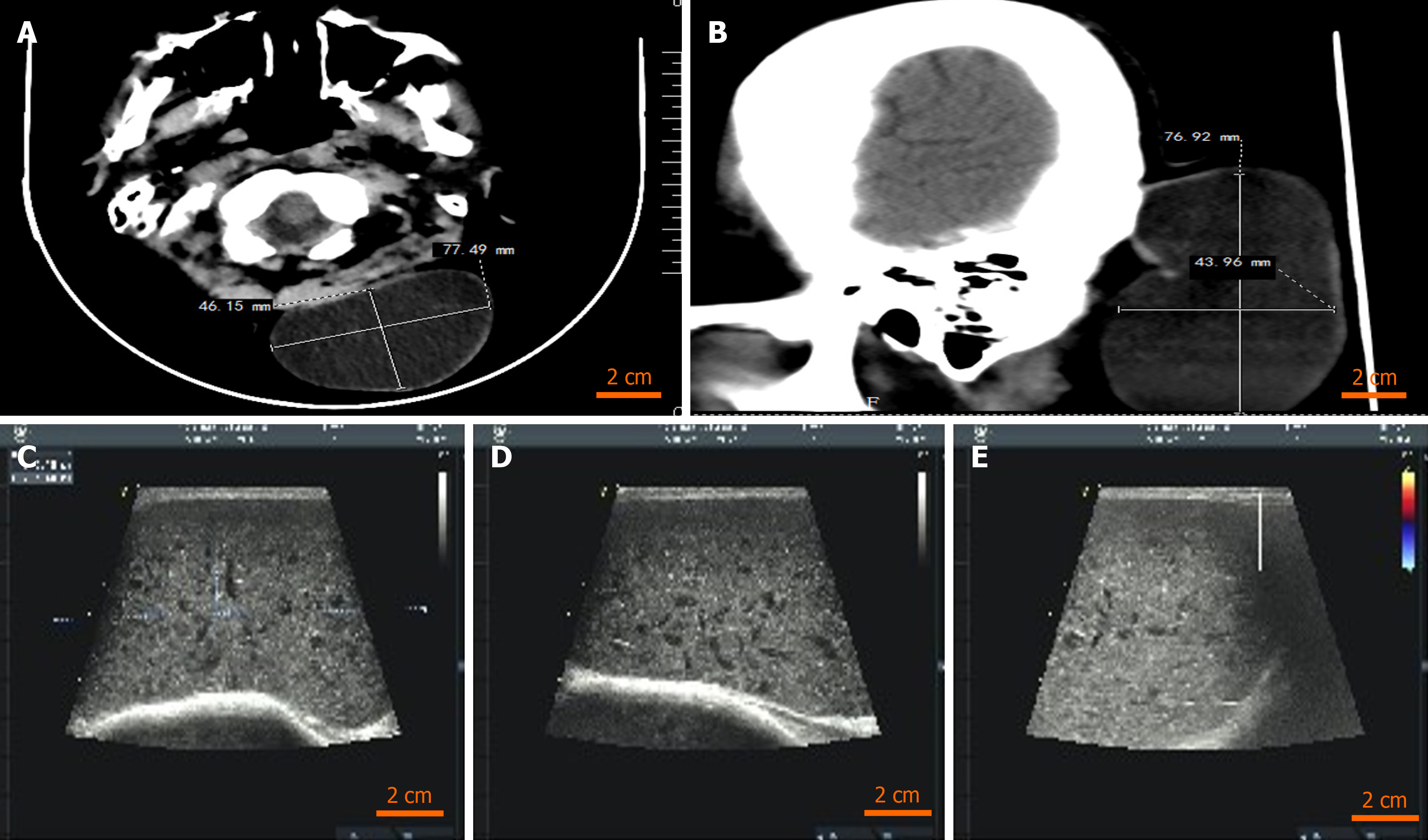
Figure 2 The computed tomography and ultrasonic image.
A: The length and width of cyst of axial computed tomography (CT); B: The length and width of cyst of sagittal CT; C: A mass can be found at the subcutaneous distance of 2.3 mm from the body surface (ultrasonic image); D: The volume of the mass is 38 mm × 87 mm × 90 mm (ultrasonic image); E: The occipital mass is a solid mass with low echo, regular shape, clear boundary, complete envelope and less uniform internal echo (ultrasonic image).
- Citation: Wei Y, Chen P, Wu H. Gigantic occipital epidermal cyst in a 56-year-old female: A case report. World J Clin Cases 2024; 12(6): 1169-1173
- URL: https://www.wjgnet.com/2307-8960/full/v12/i6/1169.htm
- DOI: https://dx.doi.org/10.12998/wjcc.v12.i6.1169


