Copyright
©The Author(s) 2024.
World J Clin Cases. Feb 26, 2024; 12(6): 1169-1173
Published online Feb 26, 2024. doi: 10.12998/wjcc.v12.i6.1169
Published online Feb 26, 2024. doi: 10.12998/wjcc.v12.i6.1169
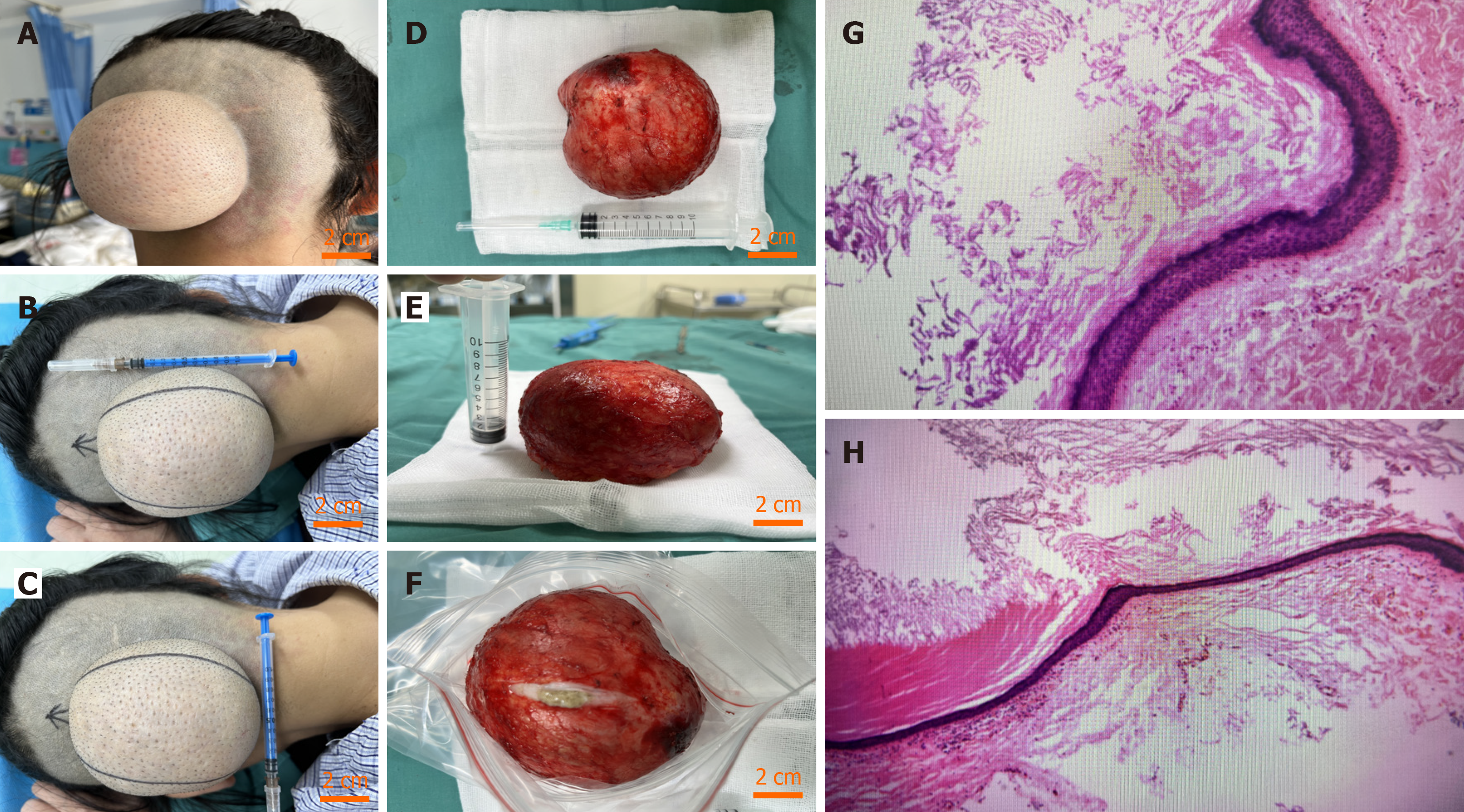
Figure 1 Preoperative manifestation, intraoperative presentation and pathology results.
A: A general view of epidermal cyst; B: The long diameter of epidermal cyst; C: The wide diameter of epidermal cyst; D: Height of epidermal cyst; E and F: Image of the cyst after incision; G: Layered cutinise (the pathological results indicated epidermal cyst); H: Squamous epithelium (the pathological results indicated epidermal cyst).
- Citation: Wei Y, Chen P, Wu H. Gigantic occipital epidermal cyst in a 56-year-old female: A case report. World J Clin Cases 2024; 12(6): 1169-1173
- URL: https://www.wjgnet.com/2307-8960/full/v12/i6/1169.htm
- DOI: https://dx.doi.org/10.12998/wjcc.v12.i6.1169


