Editorial Open Access
Copyright ©The Author(s) 2024. Published by Baishideng Publishing Group Inc. All rights reserved.
World J Clin Cases. Feb 26, 2024; 12(6): 1039-1044
Published online Feb 26, 2024. doi: 10.12998/wjcc.v12.i6.1039
Lateral clavicle fracture-plating options and considerations
Sathish Muthu, Department of Orthopaedics, Orthopaedic Research Group, Coimbatore 641045, Tamil Nadu, India
Sathish Muthu, Department of Biotechnology, Karpagam Academy of Higher Education, Coimbatore 641021, Tamil Nadu, India
Sathish Muthu, Department of Orthopaedics, Government Karur Medical College, Karur 639004, Tamil Nadu, India
Saravanan Annamalai, Department of Orthopaedics, Government Thiruvallur Medical College, Thiruvallur 631203, Tamil Nadu, India
Velmurugan Kandasamy, Department of Orthopaedics, Government Kilpauk Medical College, Chennai 600010, Tamil Nadu, India
ORCID number: Sathish Muthu (0000-0002-7143-4354).
Author contributions: Muthu S performed the conceptualization, data curation, data analysis, manuscript writing, and revision of the manuscript; Annamalai S performed the data analysis, manuscript writing, and revision of the manuscript; Kandasamy V performed data analysis, manuscript writing, and revision of the manuscript.
Conflict-of-interest statement: The authors declare having no conflicts of interest.
Open-Access: This article is an open-access article that was selected by an in-house editor and fully peer-reviewed by external reviewers. It is distributed in accordance with the Creative Commons Attribution NonCommercial (CC BY-NC 4.0) license, which permits others to distribute, remix, adapt, build upon this work non-commercially, and license their derivative works on different terms, provided the original work is properly cited and the use is non-commercial. See: https://creativecommons.org/Licenses/by-nc/4.0/
Corresponding author: Sathish Muthu, DNB, MS, Assistant Professor, Research Associate, Surgeon, Department of Orthopaedics, Orthopaedic Research Group, Ramanathapuram, Coimbatore 641045, Tamil Nadu, India. drsathishmuthu@gmail.com
Received: October 19, 2023
Peer-review started: October 19, 2023
First decision: December 19, 2023
Revised: December 21, 2023
Accepted: January 12, 2024
Article in press: January 12, 2024
Published online: February 26, 2024
Processing time: 123 Days and 18.5 Hours

Abstract

Clavicle fractures are among the most prevalent types of fractures with numerous treatment strategies that have evolved over time. In the realm of lateral-third clavicle fracture management, several surgical methods are available, with plate and screw constructs being one of the most frequently employed options. Within this construct, numerous choices exist for fixing the fracture. This editorial provides an overview of the common plate options utilized in the management of distal third clavicle fractures underscoring the critical considerations and approaches that guide clinicians in selecting the most appropriate fixation techniques, considering the complex landscape of clavicle fractures and their challenging management.

Key Words: Clavicle fracture, Surgical management, Distal clavicle plating, Superior plating, Anterior plating

Core Tip: The common options utilized in the management of the lateral end of the clavicle remain plate and screw construct. Among the myriad options available, anatomical precontoured locking plates with and without coracoclavicular suture reconstruction remain the commonly used implant. However, considering the cost and ease of molding non-locking reconstruction plates, hook plates have also been used. Among the plate positions, both superior and anterior placement provide similar clinical outcomes with their advantages and disadvantages.


INTRODUCTION

A clavicle fracture stands out as one of the most frequently occurring fractures, with an overall incidence rate of 64 cases per 100000 individuals each year[1,2]. Over time, a multitude of treatment approaches have been developed to address this issue. Extensive investigation into the therapeutic techniques for clavicle fractures has been conducted, comparing a range of treatment options from conservative to surgical management in numerous studies[3-5]. Furthermore, a mounting body of high-quality evidence from randomized controlled trials has suggested that non-surgical methods may lead to increased initial fracture displacement, higher non-union rates, and longer recovery times, fostering a growing consensus that surgical treatment may be more beneficial for individuals with clavicle fractures especially when it occurs in the lateral one-third[6-8].

Several surgical methods are available for lateral-third clavicle fracture management, with plate and screw constructs representing one of the most employed options[4]. There are a multitude of choices to fix the fracture using this construct. This editorial outlined the key plating choices employed in the management of distal third clavicle fractures and the fixation methods involved in them.

ANATOMICAL PRECONTOURED LOCKING PLATES

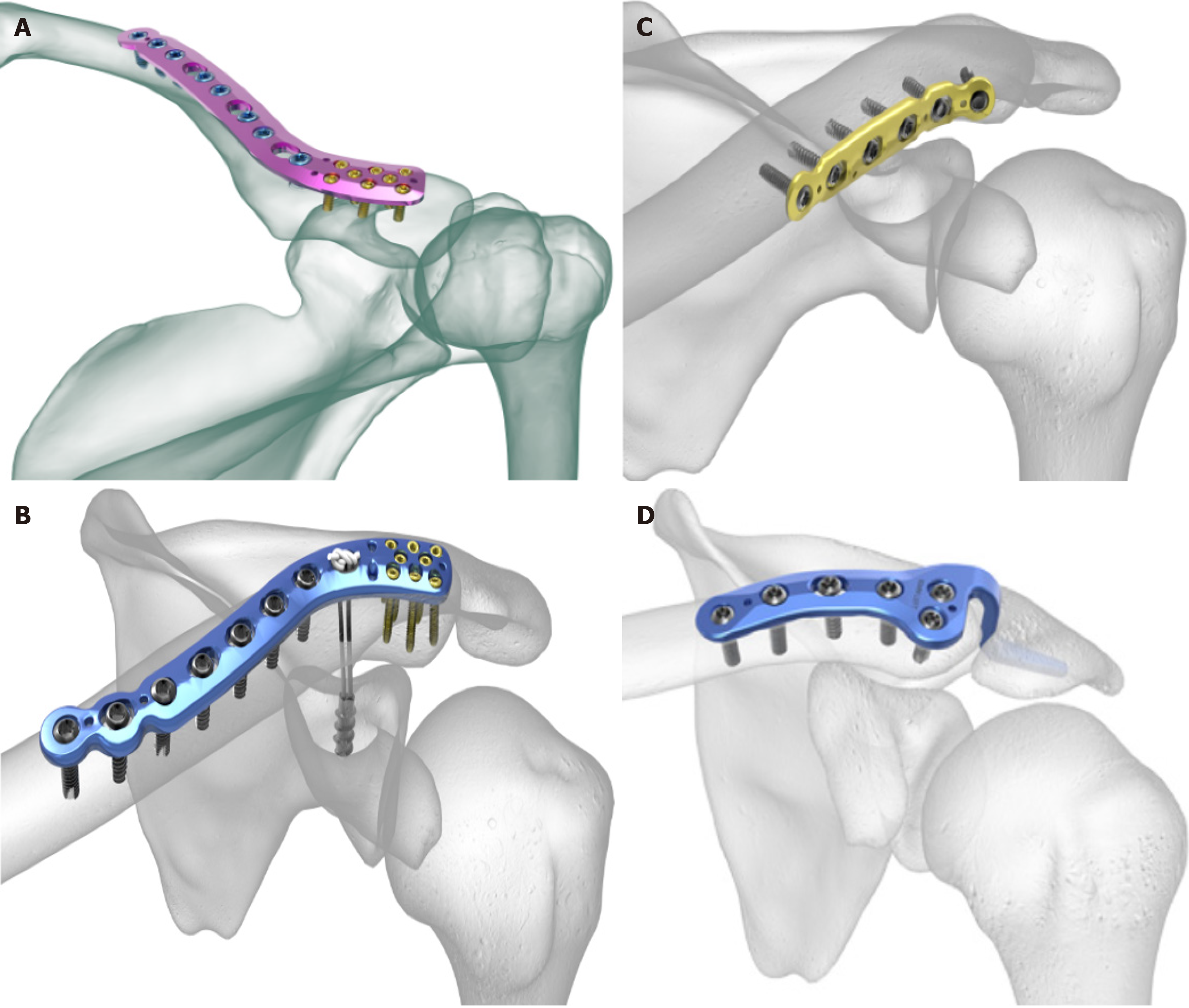
Locking plates remain one of the most utilized in the management of fractures due to their advantages, such as strong fixation resulting from the screw-plate locking mechanism and minimal contact with cortical bone, which preserves blood supply[9,10]. The current implementation of the minimally invasive technology that allows plating by percutaneous methods combined with locking plates is considered ideal for bone fixation as it minimizes periosteal stripping and promotes rapid healing[10]. The recent array of region-specific tailor-made locking plates is considered less conspicuous even after the bone union, and its implant removal rates are lower compared to the traditional plates[11]. Numerous anatomically contoured plates available in the market are equipped with lateral screw clusters, designed to secure even small lateral fragments effectively as shown in Figure 1A. In most of these cluster designs, the lateral fragment needs to be around 10 mm to 15 mm in size to accommodate three screws. Some plates come with features such as suture holes or provisions for snugly incorporating a suture button considering the additional option to add a coracoclavicular (CC) suspensory fixation as shown in Figure 1B.

Figure 1
Figure 1 Common plating options in the management of distal end of clavicle fracture. A: Anatomical precontoured locking plate in superior positioning; B: Anatomical precontoured locking plate with coracoclavicular suture construct; C: Anteroinferior positioning of dynamic compression plates; D: Hook plate.

The consideration to incorporate CC fixation along with the plated constructs is decided based on their efficacy demonstrated in various studies. For instance, in a study conducted by Furuhata et al[12], it was observed that using a locking plate as the sole treatment for injuries involving ligamentous injury demonstrated an increase in the CC distance, while those without the CC ligament injury did not show this pattern. This finding implied that locking plates on their own may not provide adequate vertical stability when CC ligaments are compromised. From this finding, one might conclude that CC fixation could be bundled with the traditional locking plates in cases with CC ligament injury. However, it is worth noting that no disparities in function scores, union rates, or complications were noted between the two groups. We also have studies reporting comparable results using either of the techniques for injuries with CC ligament injury[13]. In summary, we understand that incorporation of the CC fixation augments the biomechanics of the repair construct in distal clavicular fractures. It is indicated in cases with partial or complete CC ligament injury (Neer 2B and 5), whereas it might not be indicated in cases with intact ligaments (Neer 2A).

Celestre et al[14] conducted a study to understand the effect of localization of the plate in the management of midshaft clavicular fractures. They compared anterior vs superior locking plates and standard vs locking plates. Their findings indicated that resistance to failure due to bending along with axial compression/torsion stiffness was found at its best in the superior location of the locking plate. Hence, a similar positioning and plating option is commonly used for lateral end clavicle fractures. Locked plating has certain limitations, including the potential challenge of achieving sufficient grip when there is lateral comminution and the possibility of implant prominence. In a retrospective analysis encompassing 16 cases of distal clavicle fractures treated with plates and screws, 50% of cases required hardware removal[15]. However, there are also other studies with lower hardware removal rates[16-18]. Hence, caution should be exercised in the selection of these high-profile plates that might necessitate future surgery for implant removal.

NON-LOCKING PLATES

Apart from the precontoured anatomical locking plates for lateral end clavicle fractures, several other plate types used in surgical management include reconstruction plates, limited-contact dynamic compression plates, and T-plates with comparable clinical results[19-21]. However, clinical results comparing them with the locking plates in the fixation of the lateral end of the clavicle are limited. To enhance the rate and quality of bone union, several factors need to be considered when selecting an implant, including the location of the fracture, level of comminution, quality of the bone, demand, and compliance of the patient. Clavicle bridging plating in osteoporotic bone, while achieving stable fixation, may not provide the same level of rigidity. It is noted that in elderly patients more complications are noted in plated constructs compared to intramedullary pin fixation (Knowles pinning)[22]. Plate loosening is the drawback of dynamic compression plate fixation, especially in patients with poor bone quality. The locking plate technology mitigates this complication of the plate and screws loosening with a robust fixation construct even in osteopenic bone[22].

HOOK PLATES

Hook plates are a construct with a hook placed in the lateral aspect beneath the acromion process anchored to the medial clavicle fragment with cortical screws as shown in Figure 1C. This approach offers a means of stabilizing fracture of the distal end of the clavicle with small lateral fragments which is not sufficient for locking compression plates to act upon. In addition to the horizontal stability, the hook plates ensure vertical stability so that they can be used in fractures with CC ligament injury with coracoid fractures where CC fixation is impractical. Another additional advantage is noted in cases of sustaining type 2B fractures where there is no need for extensive dissection around the trapezoid ligament. A 98% fusion rate and comparable functional outcomes were noted in patients using hook plates and other fixation methods based on a recent meta-analysis[23].

However, there are concerns related to the use of hook plates. The hook can potentially cause erosion in the inferior aspect of the acromion, which was noted in 27% of cases[23]. There are some reported cases of acromial fractures[24,25]. As a result, it is generally recommended to remove the implant after the fracture has fully healed, adding to the overall treatment burden. Factors that increase the risk of acromion osteolysis include the placement of the hook in a position that points anteriorly by remaining posterior to the acromioclavicular joint[26], hook misalignment where it does not match with the slope of the acromion that results in point loading instead of even distribution of the load across the entire hook[27], and allowing shoulder abduction and flexion of more than 90 degrees before plate removal. Further, delayed removal of the implant after the fracture has fully healed (e.g., due to noncompliance or loss to follow-up) is a consideration with hook plates. Furthermore, it is advisable to exercise caution when contemplating the use of hook plates in cases where preexisting acromial erosion is noted as seen in cuff tear arthropathy, weak bone due to osteoporosis, or the presence of an os acromiale[28].

The mismatch noted between the hook and the acromion slope can be mitigated by pliable plates that could be manually contoured or the utilization of recent plates with a 15-degree inferior angulation[29]. The posterior aspect of the acromioclavicular joint capsule can be used as a reference point during the surgical procedure to make sure that the hook is placed in the ideal anterior position. Other noted complications with this device include arthrosis of the acromioclavicular joint (22%), fractures of the clavicle just medial to the plate (22%), and shoulder stiffness or subacromial impingement (47%)[23]. We also noted reports of tears in the rotator cuff muscles in a magnetic resonance imaging study. However, they did not find any complete tears among the 39 cases investigated[30]. Finally, another study noted that 66% of demanding athletes did not return to sports with the utilization of the hook plates[31]. Although this inference is from a single study, one could reserve the utilization of the hook plates in select cases of elite athletes rather than for everyone.

PLATE POSITIONING

One key component in the utilization of the plating for the lateral end of the clavicle is the choice of plate position. The two commonly involved plate positioning methods include anterior inferior plating and superior plating as shown in Figure 1A and D, respectively. Multiple studies have sought to compare the merits of these two approaches for clavicle fractures. However, the optimal positioning of the plate and choice of the plate type remains a subject of debate. Zlowodzki et al[3] reported an association between superior plating and increased postoperative symptoms. In contrast, Robertson et al[8] argued in favor of superior plating over anterior inferior plating, citing advantages in fracture fixation for routine activities[32]. Furthermore, previous research has highlighted the superior biomechanical stability of superior plating[14]. Conversely, evidence has suggested a reduced risk of implant prominence and injury to the neurovascular bundle with anterior inferior plating[33-35].

COMPARATIVE STUDIES

In general, comparable results were noted in studies comparing the locked plating or CC fixation or hook plates in the management of lateral clavicular fractures with respect to the bone union and final functional outcomes. Despite removing the events of hardware removal, hook plates did not perform well in early functional outcomes and complications compared to the other two[23]. When coming to the locking plates with and without the addition of the CC constructs, either comparable results or results favoring additional CC fixation were noted[16,36].

Upon analyzing 59 studies with 2284 patients in a meta-analysis comparing different distal clavicle fixation techniques, hook plates did not perform well in Constant scores compared to CC fixation. However, in comparison to locking plates and K-wire constructs no difference was noted. All the included constructs gave comparable union rates[13]. Maximum complications including hardware failure, infection, and wound complications were noted in the K-wire constructs in the form of tension banding followed by hook plates with the second-highest rate of complications. Similar results were noted in another systematic review comparing the complications with hook plates compared to locked plating and CC fixation[23]. Their recommendation was to make a surgical fixation using CC fixation alone, followed closely by a locking plate with CC fixation.

Recent case reports on using reconstruction plates in the anteroinferior position for lateral end clavicle fracture without comminution with a single screw in the lateral fragment added to the interest[37]. Recently published biomechanical studies showcased the superiority of lateral locking plates with orthogonal anteroposterior locking screw placement in the lateral fragment to negate the need for CC stabilization[38]. Further, clinical trial results demonstrated comparable clinical outcomes at 1 year in hook plate and locking plate constructs[17]. However, quicker recovery was noted in the locking plate fixation[39]. Similar results were noted between the locking and non-locking plate constructs[40].

CONCLUSION

This literature review provided recommendations on implant selection in the management of lateral end clavicle fractures. Wherever possible, utilization of fixation techniques such as CC fixation and trans-osseous suturing should be instigated to avoid complications due to hardware prominence since similar union rates were recorded compared to other plating techniques. We did not note sufficient evidence to recommend arthroscopic-assisted CC fixation or adding CC fixation to the traditional locking plates over the commonly performed open fixation techniques. However, considering the evidence from the biomechanical studies, addition of CC fixation could be considered in high-demand athletes. On the other hand, one should not consider K-wires and tension bands as their first choice due to the inferior results compared to other techniques. In fracture patterns with small lateral fragments that could not be stabilized by other means, hook plates remain an ideal candidate. However, due to the higher complication rate compared to other techniques hook plates are less suitable for routine use. Hence, the selection of the ideal fixation construct is determined by the size of the lateral fragment and the status of CC ligaments.

Footnotes

Provenance and peer review: Invited article; Externally peer reviewed.

Peer-review model: Single blind

Specialty type: Orthopedics

Country/Territory of origin: India

Peer-review report’s scientific quality classification

Grade A (Excellent): 0

Grade B (Very good): B

Grade C (Good): 0

Grade D (Fair): 0

Grade E (Poor): 0

P-Reviewer: Jiang N, China S-Editor: Chen YL L-Editor: Filipodia P-Editor: Xu ZH

References
1.  Nordqvist A, Petersson C. The incidence of fractures of the clavicle. Clin Orthop Relat Res. 1994;127-132.  [PubMed]  [DOI]  [Cited in This Article: ]
2.  Postacchini F, Gumina S, De Santis P, Albo F. Epidemiology of clavicle fractures. J Shoulder Elbow Surg. 2002;11:452-456.  [PubMed]  [DOI]  [Cited in This Article: ]  [Cited by in Crossref: 588]  [Cited by in F6Publishing: 535]  [Article Influence: 24.3]  [Reference Citation Analysis (0)]
3.  Zlowodzki M, Zelle BA, Cole PA, Jeray K, McKee MD; Evidence-Based Orthopaedic Trauma Working Group. Treatment of acute midshaft clavicle fractures: systematic review of 2144 fractures: on behalf of the Evidence-Based Orthopaedic Trauma Working Group. J Orthop Trauma. 2005;19:504-507.  [PubMed]  [DOI]  [Cited in This Article: ]  [Cited by in Crossref: 452]  [Cited by in F6Publishing: 384]  [Article Influence: 20.2]  [Reference Citation Analysis (0)]
4.  McKee RC, Whelan DB, Schemitsch EH, McKee MD. Operative versus nonoperative care of displaced midshaft clavicular fractures: a meta-analysis of randomized clinical trials. J Bone Joint Surg Am. 2012;94:675-684.  [PubMed]  [DOI]  [Cited in This Article: ]  [Cited by in Crossref: 272]  [Cited by in F6Publishing: 257]  [Article Influence: 21.4]  [Reference Citation Analysis (0)]
5.  Robinson CM, Goudie EB, Murray IR, Jenkins PJ, Ahktar MA, Read EO, Foster CJ, Clark K, Brooksbank AJ, Arthur A, Crowther MA, Packham I, Chesser TJ. Open reduction and plate fixation versus nonoperative treatment for displaced midshaft clavicular fractures: a multicenter, randomized, controlled trial. J Bone Joint Surg Am. 2013;95:1576-1584.  [PubMed]  [DOI]  [Cited in This Article: ]  [Cited by in Crossref: 280]  [Cited by in F6Publishing: 271]  [Article Influence: 24.6]  [Reference Citation Analysis (0)]
6.  Robinson CM, Court-Brown CM, McQueen MM, Wakefield AE. Estimating the risk of nonunion following nonoperative treatment of a clavicular fracture. J Bone Joint Surg Am. 2004;86:1359-1365.  [PubMed]  [DOI]  [Cited in This Article: ]  [Cited by in Crossref: 477]  [Cited by in F6Publishing: 402]  [Article Influence: 20.1]  [Reference Citation Analysis (0)]
7.  Hill JM, McGuire MH, Crosby LA. Closed treatment of displaced middle-third fractures of the clavicle gives poor results. J Bone Joint Surg Br. 1997;79:537-539.  [PubMed]  [DOI]  [Cited in This Article: ]  [Cited by in Crossref: 341]  [Cited by in F6Publishing: 335]  [Article Influence: 12.4]  [Reference Citation Analysis (0)]
8.  Robertson GA, Wood AM. Return to sport following clavicle fractures: a systematic review. Br Med Bull. 2016;119:111-128.  [PubMed]  [DOI]  [Cited in This Article: ]  [Cited by in Crossref: 40]  [Cited by in F6Publishing: 38]  [Article Influence: 4.8]  [Reference Citation Analysis (0)]
9.  Perren SM. Evolution and rationale of locked internal fixator technology. Introductory remarks. Injury. 2001;32 Suppl 2:B3-B9.  [PubMed]  [DOI]  [Cited in This Article: ]  [Cited by in Crossref: 58]  [Cited by in F6Publishing: 60]  [Article Influence: 2.6]  [Reference Citation Analysis (0)]
10.  Haidukewych GJ. Innovations in locking plate technology. J Am Acad Orthop Surg. 2004;12:205-212.  [PubMed]  [DOI]  [Cited in This Article: ]  [Cited by in Crossref: 171]  [Cited by in F6Publishing: 134]  [Article Influence: 6.7]  [Reference Citation Analysis (0)]
11.  Jiang H, Qu W. Operative treatment of clavicle midshaft fractures using a locking compression plate: comparison between mini-invasive plate osteosynthesis (MIPPO) technique and conventional open reduction. Orthop Traumatol Surg Res. 2012;98:666-671.  [PubMed]  [DOI]  [Cited in This Article: ]  [Cited by in Crossref: 37]  [Cited by in F6Publishing: 31]  [Article Influence: 2.6]  [Reference Citation Analysis (0)]
12.  Furuhata R, Matsumura N, Udagawa K, Oki S, Morioka H. Residual coracoclavicular separation after plate fixation for distal clavicle fractures: comparison between fracture patterns. JSES Int. 2021;5:840-845.  [PubMed]  [DOI]  [Cited in This Article: ]  [Cited by in Crossref: 1]  [Cited by in F6Publishing: 1]  [Article Influence: 0.3]  [Reference Citation Analysis (0)]
13.  Uittenbogaard SJ, van Es LJM, den Haan C, van Deurzen DFP, van den Bekerom MPJ. Outcomes, Union Rate, and Complications After Operative and Nonoperative Treatments of Neer Type II Distal Clavicle Fractures: A Systematic Review and Meta-analysis of 2284 Patients. Am J Sports Med. 2023;51:534-544.  [PubMed]  [DOI]  [Cited in This Article: ]  [Cited by in Crossref: 5]  [Cited by in F6Publishing: 5]  [Article Influence: 5.0]  [Reference Citation Analysis (0)]
14.  Celestre P, Roberston C, Mahar A, Oka R, Meunier M, Schwartz A. Biomechanical evaluation of clavicle fracture plating techniques: does a locking plate provide improved stability? J Orthop Trauma. 2008;22:241-247.  [PubMed]  [DOI]  [Cited in This Article: ]  [Cited by in Crossref: 92]  [Cited by in F6Publishing: 85]  [Article Influence: 5.3]  [Reference Citation Analysis (0)]
15.  Davis BP, Shybut TB, Coleman MM, Shah AA. Risk factors for hardware removal following operative treatment of middle- and distal-third clavicular fractures. J Shoulder Elbow Surg. 2021;30:e103-e113.  [PubMed]  [DOI]  [Cited in This Article: ]  [Cited by in Crossref: 1]  [Cited by in F6Publishing: 1]  [Article Influence: 0.3]  [Reference Citation Analysis (0)]
16.  Zhang C, Huang J, Luo Y, Sun H. Comparison of the efficacy of a distal clavicular locking plate versus a clavicular hook plate in the treatment of unstable distal clavicle fractures and a systematic literature review. Int Orthop. 2014;38:1461-1468.  [PubMed]  [DOI]  [Cited in This Article: ]  [Cited by in Crossref: 57]  [Cited by in F6Publishing: 62]  [Article Influence: 6.2]  [Reference Citation Analysis (0)]
17.  Orlandi TV, Rogers NS, Burger MC, King PR, Lamberts RP. A prospective randomized controlled trial comparing plating augmented with coracoclavicular fixation and hook plate fixation of displaced distal-third clavicle fractures. J Shoulder Elbow Surg. 2022;31:906-913.  [PubMed]  [DOI]  [Cited in This Article: ]  [Cited by in Crossref: 2]  [Cited by in F6Publishing: 2]  [Article Influence: 1.0]  [Reference Citation Analysis (0)]
18.  Gutman MJ, Joyce CD, Patel MS, Lazarus MD, Horneff JG. Outcomes Following Different Fixation Strategies of Neer Type IIB Distal Clavicle Fractures. Arch Bone Jt Surg. 2022;10:160-165.  [PubMed]  [DOI]  [Cited in This Article: ]  [Cited by in F6Publishing: 1]  [Reference Citation Analysis (0)]
19.  Kaipel M, Majewski M, Regazzoni P. Double-plate fixation in lateral clavicle fractures-a new strategy. J Trauma. 2010;69:896-900.  [PubMed]  [DOI]  [Cited in This Article: ]  [Cited by in Crossref: 18]  [Cited by in F6Publishing: 22]  [Article Influence: 1.6]  [Reference Citation Analysis (1)]
20.  Teimouri M, Ravanbod H, Farrokhzad A, Sabaghi J, Mirghaderi SP. Comparison of hook plate versus T-plate in the treatment of Neer type II distal clavicle fractures: a prospective matched comparative cohort study. J Orthop Surg Res. 2022;17:369.  [PubMed]  [DOI]  [Cited in This Article: ]  [Reference Citation Analysis (0)]
21.  Kingsly P, Sathish M, Ismail NDM. Comparative analysis of functional outcome of anatomical precontoured locking plate versus reconstruction plate in the management of displaced midshaft clavicular fractures. J Orthop Surg (Hong Kong). 2019;27:2309499018820351.  [PubMed]  [DOI]  [Cited in This Article: ]  [Cited by in Crossref: 4]  [Cited by in F6Publishing: 6]  [Article Influence: 1.2]  [Reference Citation Analysis (0)]
22.  Lee YS, Lin CC, Huang CR, Chen CN, Liao WY. Operative treatment of midclavicular fractures in 62 elderly patients: knowles pin versus plate. Orthopedics. 2007;30:959-964.  [PubMed]  [DOI]  [Cited in This Article: ]  [Cited by in Crossref: 71]  [Cited by in F6Publishing: 71]  [Article Influence: 4.2]  [Reference Citation Analysis (0)]
23.  Asadollahi S, Bucknill A. Hook Plate Fixation for Acute Unstable Distal Clavicle Fracture: A Systematic Review and Meta-analysis. J Orthop Trauma. 2019;33:417-422.  [PubMed]  [DOI]  [Cited in This Article: ]  [Cited by in Crossref: 31]  [Cited by in F6Publishing: 31]  [Article Influence: 6.2]  [Reference Citation Analysis (0)]
24.  Chiang CL, Yang SW, Tsai MY, Kuen-Huang Chen C. Acromion osteolysis and fracture after hook plate fixation for acromioclavicular joint dislocation: a case report. J Shoulder Elbow Surg. 2010;19:e13-e15.  [PubMed]  [DOI]  [Cited in This Article: ]  [Cited by in Crossref: 60]  [Cited by in F6Publishing: 58]  [Article Influence: 4.1]  [Reference Citation Analysis (0)]
25.  Lee SJ, Eom TW, Hyun YS. Complications and Frequency of Surgical Treatment with AO-Type Hook Plate in Shoulder Trauma: A Retrospective Study. J Clin Med. 2022;11.  [PubMed]  [DOI]  [Cited in This Article: ]  [Cited by in Crossref: 1]  [Reference Citation Analysis (0)]
26.  Shimpuku E, Uchiyama Y, Imai T, Takatori N, Watanabe M. Relationship Between Subacromial Bone Erosion and Hook Position of Clavicular Plate in Distal Clavicle Fractures. J Orthop Trauma. 2022;36:e243-e249.  [PubMed]  [DOI]  [Cited in This Article: ]  [Cited by in Crossref: 1]  [Reference Citation Analysis (0)]
27.  Kirsch JM, Blum L, Hake ME. Distal Clavicle Fractures: Open Reduction and Internal Fixation With a Hook Plate. J Orthop Trauma. 2018;32 Suppl 1:S2-S3.  [PubMed]  [DOI]  [Cited in This Article: ]  [Cited by in Crossref: 11]  [Cited by in F6Publishing: 10]  [Article Influence: 1.7]  [Reference Citation Analysis (0)]
28.  Sun Q, Cai M, Wu X. Os acromiale may be a contraindication of the clavicle hook plate: case reports and literature review. BMC Musculoskelet Disord. 2021;22:969.  [PubMed]  [DOI]  [Cited in This Article: ]  [Reference Citation Analysis (0)]
29.  Li G, Liu T, Shao X, Liu Z, Duan J, Akileh R, Cao S, Jin D. Fifteen-degree clavicular hook plate achieves better clinical outcomes in the treatment of acromioclavicular joint dislocation. J Int Med Res. 2018;46:4547-4559.  [PubMed]  [DOI]  [Cited in This Article: ]  [Cited by in Crossref: 13]  [Cited by in F6Publishing: 9]  [Article Influence: 1.5]  [Reference Citation Analysis (0)]
30.  Hackenberger J, Schmidt J, Altmann T. [The effects of hook plates on the subacromial space--a clinical and MRT study]. Z Orthop Ihre Grenzgeb. 2004;142:603-610.  [PubMed]  [DOI]  [Cited in This Article: ]  [Cited by in Crossref: 28]  [Cited by in F6Publishing: 23]  [Article Influence: 1.2]  [Reference Citation Analysis (0)]
31.  Bhatia DN, Page RS. Surgical treatment of lateral clavicle fractures associated with complete coracoclavicular ligament disruption: Clinico-radiological outcomes of acromioclavicular joint sparing and spanning implants. Int J Shoulder Surg. 2012;6:116-120.  [PubMed]  [DOI]  [Cited in This Article: ]  [Cited by in Crossref: 11]  [Cited by in F6Publishing: 12]  [Article Influence: 1.1]  [Reference Citation Analysis (0)]
32.  Hulsmans MH, van Heijl M, Houwert RM, Timmers TK, van Olden G, Verleisdonk EJ. Anteroinferior versus superior plating of clavicular fractures. J Shoulder Elbow Surg. 2016;25:448-454.  [PubMed]  [DOI]  [Cited in This Article: ]  [Cited by in Crossref: 26]  [Cited by in F6Publishing: 28]  [Article Influence: 3.5]  [Reference Citation Analysis (0)]
33.  Collinge C, Devinney S, Herscovici D, DiPasquale T, Sanders R. Anterior-inferior plate fixation of middle-third fractures and nonunions of the clavicle. J Orthop Trauma. 2006;20:680-686.  [PubMed]  [DOI]  [Cited in This Article: ]  [Cited by in Crossref: 127]  [Cited by in F6Publishing: 100]  [Article Influence: 5.6]  [Reference Citation Analysis (0)]
34.  Kloen P, Sorkin AT, Rubel IF, Helfet DL. Anteroinferior plating of midshaft clavicular nonunions. J Orthop Trauma. 2002;16:425-430.  [PubMed]  [DOI]  [Cited in This Article: ]  [Cited by in Crossref: 70]  [Cited by in F6Publishing: 71]  [Article Influence: 3.2]  [Reference Citation Analysis (0)]
35.  Sinha A, Edwin J, Sreeharsha B, Bhalaik V, Brownson P. A radiological study to define safe zones for drilling during plating of clavicle fractures. J Bone Joint Surg Br. 2011;93:1247-1252.  [PubMed]  [DOI]  [Cited in This Article: ]  [Cited by in Crossref: 53]  [Cited by in F6Publishing: 45]  [Article Influence: 3.5]  [Reference Citation Analysis (0)]
36.  Salazar BP, Chen MJ, Bishop JA, Gardner MJ. Outcomes after locking plate fixation of distal clavicle fractures with and without coracoclavicular ligament augmentation. Eur J Orthop Surg Traumatol. 2021;31:473-479.  [PubMed]  [DOI]  [Cited in This Article: ]  [Cited by in Crossref: 7]  [Cited by in F6Publishing: 8]  [Article Influence: 2.7]  [Reference Citation Analysis (0)]
37.  Zhao XL, Liu YQ, Wang JG, Liu YC, Zhou JX, Wang BY, Zhang YJ. Distal clavicle fractures treated by anteroinferior plating with a single screw: Two case reports. World J Clin Cases. 2023;11:7502-7507.  [PubMed]  [DOI]  [Cited in This Article: ]  [Reference Citation Analysis (0)]
38.  Jo OI, Almond M, Rupansinghe HS, Ackland DC, Ernstbrunner L, Ek ET. Biomechanical analysis of plating techniques for unstable lateral clavicle fractures with coracoclavicular ligament disruption (Neer type IIB). J Shoulder Elbow Surg. 2023;32:695-702.  [PubMed]  [DOI]  [Cited in This Article: ]  [Reference Citation Analysis (0)]
39.  Wang HK, Liang LS, He RG, Su YB, Mao P, Hu JZ. Comparative analysis of locking plates versus hook plates in the treatment of Neer type II distal clavicle fractures. J Int Med Res. 2020;48:300060520918060.  [PubMed]  [DOI]  [Cited in This Article: ]  [Cited by in Crossref: 2]  [Cited by in F6Publishing: 3]  [Article Influence: 0.8]  [Reference Citation Analysis (0)]
40.  Uchiyama Y, Handa A, Omi H, Hashimoto H, Shimpuku E, Imai T, Takatori N, Watanabe M. Locking versus nonlocking superior plate fixations for displaced midshaft clavicle fractures: A prospective randomized trial comparing clinical and radiografic results. J Orthop Sci. 2021;26:1094-1099.  [PubMed]  [DOI]  [Cited in This Article: ]  [Cited by in Crossref: 2]  [Cited by in F6Publishing: 2]  [Article Influence: 0.7]  [Reference Citation Analysis (0)]