Copyright
©The Author(s) 2023.
World J Clin Cases. Mar 26, 2023; 11(9): 2009-2014
Published online Mar 26, 2023. doi: 10.12998/wjcc.v11.i9.2009
Published online Mar 26, 2023. doi: 10.12998/wjcc.v11.i9.2009
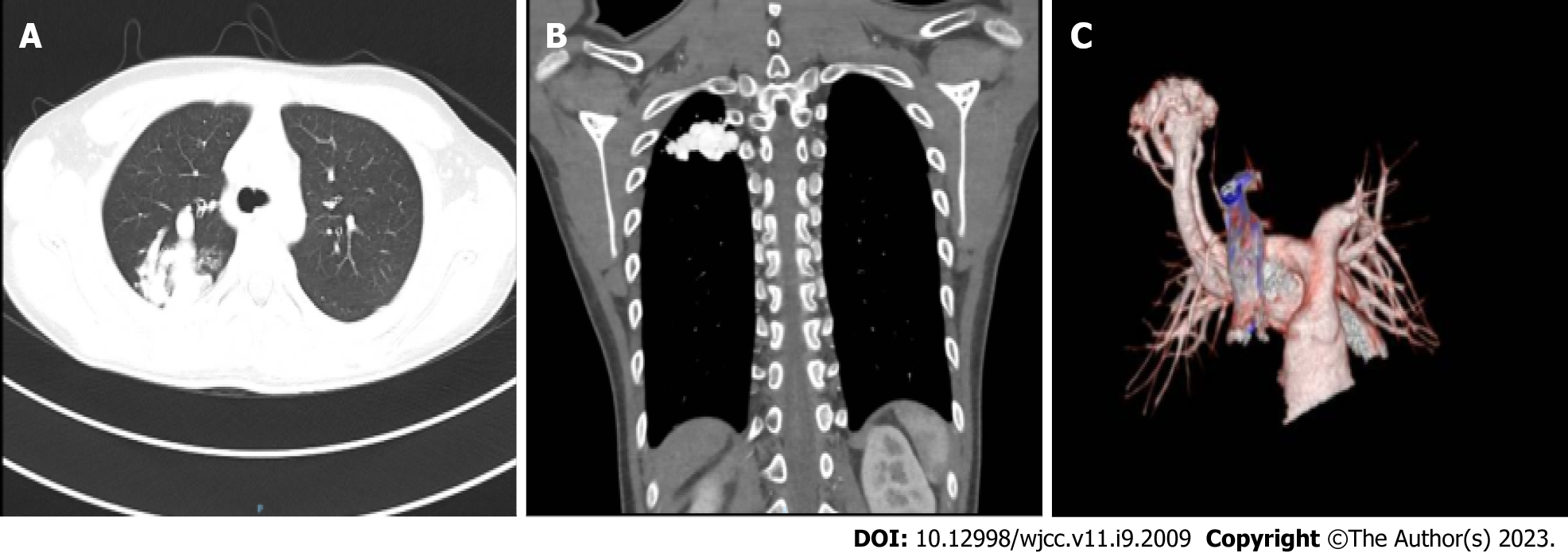
Figure 2 Cardiovascular computed tomography angiography.
A and B: Representative images showing the abnormal vascular nest in the right upper lung; C: Expansion of the right upper pulmonary artery with thickened and twisted branching vessels to form an abnormal vascular nest with direct reflux into the right upper pulmonary posterior vein. The artery finally merged into the right upper pulmonary vein.
- Citation: Zheng J, Wu QY, Zeng X, Zhang DF. Transient ischemic attack induced by pulmonary arteriovenous fistula in a child: A case report. World J Clin Cases 2023; 11(9): 2009-2014
- URL: https://www.wjgnet.com/2307-8960/full/v11/i9/2009.htm
- DOI: https://dx.doi.org/10.12998/wjcc.v11.i9.2009


