Published online Nov 26, 2023. doi: 10.12998/wjcc.v11.i33.8084
Peer-review started: September 14, 2023
First decision: October 17, 2023
Revised: October 28, 2023
Accepted: November 14, 2023
Article in press: November 14, 2023
Published online: November 26, 2023
Processing time: 62 Days and 7 Hours
Granuloma annulare (GA) has diverse clinical manifestations including papules, plaques, and nodules on the extremities that are skin-colored, pink, or purple. Approximately 15% of all GA cases are considered generalized GA.
Herein, we describe the case of a pediatric patient who initially presented with papules and later developed generalized atrophic macules. Upon examination, two different morphologic lesions were histopathologically confirmed: Epithelioid nodular GA and scattered histiocytic infiltrative GA. This patient exhibited rare clinical manifestations that differed throughout the course of the disease. The varying histopathological types and clinical manifestations of GA may be linked to the different stages of the disease.
This rare case demonstrates the different histopathological features of different stages and clinical manifestations of granuloma annulare in an infant.
Core Tip: Generalized papules were discovered in the early stages of the disease, and the histopathology was epithelioid nodular granuloma annulare (GA). Later in disease progression, the lesions transformed from papules to atrophic macules, and the corresponding histopathology displayed scattered histiocytic infiltrating GA. The clinical manifestations of this case included rare, generalized papules and atrophic maculae. The disease’s clinical manifestations varied at different stages, and the corresponding histopathological types differed as well. This suggests that the distinct histopathological types and clinical manifestations of GA may be connected to disease progression.
- Citation: Zhang DY, Zhang L, Yang QY, Li J, Jiang HC, Xie YC, Shu H. Generalized granuloma annulare in an infant clinically manifested as papules and atrophic macules: A case report. World J Clin Cases 2023; 11(33): 8084-8088
- URL: https://www.wjgnet.com/2307-8960/full/v11/i33/8084.htm
- DOI: https://dx.doi.org/10.12998/wjcc.v11.i33.8084
Granuloma annulare (GA) is a non-infectious, benign, self-limiting, granulomatous inflammation that typically affects patients aged 3–50 years, with an incidence of approximately 0.04%. Although some small, single-center studies have suggested that GA may be common in children[1-3], the etiology and pathogenesis of this condition are unknown, its clinical manifestations are diverse, and it can present in various forms. GA typically appears as papules, plaques, and nodules on the extremities, with skin-colored, pink, or violaceous hues. However, its diagnosis primarily relies on histopathological examination. The most common form of GA is localized, and generalized GA accounts for only about 15% of all cases[4]. Herein, we describe the case of an infant with the rare primary clinical manifestation of a generalized pitting macule present across the entire body that was histopathologically confirmed as GA.
The patient was a female infant (aged 3 mo and 15 d) who had visited the Department of Dermatology at Kunming Children’s Hospital in April 2022 owing to scattered papules and pitting macules on the trunk and limbs that had lasted for more than 10 d.
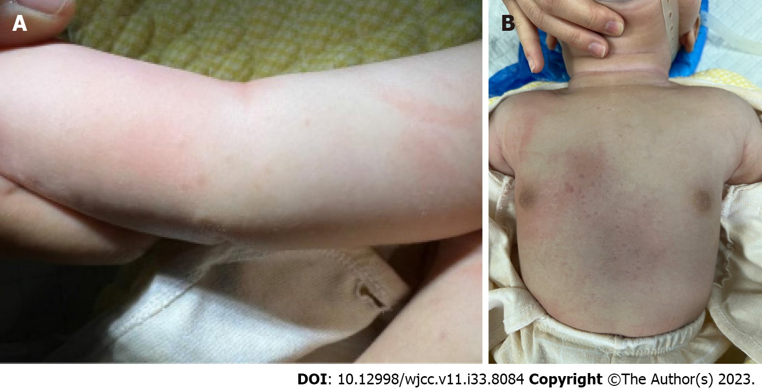
The child had originally developed papules that ranged in size from approximately 0.1–0.5 cm in diameter with a reddish–light-brown surface and a tough texture on the trunk and limbs without a clear cause. With an absence of subjective symptoms, the number of papules gradually increased and evolved into white pitting macules with a scar-like appearance and normal texture (Figures 1A and B).
No special notes.
Since the onset of the disease, the child had maintained a normal demeanor and appetite. The patient’s history included birth at term, normal postnatal growth and development, normal prenatal examination, and no maternal history of disease during pregnancy. The child had received her first doses of the Bacillus Calmette Guerin (BCG) and hepatitis B vaccines before the onset of GA, following typical vaccination after birth.
There were scattered papules on the limbs and trunk, mostly on the limbs, with a diameter of about 0.1–0.5 cm. These papules had a reddish-light-brown surface and had a tough texture with clear boundaries. The trunk also had pitting macules, which were distributed randomly and appeared as light white scars. The texture of the affected skin appeared normal. (Figures 1A and B).
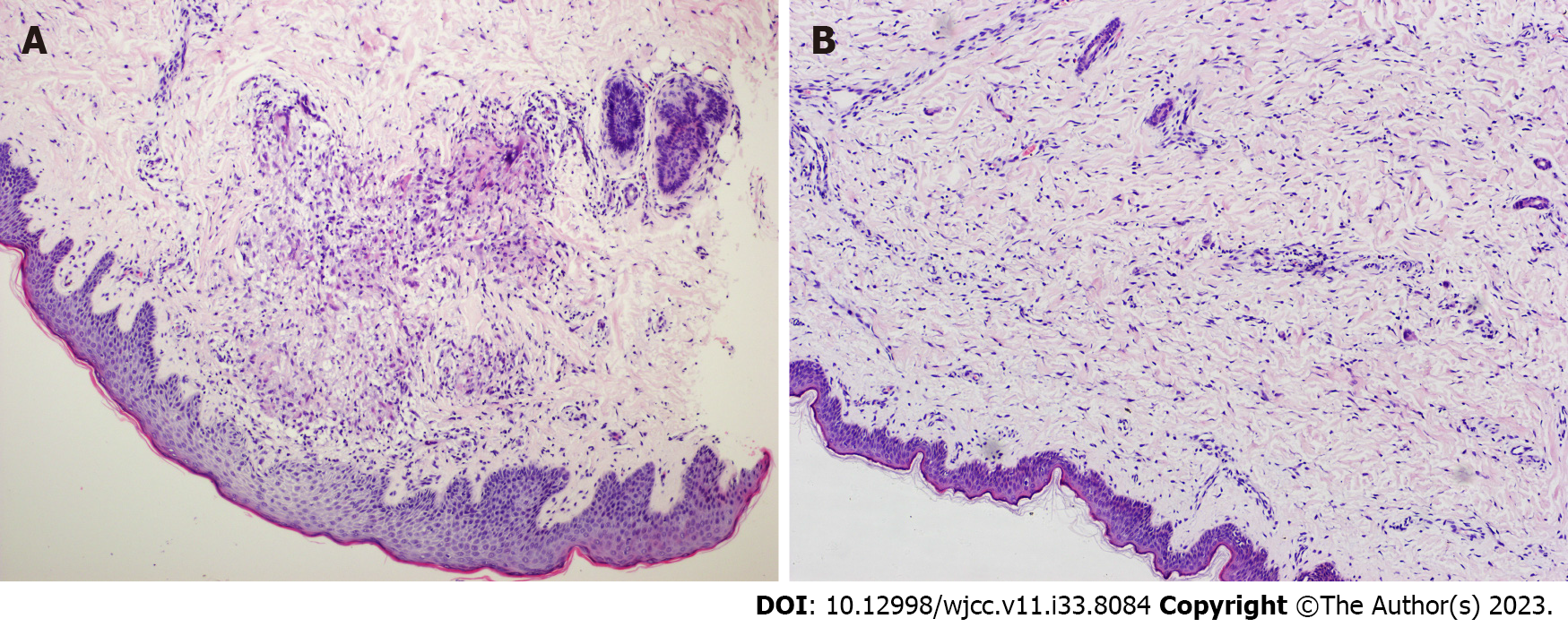
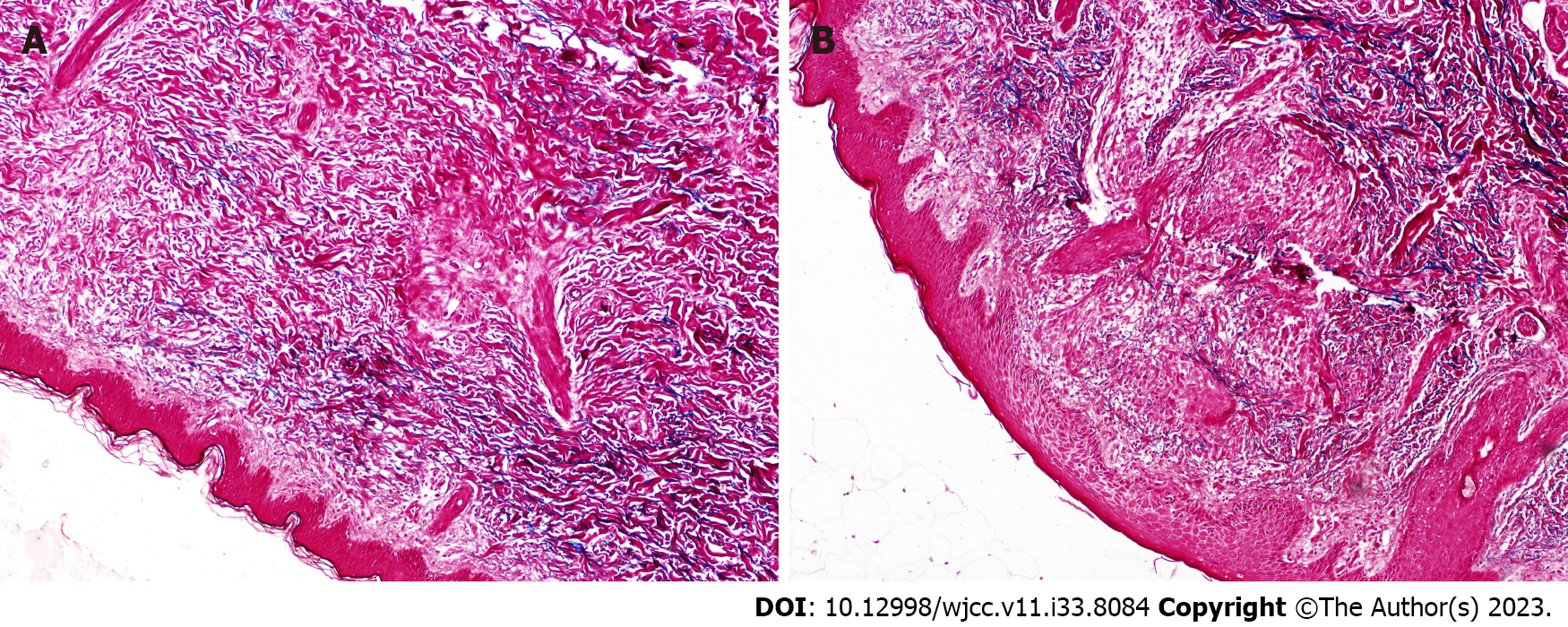
Tissue samples were obtained from each of the papules and pitting macules for pathological examination. The histopathological findings from the papular samples showed focal mild thickening of the epidermis and nested infiltration of lymphocytes, histiocytes, and multinucleated giant cells in the superficial middle dermis. Some collagen was disorganized (Figure 2A). Findings from the pitting macule samples showed approximately normal epidermis; however, the lymphocytes and histiocytes were found to have infiltrated the area between collagen fibers and around blood vessels in the superficial medial dermis, and some collagen fibers were degenerated with widened intervals (Figure 2B). Immunohistochemistry showed the following results: CD1α (+), S-100 (scattered+), langerin (-), Ki-67 (+, 20%), CD163 (+), and CD117 (-). Further, special staining of elastic fibers showed decreased or absent elastic fibers in some areas (Figure 3A and B), and negative alcian blue staining was applied.
No special notes.
According to their patterns of histiocytic infiltration[2], the papular lesions were histopathologically classified as epithelioid nodular GA, and the scar-like lesions in the depressions as scattered histiocytic infiltrative GA.
GA is considered to be a self-limiting disease in clinical practice. Therefore, after being fully informed, the parents agreed to observe the progression of the disease for a short term. No treatment was provided, and the patient was advised to return regularly for follow-up.
During follow-up, papular lesions were observed to gradually evolve into pale white, depressed, scar-like lesions that slowly subsided without scarring. On a follow-up visit in September 2022, the skin lesions had largely resolved (Supplementary Figure 1).
The etiology of GA is unclear; however, reports suggest a potential association with vaccination[5]. In this case, the previously healthy child had received the BCG and hepatitis B vaccines shortly before disease onset; thus, the relationship between GA and vaccination could not be completely ruled out.
This was a case of generalized GA with the first symptom being scattered papules on the trunk and extremities that gradually evolved into atrophic macules. The untreated skin lesions subsided spontaneously and no scarring remained after healing, which is consistent with the self-limiting nature of this disease.
GA has previously been divided into four histopathological types: Scattered histiocytic infiltrative, palisading granuloma, mixed histologic, and epithelioid nodule types[6]. As the patient in this case presented with papules in the early stage of the disease, the type was histopathologically classified as epithelioid nodular GA. While the lesions presented as atrophic macules after resolution of the papules, the child later presented with scattered histiocytic infiltrative GA (with the lesions histopathologically showing decreased cellularity). For this patient, two distinct pathological patterns corresponded to the two lesions, which represented different stages of the disease. Whether the cellular infiltration pattern and pathological type of GA were related to disease stage requires confirmation by future research.
Recent studies have shown that activation of the Th1, Th2, and JAK-STAT pathways are involved in the pathogenesis of GA[7]. In this patient, alcian blue staining was negative in both lesions; however, elastic fibers appeared to dissolve and disappear to approximately the same extent, suggesting there is breakage and phagocytosis of elastic fibers and collagen in the whole disease process of GA. Prospective analysis with larger samples is required to verify this.
However, the analysis and speculation that these events occurred in this particular case require additional prospective and baseline studies.
This suggests that there is breakage and phagocytosis of elastic fibers and collagen in the whole disease process of GA, but the specific mechanism needs more prospective analysis of large samples to verify.
The clinical manifestations of GA in children are diverse and vary in different stages of the disease, and GA should be taken into account in children who encounter such generalized and atrophic depression macula in the clinic.
Provenance and peer review: Unsolicited article; Externally peer reviewed.
Peer-review model: Single blind
Specialty type: Dermatology
Country/Territory of origin: China
Peer-review report’s scientific quality classification
Grade A (Excellent): 0
Grade B (Very good): B
Grade C (Good): 0
Grade D (Fair): 0
Grade E (Poor): 0
P-Reviewer: Manestar-Blazić T, Croatia S-Editor: Liu JH L-Editor: A P-Editor: Liu JH
| 1. | Barbieri JS, Rodriguez O, Rosenbach M, Margolis D. Incidence and Prevalence of Granuloma Annulare in the United States. JAMA Dermatol. 2021;157:824-830. [RCA] [PubMed] [DOI] [Full Text] [Cited by in Crossref: 9] [Cited by in RCA: 34] [Article Influence: 8.5] [Reference Citation Analysis (0)] |
| 2. | Joshi TP, Duvic M. Granuloma Annulare: An Updated Review of Epidemiology, Pathogenesis, and Treatment Options. Am J Clin Dermatol. 2022;23:37-50. [RCA] [PubMed] [DOI] [Full Text] [Full Text (PDF)] [Cited by in Crossref: 3] [Cited by in RCA: 59] [Article Influence: 19.7] [Reference Citation Analysis (0)] |
| 3. | Leasure AC, Damsky W, Cohen JM. Prevalence of granuloma annulare in the United States: a cross-sectional study in the All of Us Research Program. Int J Dermatol. 2022;61:e301-e302. [RCA] [PubMed] [DOI] [Full Text] [Cited by in Crossref: 1] [Cited by in RCA: 7] [Article Influence: 1.8] [Reference Citation Analysis (0)] |
| 4. | Ehret M, Lenormand C, Scrivener JN, Gusdorf L, Lipsker D, Cribier B. [Generalized granuloma annulare: A clinicopathological study]. Ann Dermatol Venereol. 2020;147:271-278. [RCA] [PubMed] [DOI] [Full Text] [Cited by in Crossref: 2] [Cited by in RCA: 2] [Article Influence: 0.4] [Reference Citation Analysis (0)] |
| 5. | García-Gil MF, Álvarez-Salafranca M, Martínez García A, Ara-Martín M. Generalized granuloma annulare after pneumococcal vaccination. An Bras Dermatol. 2021;96:59-63. [RCA] [PubMed] [DOI] [Full Text] [Full Text (PDF)] [Cited by in Crossref: 4] [Cited by in RCA: 8] [Article Influence: 1.6] [Reference Citation Analysis (0)] |
| 6. | Friedman-Birnbaum R, Weltfriend S, Munichor M, Lichtig C. A comparative histopathologic study of generalized and localized granuloma annulare. Am J Dermatopathol. 1989;11:144-148. [RCA] [PubMed] [DOI] [Full Text] [Cited by in Crossref: 24] [Cited by in RCA: 16] [Article Influence: 0.4] [Reference Citation Analysis (0)] |
| 7. | Min MS, Wu J, He H, Sanz-Cabanillas JL, Del Duca E, Zhang N, Renert-Yuval Y, Pavel AB, Lebwohl M, Guttman-Yassky E. Granuloma annulare skin profile shows activation of T-helper cell type 1, T-helper cell type 2, and Janus kinase pathways. J Am Acad Dermatol. 2020;83:63-70. [RCA] [PubMed] [DOI] [Full Text] [Cited by in Crossref: 17] [Cited by in RCA: 50] [Article Influence: 8.3] [Reference Citation Analysis (0)] |


