


Published online Oct 16, 2023. doi: 10.12998/wjcc.v11.i29.7061
Peer-review started: July 20, 2023
First decision: August 30, 2023
Revised: September 4, 2023
Accepted: September 18, 2023
Article in press: September 18, 2023
Published online: October 16, 2023
Processing time: 85 Days and 8.3 Hours
Gait is influenced by race, age, and diseases type. Reference values for gait are closely related to numerous health outcomes. To gain a comprehensive under
To establish reference values for lower extremity joint kinematics and kinetics during gait in asymptomatic adult women and men.
Spatiotemporal, kinematics and kinetics parameters were measured in 171 healthy adults (70 males and 101 females) using the computer-aided soft tissue foot model. Full curve statistical parametric mapping was performed using independent and paired-samples
Compared with females, males required more time (cycle time, double-limb support time, stance time, swing time, and stride time), and the differences were statistically significant. In addition, the step and stride lengths of males were longer. Compared to males, female cadence was faster, and statures-per-second and stride-per-minute were higher. There were no statistical differences in speed and stride width between the two groups. After adjusting for height, it was observed that women walked significantly faster than men, and they also had a higher cadence. However, in terms of step length, stride length, and stride width, both genders exhibited similarities.
We established reference values for gait speed and spatiotemporal gait parameters in Chinese university students. This contributes to a valuable database for gait assessment and evaluation of preventive or rehabilitative programs.
Core Tip: In this observational study, gait parameters of healthy Chinese university students were provided, and gender differences in gait as well as differences compared to other ethnicities were observed. These findings may contribute to localized clinical guidance and reference for the region.
- Citation: Yu JS, Zhuang C, Guo WX, Chen JJ, Wu XK, Xie W, Zhou X, Su H, Chen YX, Wang LK, Li WK, Tian K, Zhuang RJ. Reference values of gait parameters in healthy Chinese university students: A cross-sectional observational study. World J Clin Cases 2023; 11(29): 7061-7074
- URL: https://www.wjgnet.com/2307-8960/full/v11/i29/7061.htm
- DOI: https://dx.doi.org/10.12998/wjcc.v11.i29.7061
Gait is a fundamental characteristic of human walking, that can be influenced by factors such as race, age, and various diseases. Three-dimensional (3D) motion analysis systems are commonly employed for movement assessments in both clinical and research settings[1,2]. This approach requires a normal reference derived from a healthy population to distinguish it from abnormal conditions. Notably, the reference values may not be universally applicable to every race or age group[3].
In China, approximately 22.7% of college students reported experiencing sports-related injuries in the past year[4]. Lower extremity injuries, including anterior cruciate ligament rupture, acute ankle sprain, and meniscal injury, account for nearly two-thirds of all reported injuries[5-8]. Numerous studies have examined gait changes in both African American and Caucasian populations[9,10]. Gait studies on Asians primarily focused on individuals aged 60 years and above[11,12]. Currently, the reference values for gait among college students remains inconclusive, leadings to overlook early changes in gait and, an increased incidence of osteoarthritis. Three-dimensional motion analysis can serve as an early screening tool for knee osteoarthritis (KOA) and a method of evaluating rehabilitation progress[13].
Due to variations in lower-extremity kinematics and kinetics across different races, ages, and sexes, as well as the limited availability of data on Chinese adults other than the elderly population, this study aimed to investigate three-dimensional lower extremity kinematics and kinetics in healthy Chinese university students. Specifically, we aimed to enhance our understanding of country-specific gait patterns and establish normative data as a reference for lower-extremity movements in all dimensions. Additionally, these new reference values will be beneficial for identifying abnormal motion patterns related to conditions such as chronic ankle instability, deformities, and neuromuscular disorders.
According to previous reports related to gait reference values, a sample size of 30 or more is considered sufficient for normative measurements[12]. In this study, we included participants aged between 18 and 35 years with a body mass index (BMI) between 18.5 and 23.9 kg/m², excluding individuals who were underweight or overweight, based on the World Health Organization's recommended normal weight range. Research has indicated that gait patterns mature around the age of 7 years[14], and certain gait parameters, such as walking speed, tend to decline after the age of 40 years[12]. Therefore, this study focused primarily on university students whose ages fell within the age range associated with mature gait patterns. Written informed consent was obtained from all participants who completed the three health-screening questionnaires prior to participation. In addition, participants were required to engage in exercise for at least 20 minutes, at least three times per week. Individuals with a pathological gait, serious cardiovascular and cerebrovascular diseases, mental and spiritual abnormalities, pregnant and lactating women, or those with contraindications to exercise owing to other medical conditions, were excluded. The inclusion and exclusion criteria are presented in Table 1.
| Inclusion | Exclusion |
| 18-35 yr old, BMI: 18.5-23.9 kg/m² | Acute lower limb injury in the past 6 mo |
| Participation in team sport a minimum of three times a week | Cardiovascular and cerebrovascular diseases |
| FAAM ≥ 98 | History of neurological disease |
| Lysholm knee score ≥ 95 | Pregnant and lactating women |
| Harris hip score ≥ 95 | Balance or motion disorders |
All volunteers provided informed consent before undergoing the motion analysis. Gait analysis was performed using a 3D motion capture system (Qualisys Track Manager, Qualisys, Sweden), consisting of eight high-speed infrared cameras (Oqus700+, Qualisys, Sweden) at a sampling frequency of 1100 Hz. Data were collected by synchronising four Kistler force plates (9260AA; Kistler, Switzerland). Gait data were analysed using modelling and simulation software (Visual 3D Professional V6, C-Motion Incorporation, United States). Lower limbs were analyzed using the Calibrated Anatomical System Technique model[15]. A total of 32 reflective markers were attached to the participants' bony landmarks and placed on the skin surface. Elastic bandages were used to secure the marker plates on the lateral upper two-thirds of the thigh and calf with four reflective markers on each plate. Before commencing data collection, the participants were familiarised with the walking procedure. This involved practising walking at their preferred speed within a 2-meter range in front of and behind the Kistler force plate recording area. The participants completed three rounds of practice walking to minimise the potential impact of acceleration and deceleration on the results. After ensuring that the participants were familiar with the walking procedure and that all the reflective markers were detected, the data from the three best gait cycles were averaged to calculate the joint angles for each participant. Gait speed (m/s), stride length (m), and stride width (m) were assessed, and kinematic and kinetic parameters were simultaneously recorded during the gait analysis.
Data on age, sex, weight (kg), height (cm), BMI (kg/m2), cadence, speed, cycle, stance, and swing times were collected. To ensure accuracy, we calibrated and corrected the geodetic coordinate systems and positions of the eight cameras. Subsequently, static and dynamic data were collected. For the dynamic data, we selected eight consecutive gait cycles performed by the participants at their preferred walking speeds, starting from the beginning of each walking trial. Data from each gait cycle were processed to represent the 0%–100% gait cycle. Finally, we averaged the values from the eight gait cycles to evaluate the kinematics and kinetics of the ankle, knee, and hip joints in each participant.
As height affects certain gait parameters, we normalised specific gait parameters based on height. Height-adjusted gait speed (gait speed/height)[16], height-adjusted step length (step length/height)[17], height-adjusted stride length (stride length/height), and height-adjusted stride width (stride width/height) were normalised to account for height, while height-adjusted cadence was normalised using the square root of height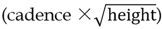 [18].
[18].
All parameters are described using means and standard deviations, and the data were analysed using SPSS 25.0. An independent sample t-test was employed for data conforming to a normal distribution, whereas the rank sum test was used for data that did not conform to a normal distribution. Statistical parametric mapping (SPM) was conducted to visualise the complete time series of angle, moment, and power assessments. Two-tailed paired t-tests were used to compare kinematics and kinetics. SPM analyses were performed in MATLAB (R2020b, The MathWorks Inc) using an open-source code (M.0.4.8, www.spm1d.org). The significance level was set at P = 0.05.
In total, 171 volunteers participated in this study, with an average age of 24.63 ± 2.19 years. Of them, 101 (59%) were female. The mean height was 1.68 ± 0.08 cm, and the mean weight was 58.59 ± 10.06 kg. The average BMI was 20.60 ± 2.15 kg/m2. The demographic data are presented in Table 2.
| Characteristics | Female | Male | Total |
| Age (yr), mean (SD) | 24.50 (2.18) | 24.81 (2.19) | 24.63 (2.19) |
| Gender (n, %) | 101 (59.06%) | 70 (40.93%) | |
| Height (m), mean (SD) | 1.62 (0.0) | 1.75 (0.05) | 1.68 (0.08) |
| Weight (kg), mean (SD) | 52.14 (5.10) | 67.90 (7.93) | 58.59 (10.06) |
| BMI (kg/m2), mean (SD) | 19.72 (1.89) | 21.87 (1.86) | 20.64 (2.16) |
| FAAM, mean (SD) | 100 (0.00) | 100 (0.00) | 100 (0.00) |
| Lysholm knee score, mean (SD) | 100 (0.00) | 100 (0.00) | 100 (0.00) |
| Harris hip score, mean (SD) | 100 (0.00) | 100 (0.00) | 100 (0.00) |
The spatiotemporal parameters of the participants are listed in Table 3. Compared with females, males required more time in terms of cycle time, double-limb support time, stance time, swing time, and stride time, with statistically significant differences. In addition, males exhibited longer steps and stride lengths. Conversely, females demonstrated faster cadence, higher steps per second, and more strides per minute than males. There were no statistically significant differences in speed or stride width between the sexes. However, after adjusting for height, women walked significantly faster than men (0.06 m/s, P < 0.05) (Table 3). Additionally, even after height adjustment, women maintained a significantly higher cadence than did men (P < 0.05), whereas the significant differences in step and stride lengths between men and women disappeared. There were no significant differences in the stride width between men and women.
| Variables | Mean (SD) | P value | |
| Male | Female | ||
| Cadence (steps/min) | 113.35 (11.05) | 118 (7.63) | < 0.05 |
| Height-adjusted cadence | 1503.74 (151) | 1539.60 (80) | < 0.05 |
| Speed (m/s) | 1.24 (0.17) | 1.22 (0.11) | 0.35 |
| Height-adjusted gait speed (cm/s) | 0.70 (0.09) | 0.76 (0.06) | < 0.05 |
| Step length (m) | 0.66 (0.09) | 0.62 (0.05) | < 0.05 |
| Height-adjusted step length (cm) | 0.37 (0.05) | 0.38 (0.02) | 0.60 |
| Stride length (m) | 1.31 (0.13) | 1.24 (0.29) | < 0.05 |
| Height-adjusted stride length (cm) | 0.75 (0.07) | 0.76 (0.05) | 0.30 |
| Stride width (m) | 0.11 (0.03) | 0.10 (0.03) | 0.07 |
| Height-adjusted stride width (cm) | 0.06 (0.01) | 0.06 (0.01) | 0.42 |
| Cycle time (sec) | 1.07 (0.09) | 1.01 (0.07) | < 0.05 |
| Double limb support time (sec) | 0.23 (0.04) | 0.21 (0.03) | < 0.05 |
| Stance time (%GC) | 0.65 (0.06) | 0.61 (0.05) | < 0.05 |
| Swing time (%GC) | 0.42 (0.04) | 0.40 (0.03) | < 0.05 |
| Statures-per second | 0.71 (0.10) | 0.77 (0.08) | < 0.05 |
| Stride time (sec) | 1.08 (0.08) | 1.02 (0.08) | < 0.05 |
| Stride-per minute | 55.94 (4.65) | 59.72 (3.40) | < 0.05 |
As shown in Figure 1, we gathered the motion parameters for the hip, knee, and ankle joints in various planes. Significant differences were observed in the joint kinematics and kinetics of the ankles, knees, and hips among all participants. The detailed kinematics and kinetics of the ankle, knee, and hip are shown in Figures 2, 3, and 4, respectively.


Regarding the hip joint, sex-based differences were significant in sagittal hip flexion from 0%–13% and from 75%–100% of the gait cycle (P = 0.030 and P = 0.007, respectively). Moreover, significant differences were observed in the frontal plane of the hip joint during the 4%–21% (P = 0.015), 57%–66% (P = 0.034), and 86%–91% (P = 0.043) gait cycles. In the transverse planes of motion, significant differences were observed between the groups from 78%–100% of the gait cycle (P = 0.009).
No significant differences were observed in peak hip extensor moment during the early stance. However, significant differences were observed between 67% and 77% of the gait cycles (P < 0.001). Significant differences in hip power were observed during 68%–77% of the gait cycle (P < 0.001). No significant differences were observed in hip abductor moment.
At the knee joint, a significant sex-based difference was observed in sagittal knee flexion from 0%–100% of the gait cycle (P < 0.001), indicating differences in the range of knee movements during walking. However, no significant differences were observed in the frontal knee adduction. In the transverse planes of motion, particularly during 90%–97% of the gait cycle, there was high variability among the participants (P = 0.028).
Regarding knee kinetics, no statistically significant differences were detected in the knee extensor moment or knee power. However, differences were observed between the two smaller intervals in knee valgus moment (P = 0.018 and P = 0.046, respectively).
At the ankle joint, no significant sex-based differences were observed in the ankle plantarflexion, dorsiflexion, inversion, or eversion. However, significant sex differences were found in the ankle eversion moment at the initial contact (P < 0.001) and from 58%–62% of the gait cycle (P = 0.013). No significant differences were detected in ankle plantarflexor moment or ankle power.
In this study, we established inclusion and exclusion criteria based on previous research on gait reference values[11,12]. Furthermore, we utilized three assessment tools to evaluate the physical activity functionality of participants. These tools include the Foot and Ankle Ability Measure, designed to assess the muscular and musculoskeletal function of the lower extremities, feet, and ankle joints[19]; the Lysholm knee score, employed to evaluate general knee joint conditions and functionality[20,21]; and the Harris Hip Score, utilized to assess hip pathology and health-related quality of life in daily activities[22,23]. All three assessment tools were validated as responsive, reliable, and effective evaluation instruments; thus, supporting the definition of a healthy population and a mature gait pattern. Building on this foundation, we investigated the spatiotemporal parameters, kinematics, and kinetics of the gait of Chinese college students. We observed that women had significantly faster cadence, higher statures-per-second, and strides-per-minute, accompanied by longer step lengths. However, women exhibited significantly shorter stride, step, cycle, double-limb support, stance, swing, and stride times than men did. No significant sex differences were observed in speed or stride width. After adjusting for height, it became evident that women walked significantly faster than men (0.06 m/s, P < 0.05) (Table 3). Importantly, even when height differences were considered, women still exhibited a significantly higher cadence than men (P < 0.05). Interestingly, the previously observed significant differences in the step and stride lengths between males and females disappeared. Regarding stride width, no significant differences were found between men and women. These findings on sex differences in cadence, gait speed, and cycle time are consistent with previous studies conducted in laboratory settings, which reported that women had a faster cadence and shorter cycle time than men[24]. Compared to individuals from the Southeast Asian region, our study found that Chinese participants had slightly faster walking speeds (men: 1.14 m/s for Southeast Asians, 1.24 m/s for Chinese; women: 1.13 m/s for Southeast Asians, 1.22 m/s for Chinese)[12]. However, sex differences in stride width tended to be similar to those observed in Southeast Asian population. Additionally, the Chinese participants in our study exhibited longer step lengths and shorter double-limb support times than the Southeast Asian population. Furthermore, they exhibited faster cadence compared to Southeast Asian and Korean populations[25]. A faster cadence can lead to increased wear of the knee joint, which may contribute to the incidence and risk factors of symptomatic KOA in Chinese women[26].
Our findings demonstrated significant sex-based differences in the kinematic and kinetic gait features of the three major lower-limb joints. These differences were primarily observed in the hip and knee joints rather than the ankle joint, indicating overall pattern differences throughout the gait cycle. Compared to previous studies, our results also showed variations in gait patterns. Previous studies have consistently shown that females exhibit lower overall transverse plane hip internal rotation angles[27], and smaller ankle plantarflexor moments throughout most stance phases[25]. In contrast, our findings revealed that females had higher overall transverse plane hip internal rotation angles and no significant differences were observed in ankle plantarflexor moments. These discrepancies may be attributed to the variations in the gait speed of the participants. Although there are differences between men and women in many gait features, the effect sizes of some of these differences are relatively small; therefore, individual features may not fully explain these significant differences. However, when these small differences were considered, an overall difference in gait cycle between healthy males and females becomes evident. Our findings are in agreement with those of previous studies that investigated sex differences in gait kinematics among older adults[28-30]. Specifically, in our study, healthy females exhibited significantly greater hip adduction during the stance phase of gait than healthy males. Our results are similar to most previous studies that reported differences in the sagittal, frontal, and transverse plane hip joint angles between younger healthy males and females during walking[31,32]. In contrast to investigations involving healthy middle-aged and older adults[25,33], our study found no significant differences in knee adduction angles, or ankle plantarflexion and inversion angles between healthy younger males and females. These contradictory findings may be attributed to subtle changes in gait associated with biological aging[34]. The mean age of the participants in the current study was 24.92 ± 2.25 years, while the aforementioned studies, included adults in their forties or sixties. Therefore, the sex-specific gait kinematic differences in healthy younger adults differ from those previously found in healthy older adults.
In this study, spatiotemporal gait parameters, as well as kinetic and kinematic parameters were analyzed to assess sex differences and movement patterns. Future studies should investigate the impact of age on gait parameters using a comprehensive curve analysis to identify group differences. Additionally, these analytical methods should be employed to investigate dynamic motion and its association with various conditions such as changes in orientation and gait patterns during single- and double-leg landing, running, and KOA. Examining movement patterns across different activities can provide a better understanding of potential disparities between men and women. This could be particularly relevant for patients with KOA because women have a higher incidence of this condition.
This study has several limitations. First, the small sample size prevented us from conducting intragroup analysis based on sex. Future studies with larger sample sizes are needed to comprehensively investigate the potential sex differences within each group. Secondly, this study only captured gait information during a single period. Future studies should consider collecting gait data from the same individuals in different age groups to examine gait across different age ranges. Finally, our study did not report any specific muscle activity during walking. Future equipment improvements will allow for a more comprehensive analysis of the lower limb muscle work and other related parameters.
This study aimed to investigate the motion parameters of the hip, knee, and ankle joints in Chinese adults aged 18–35 years. These findings highlighted the importance of considering sex-based differences in gait biomechanics in future studies. Our results indicated significant differences between men and women in the range of motion of the knee joint in the sagittal plane, whereas smaller differences were observed in the range of motion of the hip joint. No significant differences were observed in the ankle joint range of motion between males and females, suggesting a potentially distinct gait pattern for each sex. Although our study focused on Chinese college students, further population-based research is recommended to explore gait parameters across different age groups.
Gait refers to the movement patterns and rhythms of various body parts during walking, making it a crucial indicator of human movement and overall health. Currently, there is a relatively limited studies on the gait characteristics of Chinese male and female populations. To address this gap and provide more precise reference values for the region, we aimed to collect gait data from healthy Chinese university students and conduct a comprehensive analysis of sex-based differences.
Through this study, we aimed to reveal the commonalities and disparities in gait between Chinese men and women, thereby providing a more accurate foundation and reference for future clinical diagnosis and treatment. This research could have a positive impact on improving diagnosis and rehabilitation measures for relevant conditions, as well as optimizing sports training and exercise rehabilitation programs.
We collected gait data from healthy university students in China and conducted a detailed analysis of their gait characteristics, including stride length, step frequency, and gait cycle. Through this study, we will gain a deeper understanding of sex-based differences in the gait characteristics of male and female individuals in China, providing valuable data and guidance for medical and rehabilitation practices in this region. Additionally, these findings have the potential to optimize sports training and exercise rehabilitation programs, and promote overall health and physical performance.
A total of 171 volunteers participated in this study, with an average age of 24.63 ± 2.19 years. Baseline data of the participants were collected, including gender, age, height, weight, body mass index, Foot and Ankle Ability Measure, Lysholm knee score, and Harris hip score. Gait data were captured using the Qualisys three-dimensional motion capture system. Data analysis was conducted using SPSS 25.0 and MATLAB (R2020b, The MathWorks Inc).
In this study, significant differences were observed between males and females in the range of motion of the knee joint in the sagittal plane, while smaller differences were observed in the range of motion of the hip joint. No significant differences were observed in the range of motion of the ankle joint between males and females, suggesting the existence of potentially distinct gait patterns for each sex. This study provides more accurate evidence for future clinical diagnosis and treatment.
In future studies, we will increase the sample size and collect gait data from the same participants in different age groups to study changes in gait across various age ranges.
This study utilized statistical parameter mapping to analyze the gait characteristics of males and females throughout the gait cycle, providing motion parameters for the hip, knee, and ankle joints. These findings highlighted the importance of considering sex-based differences in gait biomechanics in future studies.
When guiding treatment plans, healthcare providers should be vigilant of the varying expectations of different sexes and racial or ethnic groups. Considering these expectations is crucial to enhance patient compliance and improve treatment outcomes.
We express our heartfelt gratitude to the Gait Analysis Laboratory at Zhejiang Traditional Chinese Medicine Hospital for providing the necessary resources and facilities, which greatly facilitated the progress of this study. We sincerely appreciate the cooperation, advice, and assistance of the research team members in data collection and analysis. We also extend our gratitude to the volunteers who willingly participated in this study. We are deeply thankful to the aforementioned individuals and organizations for their invaluable support and contributions, which were vital in achieving the objectives of this research.
Provenance and peer review: Unsolicited article; Externally peer reviewed.
Peer-review model: Single blind
Specialty type: Medicine, research and experimental
Country/Territory of origin: China
Peer-review report’s scientific quality classification
Grade A (Excellent): 0
Grade B (Very good): B
Grade C (Good): C
Grade D (Fair): 0
Grade E (Poor): 0
P-Reviewer: Ito S, Japan S-Editor: Lin C L-Editor: A P-Editor: Yu HG
| 1. | Tosserams A, Keijsers N, Kapelle W, Kessels RPC, Weerdesteyn V, Bloem BR, Nonnekes J. Evaluation of Compensation Strategies for Gait Impairment in Patients With Parkinson Disease. Neurology. 2022;99:e2253-e2263. [RCA] [PubMed] [DOI] [Full Text] [Full Text (PDF)] [Cited by in Crossref: 5] [Cited by in RCA: 14] [Article Influence: 4.7] [Reference Citation Analysis (0)] |
| 2. | Spiker AM, Kraszewski AP, Maak TG, Nwachukwu BU, Backus SI, Hillstrom HJ, Kelly BT, Ranawat AS. Dynamic Assessment of Femoroacetabular Impingement Syndrome Hips. Arthroscopy. 2022;38:404-416.e3. [RCA] [PubMed] [DOI] [Full Text] [Cited by in Crossref: 4] [Cited by in RCA: 9] [Article Influence: 3.0] [Reference Citation Analysis (0)] |
| 3. | Hill CN, Reed W, Schmitt D, Arent SM, Sands LP, Queen RM. Factors contributing to racial differences in gait mechanics differ by sex. Gait Posture. 2022;95:277-283. [RCA] [PubMed] [DOI] [Full Text] [Cited by in RCA: 4] [Reference Citation Analysis (0)] |
| 4. | Gao Y, Cai W, Gao L, Wang J, Liang J, Kwok H, Jia C, Li L. Physical activity-related injuries among university students: a multicentre cross-sectional study in China. BMJ Open. 2018;8:e021845. [RCA] [PubMed] [DOI] [Full Text] [Full Text (PDF)] [Cited by in Crossref: 13] [Cited by in RCA: 18] [Article Influence: 2.6] [Reference Citation Analysis (0)] |
| 5. | Adams BG, Houston MN, Cameron KL. The Epidemiology of Meniscus Injury. Sports Med Arthrosc Rev. 2021;29:e24-e33. [RCA] [PubMed] [DOI] [Full Text] [Cited by in Crossref: 30] [Cited by in RCA: 69] [Article Influence: 17.3] [Reference Citation Analysis (0)] |
| 6. | Boden BP, Torg JS, Knowles SB, Hewett TE. Video analysis of anterior cruciate ligament injury: abnormalities in hip and ankle kinematics. Am J Sports Med. 2009;37:252-259. [RCA] [PubMed] [DOI] [Full Text] [Cited by in Crossref: 268] [Cited by in RCA: 290] [Article Influence: 18.1] [Reference Citation Analysis (0)] |
| 7. | Doherty C, Delahunt E, Caulfield B, Hertel J, Ryan J, Bleakley C. The incidence and prevalence of ankle sprain injury: a systematic review and meta-analysis of prospective epidemiological studies. Sports Med. 2014;44:123-140. [RCA] [PubMed] [DOI] [Full Text] [Cited by in Crossref: 692] [Cited by in RCA: 562] [Article Influence: 51.1] [Reference Citation Analysis (0)] |
| 8. | Trease L, Wilkie K, Lovell G, Drew M, Hooper I. Epidemiology of injury and illness in 153 Australian international-level rowers over eight international seasons. Br J Sports Med. 2020;54:1288-1293. [RCA] [PubMed] [DOI] [Full Text] [Cited by in Crossref: 8] [Cited by in RCA: 22] [Article Influence: 4.4] [Reference Citation Analysis (0)] |
| 9. | Bay AA, Ramachandran S, Ni L, Prusin T, Hackney ME. Differences in Balance Confidence, Fear of Falling, and Fall Risk Factors Among White and Black Community-Dwelling Older Adults. J Geriatr Phys Ther. 2023;46:122-131. [RCA] [PubMed] [DOI] [Full Text] [Cited by in RCA: 1] [Reference Citation Analysis (0)] |
| 10. | Hill CN, Reed W, Schmitt D, Sands LP, Queen RM. Racial differences in gait mechanics. J Biomech. 2020;112:110070. [RCA] [PubMed] [DOI] [Full Text] [Cited by in Crossref: 3] [Cited by in RCA: 12] [Article Influence: 2.4] [Reference Citation Analysis (0)] |
| 11. | Kawai H, Taniguchi Y, Seino S, Sakurai R, Osuka Y, Obuchi S, Watanabe Y, Kim H, Inagaki H, Kitamura A, Awata S, Shinkai S. Reference values of gait parameters measured with a plantar pressure platform in community-dwelling older Japanese adults. Clin Interv Aging. 2019;14:1265-1276. [RCA] [PubMed] [DOI] [Full Text] [Full Text (PDF)] [Cited by in Crossref: 11] [Cited by in RCA: 21] [Article Influence: 3.5] [Reference Citation Analysis (0)] |
| 12. | Lau LK, Wee SL, Pang WJB, Chen KK, Abdul Jabbar K, Yap PLK, Mallya JU, Ng DHM, Tan QLL, Seah WT, Ng TP. Reference Values of Gait Speed and Gait Spatiotemporal Parameters for a South East Asian Population: The Yishun Study. Clin Interv Aging. 2020;15:1753-1765. [RCA] [PubMed] [DOI] [Full Text] [Full Text (PDF)] [Cited by in Crossref: 10] [Cited by in RCA: 25] [Article Influence: 5.0] [Reference Citation Analysis (0)] |
| 13. | Bucinskas V, Dzedzickis A, Rozene J, Subaciute-Zemaitiene J, Satkauskas I, Uvarovas V, Bobina R, Morkvenaite-Vilkonciene I. Wearable Feet Pressure Sensor for Human Gait and Falling Diagnosis. Sensors (Basel). 2021;21. [RCA] [PubMed] [DOI] [Full Text] [Full Text (PDF)] [Cited by in Crossref: 5] [Cited by in RCA: 11] [Article Influence: 2.8] [Reference Citation Analysis (0)] |
| 14. | Hillman SJ, Stansfield BW, Richardson AM, Robb JE. Development of temporal and distance parameters of gait in normal children. Gait Posture. 2009;29:81-85. [RCA] [PubMed] [DOI] [Full Text] [Cited by in Crossref: 42] [Cited by in RCA: 43] [Article Influence: 2.7] [Reference Citation Analysis (0)] |
| 15. | Cappozzo A, Catani F, Croce UD, Leardini A. Position and orientation in space of bones during movement: anatomical frame definition and determination. Clin Biomech (Bristol, Avon). 1995;10:171-178. [RCA] [PubMed] [DOI] [Full Text] [Cited by in Crossref: 1147] [Cited by in RCA: 1081] [Article Influence: 36.0] [Reference Citation Analysis (0)] |
| 16. | Seino S, Shinkai S, Fujiwara Y, Obuchi S, Yoshida H, Hirano H, Kim HK, Ishizaki T, Takahashi R; TMIG-LISA Research Group. Reference values and age and sex differences in physical performance measures for community-dwelling older Japanese: a pooled analysis of six cohort studies. PLoS One. 2014;9:e99487. [RCA] [PubMed] [DOI] [Full Text] [Full Text (PDF)] [Cited by in Crossref: 81] [Cited by in RCA: 106] [Article Influence: 9.6] [Reference Citation Analysis (0)] |
| 17. | Morio Y, Izawa KP, Omori Y, Katata H, Ishiyama D, Koyama S, Yamano Y. The Relationship between Walking Speed and Step Length in Older Aged Patients. Diseases. 2019;7. [RCA] [PubMed] [DOI] [Full Text] [Full Text (PDF)] [Cited by in Crossref: 15] [Cited by in RCA: 22] [Article Influence: 3.7] [Reference Citation Analysis (0)] |
| 18. | Eppeland SG, Myklebust G, Hodt-Billington C, Moe-Nilssen R. Gait patterns in subjects with rheumatoid arthritis cannot be explained by reduced speed alone. Gait Posture. 2009;29:499-503. [RCA] [PubMed] [DOI] [Full Text] [Cited by in Crossref: 30] [Cited by in RCA: 30] [Article Influence: 1.9] [Reference Citation Analysis (0)] |
| 19. | Martin RL, Irrgang JJ, Burdett RG, Conti SF, Van Swearingen JM. Evidence of validity for the Foot and Ankle Ability Measure (FAAM). Foot Ankle Int. 2005;26:968-983. [RCA] [PubMed] [DOI] [Full Text] [Cited by in Crossref: 622] [Cited by in RCA: 735] [Article Influence: 36.8] [Reference Citation Analysis (0)] |
| 20. | Lysholm J, Gillquist J. Evaluation of knee ligament surgery results with special emphasis on use of a scoring scale. Am J Sports Med. 1982;10:150-154. [RCA] [PubMed] [DOI] [Full Text] [Cited by in Crossref: 1767] [Cited by in RCA: 1843] [Article Influence: 42.9] [Reference Citation Analysis (0)] |
| 21. | Chamorro-Moriana G, Perez-Cabezas V, Espuny-Ruiz F, Torres-Enamorado D, Ridao-Fernández C. Assessing knee functionality: Systematic review of validated outcome measures. Ann Phys Rehabil Med. 2022;65:101608. [RCA] [PubMed] [DOI] [Full Text] [Cited by in RCA: 12] [Reference Citation Analysis (0)] |
| 22. | Söderman P, Malchau H, Herberts P. Outcome of total hip replacement: a comparison of different measurement methods. Clin Orthop Relat Res. 2001;163-172. [RCA] [PubMed] [DOI] [Full Text] [Cited by in Crossref: 46] [Cited by in RCA: 52] [Article Influence: 2.2] [Reference Citation Analysis (0)] |
| 23. | Lieberman JR, Dorey F, Shekelle P, Schumacher L, Kilgus DJ, Thomas BJ, Finerman GA. Outcome after total hip arthroplasty. Comparison of a traditional disease-specific and a quality-of-life measurement of outcome. J Arthroplasty. 1997;12:639-645. [RCA] [PubMed] [DOI] [Full Text] [Cited by in Crossref: 72] [Cited by in RCA: 74] [Article Influence: 2.6] [Reference Citation Analysis (0)] |
| 24. | Obuchi SP, Kawai H, Murakawa K. Reference value on daily living walking parameters among Japanese adults. Geriatr Gerontol Int. 2020;20:664-669. [RCA] [PubMed] [DOI] [Full Text] [Full Text (PDF)] [Cited by in Crossref: 6] [Cited by in RCA: 21] [Article Influence: 4.2] [Reference Citation Analysis (0)] |
| 25. | Cho SH, Park JM, Kwon OY. Gender differences in three dimensional gait analysis data from 98 healthy Korean adults. Clin Biomech (Bristol, Avon). 2004;19:145-152. [RCA] [PubMed] [DOI] [Full Text] [Cited by in Crossref: 184] [Cited by in RCA: 181] [Article Influence: 8.6] [Reference Citation Analysis (0)] |
| 26. | Ren Y, Hu J, Tan J, Tang X, Li Q, Yang H, Liu C, He Q, Zou K, Sun X, Tan B. Incidence and risk factors of symptomatic knee osteoarthritis among the Chinese population: analysis from a nationwide longitudinal study. BMC Public Health. 2020;20:1491. [RCA] [PubMed] [DOI] [Full Text] [Full Text (PDF)] [Cited by in Crossref: 14] [Cited by in RCA: 34] [Article Influence: 6.8] [Reference Citation Analysis (0)] |
| 27. | Bruening DA, Frimenko RE, Goodyear CD, Bowden DR, Fullenkamp AM. Sex differences in whole body gait kinematics at preferred speeds. Gait Posture. 2015;41:540-545. [RCA] [PubMed] [DOI] [Full Text] [Cited by in Crossref: 97] [Cited by in RCA: 116] [Article Influence: 11.6] [Reference Citation Analysis (0)] |
| 28. | Boyer KA, Beaupre GS, Andriacchi TP. Gender differences exist in the hip joint moments of healthy older walkers. J Biomech. 2008;41:3360-3365. [RCA] [PubMed] [DOI] [Full Text] [Cited by in Crossref: 50] [Cited by in RCA: 56] [Article Influence: 3.3] [Reference Citation Analysis (0)] |
| 29. | Ko SU, Tolea MI, Hausdorff JM, Ferrucci L. Sex-specific differences in gait patterns of healthy older adults: results from the Baltimore Longitudinal Study of Aging. J Biomech. 2011;44:1974-1979. [RCA] [PubMed] [DOI] [Full Text] [Full Text (PDF)] [Cited by in Crossref: 142] [Cited by in RCA: 112] [Article Influence: 8.0] [Reference Citation Analysis (0)] |
| 30. | Nigg BM, G KE, Federolf P, Landry SC. Gender differences in lower extremity gait biomechanics during walking using an unstable shoe. Clin Biomech (Bristol, Avon). 2010;25:1047-1052. [RCA] [PubMed] [DOI] [Full Text] [Cited by in Crossref: 23] [Cited by in RCA: 26] [Article Influence: 1.7] [Reference Citation Analysis (0)] |
| 31. | Hurd WJ, Chmielewski TL, Axe MJ, Davis I, Snyder-Mackler L. Differences in normal and perturbed walking kinematics between male and female athletes. Clin Biomech (Bristol, Avon). 2004;19:465-472. [RCA] [PubMed] [DOI] [Full Text] [Cited by in Crossref: 58] [Cited by in RCA: 59] [Article Influence: 2.8] [Reference Citation Analysis (0)] |
| 32. | Phinyomark A, Hettinga BA, Osis ST, Ferber R. Gender and age-related differences in bilateral lower extremity mechanics during treadmill running. PLoS One. 2014;9:e105246. [RCA] [PubMed] [DOI] [Full Text] [Full Text (PDF)] [Cited by in Crossref: 54] [Cited by in RCA: 67] [Article Influence: 6.1] [Reference Citation Analysis (0)] |
| 33. | Hollman JH, McDade EM, Petersen RC. Normative spatiotemporal gait parameters in older adults. Gait Posture. 2011;34:111-118. [RCA] [PubMed] [DOI] [Full Text] [Full Text (PDF)] [Cited by in Crossref: 599] [Cited by in RCA: 522] [Article Influence: 37.3] [Reference Citation Analysis (0)] |
| 34. | Fukuchi RK, Eskofier BM, Duarte M, Ferber R. Support vector machines for detecting age-related changes in running kinematics. J Biomech. 2011;44:540-542. [RCA] [PubMed] [DOI] [Full Text] [Cited by in Crossref: 45] [Cited by in RCA: 36] [Article Influence: 2.4] [Reference Citation Analysis (0)] |