Published online Jun 26, 2023. doi: 10.12998/wjcc.v11.i18.4334
Peer-review started: February 6, 2023
First decision: May 8, 2023
Revised: May 14, 2023
Accepted: May 24, 2023
Article in press: May 24, 2023
Published online: June 26, 2023
Processing time: 140 Days and 14.6 Hours
Traumatic radial head dislocation (RHD) is a well-described injury in the pediatric population. It is usually associated with an injury to the ulna in Monteggia fracture-dislocation, although it can occur as an isolated injury. Traumatic RHD with ipsilateral radial shaft fracture has rarely been reported. Delayed RHD secondary to the malunion of an isolated radial shaft fracture is extremely rare.
We report a 9-year-old boy with limited pronation of the right elbow. The patient was diagnosed with delayed RHD associated with the malunion of a distal radial fracture. Since the annular ligament was disrupted with forearm rotation causing subluxation of the radial head, a modified double-strip Bell Tawse procedure was performed to reconstruct the annular ligament without corrective osteotomy for the malunited site. Four years after surgery, the angulation deformity of the distal radius was corrected with the restoration of the normal curvature of the radius. There was no recurrence of RHD.
Annular ligament reconstruction without corrective osteotomy could reduce RHD and restore the normal curve of the radial shaft in children with delayed dislocation of the radial head associated with malunion of the radial shaft. Annular reconstruction using double triceps tendon strips might be useful for maintaining a more stable reduction by augmenting anterolateral parts.
Core Tip: Delayed radial head dislocation secondary to the malunion of an isolated radial shaft fracture is extremely rare. Corrective wedge osteotomy was performed in all previously reported cases to restore the normal curve of the radial shaft. The authors report a case in which the normal curve of the radial shaft was restored with a modified double-strip Bell Tawse procedure without corrective wedge osteotomy.
- Citation: Kim KB, Wang SI. Delayed dislocation of the radial head associated with malunion of distal radial fracture: A case report. World J Clin Cases 2023; 11(18): 4334-4340
- URL: https://www.wjgnet.com/2307-8960/full/v11/i18/4334.htm
- DOI: https://dx.doi.org/10.12998/wjcc.v11.i18.4334
Traumatic radial head dislocation (RHD) with ipsilateral radial shaft fracture has rarely been reported[1,2]. Delayed RHD secondary to the malunion of an isolated radial shaft fracture is extremely rare. Only four such cases have been reported in the English-language literature[3-6]. Thus, currently, there are no clear guidelines on surgical methods that can be successfully used for RHD with the malunion of a radial shaft fracture.
Corrective wedge osteotomy was performed in all previous cases to restore normal radial curvature (Table 1).
| Ref. | Gender (boy/girl) | Age (yr) | Fracture site of radius | Initial treatment | Time interval between injury and operation (mo) | Treatment method | Metal removal |
| Kim et al (present case) | Boy | 9 | Distal 1/3 | CR and cast immobilization | 3 | Annular ligament reconstruction | (-) |
| Yasutomi et al[3], 2000 | Boy | 15 | Mid 1/3 | CR and cast immobilization | 36 | Corrective osteotomy | (+) |
| Yamazaki et al[4], 2007 | Girl | 12 | Proximal 1/3 | CR and cast immobilization | 6 | Corrective osteotomy+annular ligament repair | (+) |
| Wang et al[5], 2022 | Boy | 12 | Proximal 1/3 | CR and cast immobilization | 16 | Corrective osteotomy+annular ligament reconstruction | (+) |
| Haines et al[6], 2020 | Girl | 2 | Proximal 1/3 | CR and cast immobilization | 10 | Corrective osteotomy | (+) |
Herein, we report a case of delayed RHD associated with the malunion of a distal radial fracture in a 9-year-old boy. In the present case, the modified double-strip Bell Tawse procedure without corrective wedge osteotomy reduced the RHD and restored the normal curve of the radius shaft.
A 9-year-old boy visited our clinic complaining of pain and pronation limitation of the left forearm.
The patient sustained an angulated left distal radial fracture without a concomitant injury after falling from a height of three meters three months earlier. His fracture was immobilized with an above-elbow plaster cast for six weeks after closed reduction at a local clinic.
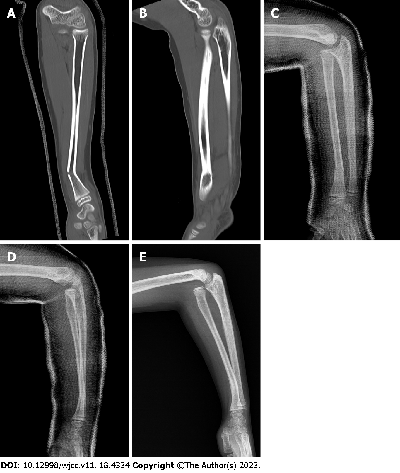
Computed tomography (CT) images of the left forearm performed at the time of injury showed a fracture with angulation of the radius at the distal third (Figure 1A). There was no dislocation of the radial head (Figure 1B). Two weeks after the injury, there was no evidence of displacement progression in the radial fracture, although mild subluxation of the radial head was observed radiographically.
The patient had no significant medical history. No specific genetic disease was found in the family history.
Physical examination revealed limited pronation of the last 35° in the forearm.
Malunion with a posterior convex deformity of 22° was observed at the radial fracture site on the radiograph taken at our clinic three months after the injury.
The patient was diagnosed with a delayed dislocation of the radial head associated with the malunion of a distal radial fracture (Figure 1E).
Although the patient was asymptomatic except for limitations in pronation, as his skeleton matures, radial head dislocation could cause long-term problems with forearm rotation and elbow function. Since remodeling ability remained in the malunion three months after injury in this 9-year-old child, good results could be obtained without corrective osteotomy as long as the reduction of the radial head and sufficient stability of the radiocapitellar joint could be maintained.
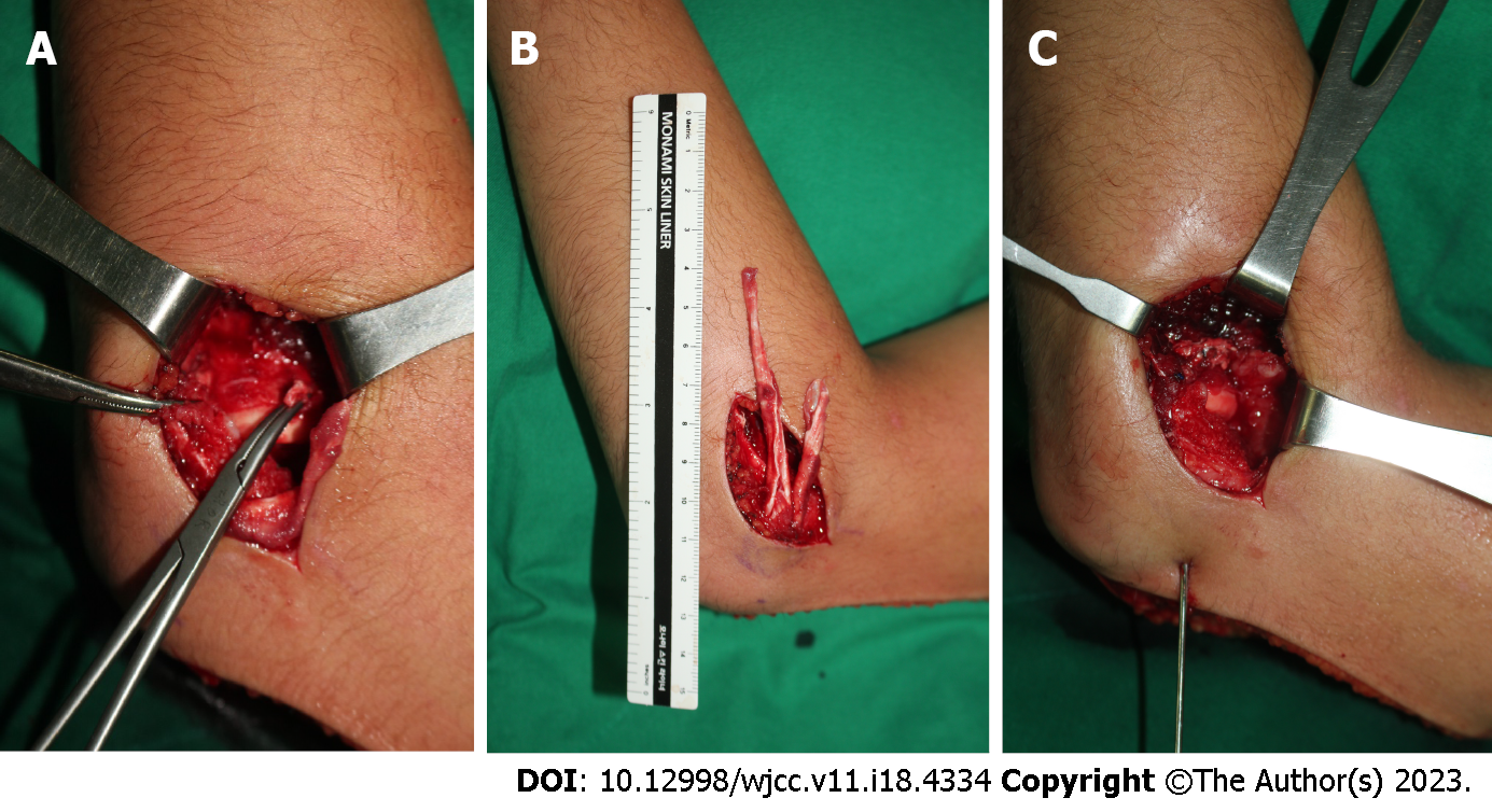
The elbow joint was approached from the lateral aspect. Soft tissue, such as the joint capsule, was impinged between the capitellum and the radial head. The annular ligament was ruptured, making it difficult to recognize (Figure 2A). After the capsular block was removed, the radial head was reduced. However, rotation of the forearm continued to cause subluxation of the radial head. Thus, an 8 cm × 1 cm strip of lateral triceps fascia and another 5 cm × 1 cm strip were harvested using a posterior incision, leaving the distal portion attached to the proximal ulna. This distal attachment was dissected subperiosteally to the level of the annular ligament insertion on the ulna to provide appropriate alignment of the reconstructed ligament (Figure 2B). After passing this double-strip down to anconeus, the long fascial strip was passed around the neck of the radius from behind to forward and sutured back to itself and the ulnar periosteum. The short strip was additionally augmented to the anterior part of the long strip to provide stability to the radiocapitellar joint (Figure 2C). Finally, reduction of the radial head was performed using transarticular Kirschner wires with the elbow at 90° of flexion and the forearm in neutral rotation. Kirschner wires were removed after cast immobilization for three weeks, and active elbow flexion-extension was started. Active forearm rotation was permitted five weeks after surgery.
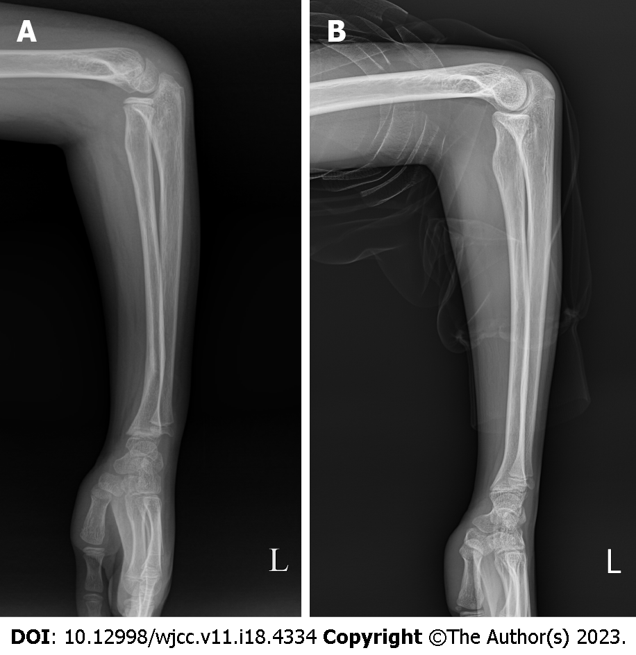
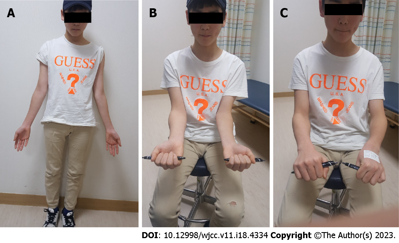
The RHD was well reduced within the radiocapitellar joint in the lateral forearm radiograph taken three months after surgery. However, angulation of the distal radius remained (Figure 3A). The angulation deformity of the distal radius was corrected four years after surgery with the restoration of the normal curvature of the radius. There was no recurrence of RHD (Figure 3B). The flexion of the right elbow was 130° with extension at 0°. Supination was 85°, and pronation was 80° for the left forearm (Figure 4).
RHD is a well-described injury in the pediatric population. It is usually associated with an injury to the ulna in a Monteggia fracture-dislocation. Although rare, traumatic RHD with ipsilateral radial shaft fracture can occur when excessive pronation force is applied while the radial head is dislocated[7]. Many traumatic dislocations of the radial head are missed during the acute phase of treatment. Weisman et al[8] reported that the diagnosis of traumatic dislocation of the radial head was delayed in 10 (9.1%) of 110 children treated with these injuries. In eight children, the dislocation was overlooked on the initial radiographs. In two children, the radial head was reduced on the initial elbow radiographs. It was dislocated 10 d later in one child and 21 d later in the other. In the present study, we could not obtain initial elbow radiographs of the patient taken at a local clinic. The radial head was well positioned at the radiocapitella joint without subluxation or dislocation on CT images at the time of injury (Figure 1A and B). However, radiographic evaluation of the elbow using the radiocapitellar line showed subluxation of the radial head two weeks after the injury (Figure 1C and D). Radiographs taken 12 wk after the injury showed dislocation (Figure 1E). The most likely explanation was that the radial head might have dislocated at the time of impact and spontaneously reduced by the time the first radiographs were obtained. It was then re-dislocated while the arm was in a cast.
Delayed RHD associated with the malunion of an isolated radial shaft fracture is extremely rare. Only four such cases have been reported in the English-language literature[3-6]. Currently, there are no clear guidelines on surgical methods that can be successfully used for delayed RHD with the malunion of a radial shaft fracture. A study has performed corrective osteotomy to restore the normal curve of forearm bones and prevent dislocation of the radial head in a 15-year-old boy[3]. Yamazaki et al[4] have performed a corrective osteotomy of radial shaft and repair of the annular ligament in six months after the injury. In another report, a 12-year-old boy underwent corrective osteotomy with annular ligament reconstruction at 16 mo after injury[5]. In previously reported cases, corrective wedge osteotomy was performed in all cases to restore the normal curvature of the radius, with the repair or reconstruction of the annular ligament performed in two cases for RHD (Table 1) [3-6]. However, in the present case, the question was whether corrective osteotomy should be additionally performed in a situation where open reduction and repair or reconstruction of the annular ligament was considered. Although Sinikumpu et al[9] reported that operative treatment could be considered for angulation of more than 15° in forearm shaft fractures in children older than 8 years old, we judged that there was still the ability for spontaneous remodeling at the fracture site without corrective osteotomy on radiographs taken 12 wk after injury in our patient. Therefore, this lesion was treated with only annular ligament reconstruction using triceps strips without corrective osteotomy of the radial shaft. The reduction of the radial head was well-maintained four years postoperatively, and the normal curve of the radial shaft was restored.
Several resources have been used for reconstructing the annular ligament, including grafts from fascial strips of the forearm, fascia lata, tendon of palmaris longus, triceps tendon, and extensor aponeurosis[10]. However, each of these tissues has drawbacks. The forearm fascia and tendon of the palmaris longus are too weak to restrict the radius, and an additional incision is required for harvesting fascia lata.
The Bell Tawse procedure was originally introduced to treat a malunited anterior Monteggia fracture[11]. Bell Tawse described reconstruction of the annular ligament by turning down a strip of the triceps tendon, leaving it attached to the ulna, passing it around the neck of the radius from behind to forward, and securing it through a drilled hole in the ulna. Since its original description, several variations of the Bell Tawse procedure have been published. Lloyd-Roberts et al[12] preferred reconstruction of the annular ligament using a lateral slip of the triceps tendon because it confined the operation to one surgical field. In addition, preservation of the normal ulnar attachment inspires confidence in the viability of the refashioned ligament. Hurst et al[13] suggested that it is important to strip the tendon of the proximal olecranon with a 2- to 3-cm segment of the dorsal periosteum so that the attachment site of the newly reconstructed ligament will be parallel to the radial neck, not proximal to it. This alignment more closely approximates the normal anatomy of the annular ligament, unlike fixation to the olecranon. We also prefer to reconstruct the annular ligament using the triceps tendon. During the procedure, we dissected the distal attachment subperiosteally to the level of the annular ligament insertion in the ulna, as suggested by Hurst et al[13]. However, it is difficult to always obtain a good quality fascia strip of sufficient length for successful reconstruction since re-dislocation may occur as the graft material becomes loose. We have recently used a double-strip procedure that includes the lateral portion of the triceps tendon. First, the long strip was passed around the neck of the radius from behind to forward and sutured to itself and the ulnar periosteum. Next, the short strip was additionally reinforced in the anterior part of the long strip. This modified double-strip procedure is thought to provide greater stability to the radiocapitellar joint than a single strip, although biomechanical studies need to be performed in the future. Therefore, we could prevent limitations in the range of motion of the forearm by removing the transarticular Kirschner wire after three weeks and allowing active forearm rotation from four weeks after surgery.
In the present case, annular ligament reconstruction alone without corrective wedge osteotomy reduced RHD and restored the normal curve of the radial shaft. A modified double-strip Bell Tawse procedure might be useful for maintaining a more stable reduction through the plication of anterolateral parts.
Provenance and peer review: Unsolicited article; Externally peer reviewed.
Peer-review model: Single blind
Specialty type: Orthopedics
Country/Territory of origin: South Korea
Peer-review report’s scientific quality classification
Grade A (Excellent): A
Grade B (Very good): 0
Grade C (Good): 0
Grade D (Fair): 0
Grade E (Poor): 0
P-Reviewer: Kumar GM Y, India S-Editor: Chen YL L-Editor: A P-Editor: Chen YL
| 1. | Simpson JM, Andreshak TG, Patel A, Jackson WT. Ipsilateral radial head dislocation and radial shaft fracture. A case report. Clin Orthop Relat Res. 1991;205-208. [PubMed] [DOI] [Full Text] |
| 2. | Adhikari A, Acharya S, Bhandari R. Radial Head Dislocation with Ipsilateral Proximal Shaft of Radius Fracture: A Case Report. JNMA J Nepal Med Assoc. 2020;58:416-418. [RCA] [PubMed] [DOI] [Full Text] [Full Text (PDF)] [Cited by in Crossref: 1] [Cited by in RCA: 1] [Article Influence: 0.2] [Reference Citation Analysis (0)] |
| 3. | Yasutomi T, Nakatsuchi Y, Koike H. Anterior dislocation of the radial head secondary to malunion of the forearm bones. J Shoulder Elbow Surg. 2000;9:536-540. [RCA] [PubMed] [DOI] [Full Text] [Cited by in Crossref: 8] [Cited by in RCA: 8] [Article Influence: 0.3] [Reference Citation Analysis (0)] |
| 4. | Yamazaki H, Kato H, Yasutomi T, Murakami N, Hata Y. Delayed radial head dislocation associated with malunion of radial shaft fracture: a case report. J Shoulder Elbow Surg. 2007;16:e18-e21. [RCA] [PubMed] [DOI] [Full Text] [Cited by in Crossref: 3] [Cited by in RCA: 3] [Article Influence: 0.2] [Reference Citation Analysis (0)] |
| 5. | Wang SI, Lee SC. Delayed anterolateral radial head dislocation secondary to radial shaft fracture malunion: A case report. Medicine (Baltimore). 2022;101:e28661. [RCA] [PubMed] [DOI] [Full Text] [Full Text (PDF)] [Reference Citation Analysis (0)] |
| 6. | Haines S, Amirfeyz R. Corrective osteotomy for a malunited proximal radius fracture causing radio-capitellar dislocation in a paediatric patient: A case report. Shoulder Elbow. 2020;12:368-372. [RCA] [PubMed] [DOI] [Full Text] [Reference Citation Analysis (0)] |
| 7. | Jadaan M, Jain SK, Ahmed A, Khayyat G. Ipsilateral Radial Head Dislocation and Radial Shaft Fracture in a Child-A Case Report. J Orthop Case Rep. 2014;4:9-11. [RCA] [PubMed] [DOI] [Full Text] [Full Text (PDF)] [Cited by in RCA: 2] [Reference Citation Analysis (0)] |
| 8. | Weisman DS, Rang M, Cole WG. Tardy displacement of traumatic radial head dislocation in childhood. J Pediatr Orthop. 1999;19:523-526. [RCA] [PubMed] [DOI] [Full Text] [Cited by in Crossref: 1] [Cited by in RCA: 1] [Article Influence: 0.0] [Reference Citation Analysis (0)] |
| 9. | Sinikumpu JJ, Serlo W. The shaft fractures of the radius and ulna in children: current concepts. J Pediatr Orthop B. 2015;24:200-206. [RCA] [PubMed] [DOI] [Full Text] [Cited by in Crossref: 19] [Cited by in RCA: 24] [Article Influence: 2.4] [Reference Citation Analysis (0)] |
| 10. | Tan L, Li YH, Sun DH, Zhu D, Ning SY. Modified technique for correction of isolated radial head dislocation without apparent ulnar bowing: a retrospective case study. Int J Clin Exp Med. 2015;8:18197-18202. [PubMed] |
| 11. | Bell Tawse AJ. The treatment of malunited anterior Monteggia fractures in children. J Bone Joint Surg Br. 1965;47:718-723. [RCA] [PubMed] [DOI] [Full Text] [Cited by in Crossref: 138] [Cited by in RCA: 88] [Article Influence: 1.5] [Reference Citation Analysis (0)] |
| 12. | Lloyd-Roberts GC, Bucknill TM. Anterior dislocation of the radial head in children: aetiology, natural history and management. J Bone Joint Surg Br. 1977;59-B:402-407. [RCA] [PubMed] [DOI] [Full Text] [Cited by in Crossref: 147] [Cited by in RCA: 115] [Article Influence: 2.4] [Reference Citation Analysis (1)] |
| 13. | Hurst LC, Dubrow EN. Surgical treatment of symptomatic chronic radial head dislocation: a neglected Monteggia fracture. J Pediatr Orthop. 1983;3:227-230. [RCA] [PubMed] [DOI] [Full Text] [Cited by in Crossref: 53] [Cited by in RCA: 32] [Article Influence: 0.8] [Reference Citation Analysis (1)] |


