Published online Jun 16, 2023. doi: 10.12998/wjcc.v11.i17.4194
Peer-review started: April 18, 2023
First decision: April 26, 2023
Revised: May 1, 2023
Accepted: May 19, 2023
Article in press: May 19, 2023
Published online: June 16, 2023
Processing time: 54 Days and 11.8 Hours
Hourglass-like constriction neuropathy is a rare neurological disorder. The main clinical manifestation is peripheral nerve injury with no apparent cause, and the pathomorphological change is an unexplained narrowing of the diseased nerve. The diagnosis and treatment of the disease are challenging and there is no accepted diagnostic or therapeutic approach.
This report describes a rare hourglass constriction of the anterior interosseous nerve in the left forearm in a 47-year-old healthy male who was treated surgically and gradually recovered function over a 6-mo follow-up period.
Hourglass-like constriction neuropathy is a rare disorder. With the development of medical technology, more examinations are now available for diagnosis. This case aims to highlight the rare manifestations of Hourglass-like constriction neuropathy and provides a reference for enriching the clinical diagnosis and treatment experience.
Core Tip: The main aim of this article is to report a case of Hourglass-like constriction of the anterior interosseous nerve of the left forearm with serious consequences. The effect of hand and foot microsurgery on the pathological tissue and parallel nerve anastomosis was effective, which provides further success for clinical treatment.
- Citation: He R, Yu JL, Jin HL, Ng L, Wang JC, Li X, Gai TT, Zhou Y, Li DP. Hourglass-like constriction of the anterior interosseous nerve in the left forearm: A case report. World J Clin Cases 2023; 11(17): 4194-4201
- URL: https://www.wjgnet.com/2307-8960/full/v11/i17/4194.htm
- DOI: https://dx.doi.org/10.12998/wjcc.v11.i17.4194
Hourglass-like constriction neuropathy is a complex neurological disorder, the cause is unknown, but is typically characterized by hourglass constriction of nerve trunks (nerve branches). The clinical features are characterized by sudden onset of pain followed by dysfunction of the motor or sensory functions innervated by the corresponding nerve[1-3]. Hourglass-like constriction neuropathy has not been studied separately for its epidemiology as most scholars consider it to be the result of an altered structural pathology[4].
The preoperative diagnosis of this disease is challenging, and the definitive diagnosis is usually made after surgical exploration. Ultrasound and magnetic resonance neurography (MRN) are useful in diagnosing nerve hourglass constriction, but are usually not visualized by conventional magnetic resonance imaging (MRI)[5]. Due to the rarity of the disease and the limitations of the investigations, in general, many physicians will often misdiagnose it as neuritis or some localized nerve entrapment syndrome based on its signs and symptoms, but they are different. The latter is often misdiagnosed because their symptoms are particularly similar, mostly due to aseptic inflammation or viral infection and mechanical compression, whereas the former is more often a neurological organic lesion. On the other hand, the treatment of these diseases can be very challenging and although there are many surgical options, such as epineurectomy and neurolysis, resection and neurorrhaphy or nerve grafting, the results are not satisfactory[6,7].
We report here a case of hourglass-like constriction of the anterior interosseous nerve in the left forearm, which recovered after surgical treatment. In addition to this, we have retrospectively analyzed cases that have been reported as hourglass-like constriction treatments in the last decade to analyze their efficacy.
The patient, a 47-year-old male piano tuner, complained of weakness in his left hand, with no apparent traumatic causes of flexion or extension of the distal phalanges of his left thumb for two months.
Over the past 2 mo, his condition has progressively deteriorated with weakness in his left hand, as well as ancient flexion weakness in the distal left thumb and significant restriction of movement.
The patient has no family history of alcohol or tobacco addiction and was in good overall mental condition.
No significant medical history.
Left thumb long flexor tendon strength grade 4, left index finger deep flexor tendon strength grade 0, left thumb short flexor strength grade 5, left index finger short flexor strength grade 5, no significant abnormalities of superficial skin sensation in the left hand and left forearm. The palmar aspect of the left wrist is flatter than that of the right wrist (anterior rotator muscle), and there are no significant abnormalities in finger movement or blood flow in the remaining fingers.
None.
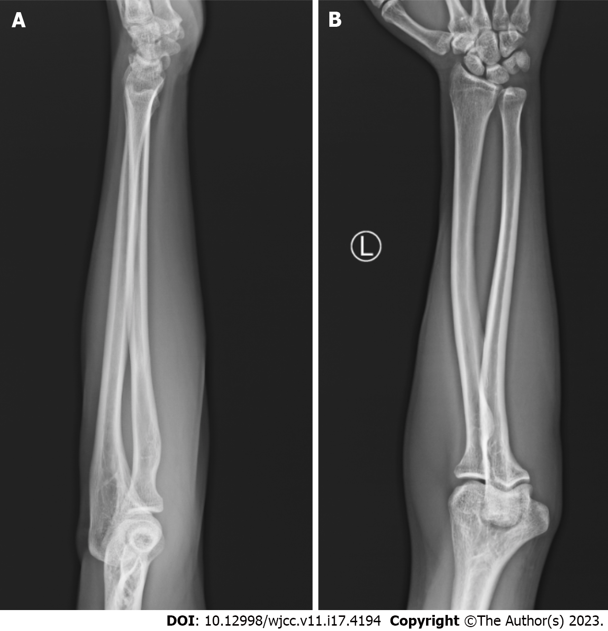
Digital radiography examination: The left ulnar radius is regular in shape with continuous bone cortex and clear bone trabeculae, with no obvious signs of bone destruction. The distal ulnar flexor and flexor carpal joint gaps were moderate and there were no obvious abnormalities in the surrounding soft tissues (Figure 1).
Colour ultrasonic Doppler examination: Widening of the median nerve cross-section over the left elbow, left side wide (0.48 cm × 0.29 cm), hypoechoic, star grid-like changes, the left side of the median nerve at the elbow is about 0.54 cm × 0.29 cm wide (right side is about 0.52 cm × 0.27 cm wide) locally hypoechoic, local nerve bundle thickness varies, one obvious narrowing is visible, local nerve bundle width is about 0.08 cm, proximal segment is 0.17 cm, the distal segment is 0.14 cm. The body surface was marked and the median nerve at the wrist was approximately 0.62 cm × 0.23 cm wide on the left (0.76 cm × 0.38 cm wide on the right).
Hourglass-like constriction of the anterior interosseous nerve in the left forearm.
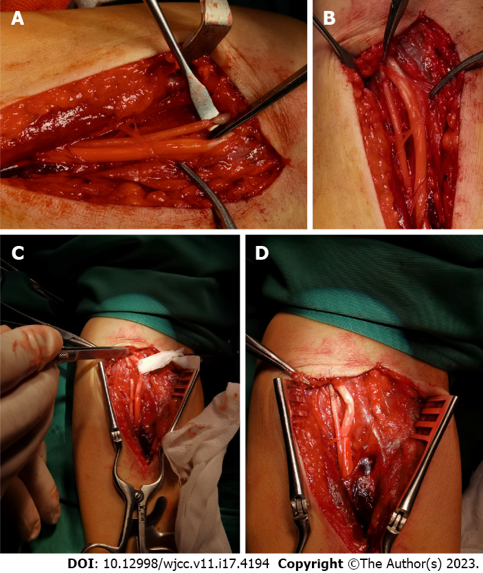
The surgical treatment was carried out by microsurgeons. The patient was placed in a supine position, prepared routinely for surgery, and a tourniquet was applied. Based on the previous color ultrasonic doppler examination, a longitudinal incision of approximately 10 cm was made on the proximal palmar side of the left forearm as the surgical incision approach. The skin and subcutaneous tissue were incised in accordance with the procedure and the biceps tendon sheath was exposed and incised. The main trunk of the median nerve was surgically exposed between the pronator teres and the radial carpal flexor, dissected along it to the anterior interosseous nerve, and the pronator teres were drawn radially to reveal the superficial flexor fibre arch. The interosseous anterior nerve was then loosened to free the epineurium. The continuous research for the lesion location, and around 1 cm after the anterior interosseous nerve, it separates from the trunk of the median nerve. An incomplete dissection of the interosseous anterior nerve was seen at approximately 1 cm from the main branch of the median nerve, the epineurium was continuous, and the nerve bundle (axon removed) was almost completely dissected, with the connection accounting for approximately 20% of the total diameter and scarring degeneration at the dissection site. Approximately 3 mm of diseased tissue is excised from the neuropathy site and the nerve repair anastomosis is then performed (Figure 2). After flushing the incision site and stopped the bleeding, the nerve anastomosis is wrapped with a collagen sponge and the surgical incision is closed layer by layer. External fixation in plaster was performed. We advised the patient to try to start functional exercises for the distal fingers, such as forceful fist clenching, as soon as the pain and swelling had subsided three days after surgery.
Two months after the operation, the cast was removed, and the functional exercise of the affected limb was further strengthened. At 6 mo postoperative follow-up, there were significant improvements in symptoms and return of voluntary movement.
Englert first described the hourglass constriction of nerves in 1978, but did not elaborate on its pathogenesis[8]. In recent years, many medical scholars have put forward numerous hypotheses and theories regarding the pathogenesis of the disease, mainly divided into: The external structural compression theory[6], the repetitive motion theory[9], the nerve torsional displacement theory[6,10], the inflammatory response theory[11,12] and the inflammatory response with repetitive motion theory[13], but ultimately no unified and accepted conclusion has been reached.
However, the morphological features of the nerve lesions are more consistent, with the main nerve trunk or its nerve branches showing significant narrowing in the form of bundle wraps. Outer nerve membrane continuity may be present, but most of the nerve bundles have been dissected[14]. The disease’s main symptom is a sudden onset of pain in the corresponding innervated area, followed by flaccid paralysis or restriction of movement in the affected muscles[1,15]. The presentation of this lesion is very similar to that of conditions such as spontaneous peripheral nerve palsy, which can be difficult to distinguish clinically and is therefore often overlooked, leading to delays in treatment and compromising recovery[1,16].
Prior to the development of MRN and high frequency ultrasound imaging, the clinical tendency was to attribute the cause of dysfunctional finger movements as well as muscle weakness to spontaneous peripheral nerve palsy, as at that time hourglass-like constrictions could only be diagnosed by surgical exploration. With advances in medical imaging, more patients diagnosed with spontaneous peripheral nerve palsy are being found to have hourglass-like constriction lesions in their nerves[17,18]. In addition, the MRN examination helps to identify the exact location of the neuropathy before surgery is required, so that surgery can be performed with less time, smaller incision areas and more precise treatment[19]. Certainly, high frequency ultrasonography is a reliable, convenient and non-invasive diagnostic imaging method for accurately locating the hourglass-like constriction neuropathy and extent of neuropathy in the anterior interosseous nerve[20].
The treatment of this disease is still somewhat controversial, with a general preference for conservative treatment first, and surgical intervention being beneficial in selected patients who do not recover promptly within 3 mo and have hourglass-like lesions confirmed on preoperative examination[1]. In addition, the use of nerve grafting is definitive for patients with nerve defects greater than 2 cm[6]. Failure to perform surgical treatment in a timely manner is thought to prevent the regeneration of nerve axons and affect the patient’s recovery of function.
We reviewed cases of peripheral nerve disease due to hourglass-like constriction over the last 10 years (Table 1). We noted that the proportion of patients presenting with hourglass-like constriction was much higher in men than in women[6,7,21,22]. This may be due to differences in daily activities between men and women, with men engaging in more repetitive physical activities that are more likely to lead to nerve entrapment and compression[23], but whether this leads to hourglass-like constriction needs to be further explored. In addition, in our retrospective study, we found that there is no age limit for this type of disease, which can occur in children as well as in the elderly, and that the disease is mostly found in the motor nerves of the upper limbs, which we hypothesize is related to the fact that the nerves of the upper limbs mostly innervate the limbs for delicate manipulations and their neuroanatomical location. In the treatment of hourglass lesions, we have found that conservative hormonal treatment is not particularly effective, whereas surgical release of the nerve and resection of the lesion with anastomosis seems to work well, and the prognosis is significantly better in young people than in older people[24]. Therefore, we report here a case of hourglass-like constriction of the anterior interosseous nerve in the left forearm, which was treated surgically by our excision of the diseased tissue and parallel nerve anastomosis, followed by a 6-mo continuous follow-up period during which the patient was asked to strengthen his functional exercises, resulting in a gradual recovery of his symptoms. This has provided more experience in clinical treatment and has enriched the success stories of effective surgical treatment.
| Ref. | Gender/age (yr) | Symptoms | Nerve explored | Imaging | Inspection result | Treatment options | Prognosis |
| Nakagawa et al[2], 2018, Japan | Male/9 | Pain in the left arm with severe paralysis when extending the wrist, thumb and fingers | Brachial Plexusin the Posterior Cord | MRI, ultrasound | There is mild diffuse enlargement and high intensity of the left brachial plexus nerve | Surgical nerve exploration, nerve release, neuropathy excision | Strength starts to return 3 wk after the operation; 6 mo after the operation, there is no restriction in daily activities |
| Kim et al[19], 2019, South Korea | Female/26 | Pain in the scapula and difficulty in raising the left arm, relief of pain within 10 d, weakness in the shoulder | Suprascapular nerve | MRN | Multiple hourglass-like contractions were found in the suprascapular nerve with no enlargement or signal constriction | Oral steroids, topical steroid injections | Muscle strength approaches normal levels after 10 mo |
| Kim et al[19], 2019, South Korea | Male/42 | Pain in the scapula and deltoid area, weakness in the left shoulder | Suprascapular nerve | MRN | Focal contraction of the suprascapular nerve | Oral prednisolone, injectable steroids | Back to normal after 15 mo |
| Kim et al[19], 2019, South Korea | Male/52 | Weakness in the left shoulder and drooping of the left wrist | Suprascapular nerve, radial nerve | MRN | Diffuse swelling of the C6 nerve and two focal contractions of the suprascapular nerve | Intravenous steroids | The shoulder joint recovered after 3 mo with no improvement in the muscles innervated by the radial nerve |
| Kim et al[3], 2020, South Korea | Male/47 | Pain in the elbow and back of the forearm, with drooping of the left wrist | Radial nerve | MRN | Focal contraction of the left radial nerve | Oral prednisolone, injectable steroids | No improvement in symptoms after 6 mo |
| Kim et al[3], 2020, South Korea | Male/19 | Drooping left wrist, dorsal sensory deficit of left wrist | Radial nerve | MRN | Focal contraction of left radial nerve at 2 | Surgical nerve release | No improvement in symptoms after 6 mo |
| Krishnan et al[25], 2020, United States | Female/58 | Pain in the right shoulder with weakness in abduction | Suprascapular, axillary, phrenic nerves | MRN, ultrasound | Focal contraction at 2 phrenic nerves | Surgical nerve release | 6 mo after surgery, symptoms improved |
| Loizides et al[26], 2015, Austria | Male/26 | Radial deviation of the wrist during wrist extension; impaired extension of the metacarpophalangeal joint; impaired extension of the fingers at the metacarpophalangeal joint; impaired abduction and adduction of the thumb | Radial nerve | Ultrasound | Focal contraction of radial nerve in 3 places | Surgical nerve release | 3 mo after the operation, there was a marked improvement in symptoms |
| Kodama et al[27], 2015, Japan | Male/37 | Anterior interosseous nerve | Ultrasound | Focal contraction at 3 anterior interosseous nerves | Surgical nerve release | Significant improvement in symptoms 5 mo after surgery |
However, there are still some limitations to this study, the main cause of the problem is due to the insufficient volume of literature and its clinical cases. For example, in this study we only analyzed the reported literature, most of which had significant treatment outcomes, but we believe that most of the cases with poor treatment outcomes were not reported, so the limited amount of data in the literature may lead us to conclude that the results are not factually accurate; on the other hand, we reviewed the literature from the last 10 years and did not combine it with previous studies, which may also have an impact on our summary and may also be less accurate in terms of our summary of treatment modalities and efficacy. More comprehensive clinical studies are needed in the future to confirm the effectiveness of their treatment modalities and their efficacy.
Hourglass-like constriction neuropathy is a rare disorder, probably due to the lack of advanced detection tools in the past, which has led to a lack of awareness of these disorders. With the development of medical technology, more and more examinations are now available for diagnosis and when we suspect that a patient may suffer from hourglass-like constriction neuropathy, the relevant examinations should be completed in time for early surgical treatment.
Provenance and peer review: Unsolicited article; Externally peer reviewed.
Peer-review model: Single blind
Specialty type: Medicine, research and experimental
Country/Territory of origin: China
Peer-review report’s scientific quality classification
Grade A (Excellent): 0
Grade B (Very good): 0
Grade C (Good): C, C, C
Grade D (Fair): D
Grade E (Poor): 0
P-Reviewer: Al-Ani RM, Iraq; Gupta L, Indonesia S-Editor: Wang JJ L-Editor: A P-Editor: Wang JJ
| 1. | Wang Y, Liu T, Song L, Zhang Z, Zhang Y, Ni J, Lu L. Spontaneous peripheral nerve palsy with hourglass-like fascicular constriction in the upper extremity. J Neurosurg. 2019;131:1876-1886. [RCA] [PubMed] [DOI] [Full Text] [Cited by in Crossref: 20] [Cited by in RCA: 21] [Article Influence: 3.5] [Reference Citation Analysis (0)] |
| 2. | Nakagawa Y, Hirata H. Hourglass-Like Constriction of the Brachial Plexus in the Posterior Cord: A Case Report. Neurosurgery. 2018;82:E1-E5. [RCA] [PubMed] [DOI] [Full Text] [Cited by in Crossref: 12] [Cited by in RCA: 13] [Article Influence: 1.6] [Reference Citation Analysis (0)] |
| 3. | Kim DH, Sung DH, Chang MC. Diagnosis of Hourglass-Like Constriction Neuropathy of the Radial Nerve Using High-Resolution Magnetic Resonance Neurography: A Report of Two Cases. Diagnostics (Basel). 2020;10. [RCA] [PubMed] [DOI] [Full Text] [Full Text (PDF)] [Cited by in Crossref: 13] [Cited by in RCA: 6] [Article Influence: 1.2] [Reference Citation Analysis (1)] |
| 4. | Gstoettner C, Mayer JA, Rassam S, Hruby LA, Salminger S, Sturma A, Aman M, Harhaus L, Platzgummer H, Aszmann OC. Neuralgic amyotrophy: a paradigm shift in diagnosis and treatment. J Neurol Neurosurg Psychiatry. 2020;91:879-888. [RCA] [PubMed] [DOI] [Full Text] [Cited by in Crossref: 37] [Cited by in RCA: 73] [Article Influence: 14.6] [Reference Citation Analysis (0)] |
| 5. | Arányi Z, Csillik A, Dévay K, Rosero M, Barsi P, Böhm J, Schelle T. Ultrasonographic identification of nerve pathology in neuralgic amyotrophy: Enlargement, constriction, fascicular entwinement, and torsion. Muscle Nerve. 2015;52:503-511. [RCA] [PubMed] [DOI] [Full Text] [Cited by in Crossref: 69] [Cited by in RCA: 99] [Article Influence: 9.9] [Reference Citation Analysis (0)] |
| 6. | Qi W, Shen Y, Qiu Y, Jiang S, Yu Y, Yin H, Xu W. Surgical treatment of hourglass-like radial nerve constrictions. Neurochirurgie. 2021;67:170-175. [RCA] [PubMed] [DOI] [Full Text] [Cited by in RCA: 9] [Reference Citation Analysis (0)] |
| 7. | Wu P, Yang JY, Chen L, Yu C. Surgical and conservative treatments of complete spontaneous posterior interosseous nerve palsy with hourglass-like fascicular constrictions: a retrospective study of 41 cases. Neurosurgery. 2014;75:250-7; discussion 257. [RCA] [PubMed] [DOI] [Full Text] [Cited by in Crossref: 27] [Cited by in RCA: 27] [Article Influence: 2.5] [Reference Citation Analysis (0)] |
| 8. | Englert HM. [Partial fascicular median-nerve atrophy of unknown origin]. Handchirurgie. 1976;8:61-62. [PubMed] |
| 9. | Vastamäki M. Prompt interfascicular neurolysis for the successful treatment of hourglass-like fascicular nerve compression. Scand J Plast Reconstr Surg Hand Surg. 2002;36:122-124. [RCA] [PubMed] [DOI] [Full Text] [Cited by in Crossref: 9] [Cited by in RCA: 9] [Article Influence: 0.4] [Reference Citation Analysis (0)] |
| 10. | Yasunaga H, Shiroishi T, Ohta K, Matsunaga H, Ota Y. Fascicular torsion in the median nerve within the distal third of the upper arm: three cases of nontraumatic anterior interosseous nerve palsy. J Hand Surg Am. 2003;28:206-211. [RCA] [PubMed] [DOI] [Full Text] [Cited by in Crossref: 44] [Cited by in RCA: 43] [Article Influence: 2.0] [Reference Citation Analysis (0)] |
| 11. | Yamamoto S, Nagano A, Mikami Y, Tajiri Y. Multiple constrictions of the radial nerve without external compression. J Hand Surg Am. 2000;25:134-137. [RCA] [PubMed] [DOI] [Full Text] [Cited by in Crossref: 16] [Cited by in RCA: 20] [Article Influence: 0.8] [Reference Citation Analysis (0)] |
| 12. | Omura T, Nagano A, Murata H, Takahashi M, Ogihara H, Omura K. Simultaneous anterior and posterior interosseous nerve paralysis with several hourglass-like fascicular constrictions in both nerves. J Hand Surg Am. 2001;26:1088-1092. [RCA] [PubMed] [DOI] [Full Text] [Cited by in Crossref: 27] [Cited by in RCA: 23] [Article Influence: 1.0] [Reference Citation Analysis (0)] |
| 13. | Lundborg G. Commentary: hourglass-like fascicular nerve compressions. J Hand Surg Am. 2003;28:212-214. [RCA] [PubMed] [DOI] [Full Text] [Cited by in Crossref: 37] [Cited by in RCA: 50] [Article Influence: 2.3] [Reference Citation Analysis (0)] |
| 14. | Komatsu M, Nukada H, Hayashi M, Ochi K, Yamazaki H, Kato H. Pathological Findings of Hourglass-Like Constriction in Spontaneous Posterior Interosseous Nerve Palsy. J Hand Surg Am. 2020;45:990.e1-990.e6. [RCA] [PubMed] [DOI] [Full Text] [Cited by in Crossref: 4] [Cited by in RCA: 13] [Article Influence: 2.6] [Reference Citation Analysis (0)] |
| 15. | Pan Y, Wang S, Zheng D, Tian W, Tian G, Ho PC, Cheng HS, Zhong Y. Hourglass-like constrictions of peripheral nerve in the upper extremity: a clinical review and pathological study. Neurosurgery. 2014;75:10-22. [RCA] [PubMed] [DOI] [Full Text] [Cited by in Crossref: 64] [Cited by in RCA: 67] [Article Influence: 6.1] [Reference Citation Analysis (0)] |
| 16. | Seror P. Neuralgic amyotrophy. An update. Joint Bone Spine. 2017;84:153-158. [RCA] [PubMed] [DOI] [Full Text] [Cited by in Crossref: 50] [Cited by in RCA: 62] [Article Influence: 6.9] [Reference Citation Analysis (0)] |
| 17. | Sneag DB, Rancy SK, Wolfe SW, Lee SC, Kalia V, Lee SK, Feinberg JH. Brachial plexitis or neuritis? MRI features of lesion distribution in Parsonage-Turner syndrome. Muscle Nerve. 2018;58:359-366. [RCA] [PubMed] [DOI] [Full Text] [Cited by in Crossref: 40] [Cited by in RCA: 71] [Article Influence: 10.1] [Reference Citation Analysis (0)] |
| 18. | Pan YW, Wang S, Tian G, Li C, Tian W, Tian M. Typical brachial neuritis (Parsonage-Turner syndrome) with hourglass-like constrictions in the affected nerves. J Hand Surg Am. 2011;36:1197-1203. [RCA] [PubMed] [DOI] [Full Text] [Cited by in Crossref: 51] [Cited by in RCA: 64] [Article Influence: 4.6] [Reference Citation Analysis (0)] |
| 19. | Kim DH, Kim J, Sung DH. Hourglass-like constriction neuropathy of the suprascapular nerve detected by high-resolution magnetic resonance neurography: report of three patients. Skeletal Radiol. 2019;48:1451-1456. [RCA] [PubMed] [DOI] [Full Text] [Cited by in Crossref: 8] [Cited by in RCA: 12] [Article Influence: 2.0] [Reference Citation Analysis (0)] |
| 20. | Wang T, Qi H, Wang D, Wang Z, Bao S, Teng J. The role of ultrasonography in diagnosing hourglass-like fascicular constriction(s) of the anterior interosseous nerve. Acta Radiol. 2022;63:1528-1534. [RCA] [PubMed] [DOI] [Full Text] [Reference Citation Analysis (0)] |
| 21. | Vigasio A, Marcoccio I. Hourglass-like constriction of the suprascapular nerve: a contraindication for minimally invasive surgery. J Shoulder Elbow Surg. 2018;27:e29-e37. [RCA] [PubMed] [DOI] [Full Text] [Cited by in Crossref: 8] [Cited by in RCA: 13] [Article Influence: 1.9] [Reference Citation Analysis (0)] |
| 22. | Nakashima Y, Sunagawa T, Shinomiya R, Ochi M. High-resolution ultrasonographic evaluation of "hourglass-like fascicular constriction" in peripheral nerves: a preliminary report. Ultrasound Med Biol. 2014;40:1718-1721. [RCA] [PubMed] [DOI] [Full Text] [Cited by in Crossref: 24] [Cited by in RCA: 24] [Article Influence: 2.2] [Reference Citation Analysis (0)] |
| 23. | Silver S, Ledford CC, Vogel KJ, Arnold JJ. Peripheral Nerve Entrapment and Injury in the Upper Extremity. Am Fam Physician. 2021;103:275-285. [PubMed] |
| 24. | Ochi K, Horiuchi Y, Tazaki K, Takayama S, Nakamura T, Ikegami H, Matsumura T, Toyama Y. Surgical treatment of spontaneous posterior interosseous nerve palsy: a retrospective study of 50 cases. J Bone Joint Surg Br. 2011;93:217-222. [RCA] [PubMed] [DOI] [Full Text] [Cited by in Crossref: 29] [Cited by in RCA: 35] [Article Influence: 2.5] [Reference Citation Analysis (0)] |
| 25. | Krishnan KR, Wolfe SW, Feinberg JH, Nwawka OK, Sneag DB. Imaging and treatment of phrenic nerve hourglass-like constrictions in neuralgic amyotrophy. Muscle Nerve. 2020;62:E81-E82. [RCA] [PubMed] [DOI] [Full Text] [Cited by in Crossref: 4] [Cited by in RCA: 10] [Article Influence: 2.0] [Reference Citation Analysis (0)] |
| 26. | Loizides A, Baur EM, Plaikner M, Gruber H. Triple hourglass-like fascicular constriction of the posterior interosseous nerve: a rare cause of PIN syndrome. Arch Orthop Trauma Surg. 2015;135:635-637. [RCA] [PubMed] [DOI] [Full Text] [Cited by in Crossref: 12] [Cited by in RCA: 13] [Article Influence: 1.3] [Reference Citation Analysis (0)] |
| 27. | Kodama A, Sunagawa T, Ochi M. Early treatment of anterior interosseous nerve palsy with hourglass-like fascicular constrictions by interfascicular neurolysis due to early diagnosis using ultrasonography: A case report. J Hand Surg Eur Vol. 2015;40:642-643. [RCA] [PubMed] [DOI] [Full Text] [Cited by in Crossref: 5] [Cited by in RCA: 5] [Article Influence: 0.5] [Reference Citation Analysis (0)] |


