Published online May 16, 2023. doi: 10.12998/wjcc.v11.i14.3256
Peer-review started: October 9, 2022
First decision: January 17, 2023
Revised: January 29, 2023
Accepted: April 7, 2023
Article in press: April 7, 2023
Published online: May 16, 2023
Processing time: 219 Days and 4 Hours
A foreign body in the digestive tract is a common disease in the clinic. However, it is rare for a foreign body to migrate into the liver. Most patients are diagnosed before or after perforation of the digestive tract. Laparoscopic removal of intrahepatic foreign bodies is an effective treatment method.
A 55-year-old male patient was admitted to the hospital due to fever for 3 d, in addition to pain and discomfort in the right side of his waist. After admission, abdominal computed tomography showed a foreign body in the liver, and gastroscopy did not indicate obvious erosion or ulcers. The patient then underwent laparoscopic surgery. During the operation, an abscess was seen near the gastric antrum and between the caudate lobes of the liver. It was approximately 30 mm × 31 mm × 23 mm in size. The abscess was cut open, and a fish bone was found inside. The fish bone had penetrated the liver and was successfully removed. It was confirmed that the fish bone migrated from the stomach to the liver.
Although intrahepatic foreign bodies are rare, they should be diagnosed and treated as early as possible to avoid serious complications such as intrahepatic abscess, which may lead to liver resection and even life-threatening events.
Core Tip: Foreign bodies migrating into the liver are rare, but they may lead to liver resection and even life-threatening events. They should be diagnosed and treated as early as possible. We report a patient with a fish bone that migrated from the stomach to the liver and was successfully removed by laparoscopic surgery in the early stage. Early management is a prerequisite to ensure treatment efficacy.
- Citation: Dai MG, Zheng JJ, Yang J, Ye B. Intragastric fish bones migrate into the liver: A case report. World J Clin Cases 2023; 11(14): 3256-3260
- URL: https://www.wjgnet.com/2307-8960/full/v11/i14/3256.htm
- DOI: https://dx.doi.org/10.12998/wjcc.v11.i14.3256
Foreign bodies in the digestive tract are common clinical diseases. Most foreign bodies enter the digestive tract consciously, or the patients are aware of foreign body ingestion. Therefore, this can be removed through endoscopy in a timely manner. Perforation of hollow viscus by a foreign body is rare, representing 1% of cases of accidental foreign body ingestion. A few sharp foreign bodies can cause perforation, bleeding, or obstruction of the digestive tract. However, sharp foreign bodies can enter the digestive tract and pass through the stomach and duodenal mucosa and enter the liver, but this is even less common. We report a patient with a fish bone that migrated from the stomach to the liver and was successfully removed by laparoscopic surgery in the early stage, which avoided liver resection. There was no serious infection, bleeding, or other complications.
A 55-year-old male was hospitalized due to fever for 3 d.
The patient developed fever 3 d previously; the highest temperature was 39 °C. The patient also experienced paroxysmal dull pain and discomfort in the right waist, without chills, shivering, or any other digestive tract and respiratory tract symptoms. He self-administered antipyretic drugs, but his temperature did not significantly decrease. Therefore, he came to the hospital for treatment.
The patient’s past medical history included hypertension, kidney stones, and gout but no history of the digestive system or other system diseases.
The patient’s personal and family history revealed no information relevant to the current case.
Upon initial evaluation, the patient was 170 cm tall and weighed 65 kg. The patient’s temperature was 38.2 °C, heart rate was 90 bpm, and blood pressure (measured with an electronic cuff) was 146/96 mmHg. Heart and lung auscultation was normal, the abdomen was soft, without tenderness, rebound pain, or muscle tension, and percussion pain in the renal area was negative. The remaining examination showed no obvious positive signs.
Laboratory examination results were as follows: C-reactive protein, 153.31 mg/L; white blood cells, 11.93 × 10⁹/L (normal: 3.5-9.5 × 10⁹/L); procalcitonin, 1.04 ng/mL (normal: < 0.05 ng/mL); and glutamic pyruvic transaminase, 57 U/L (normal: 9-50 U/L). All other tests were normal.
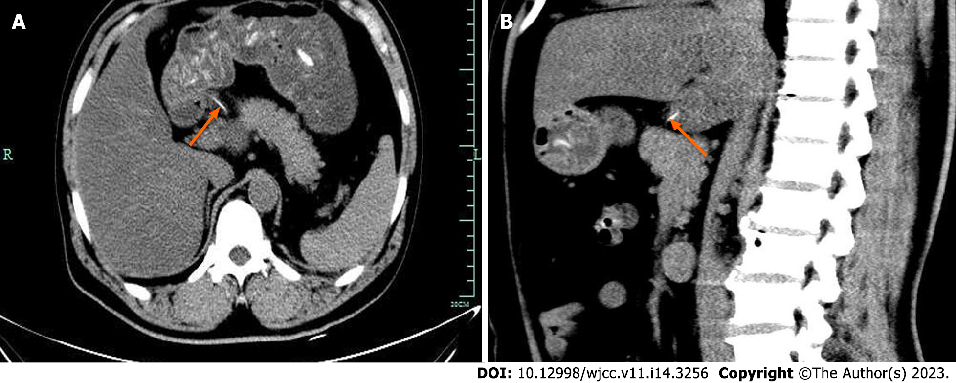
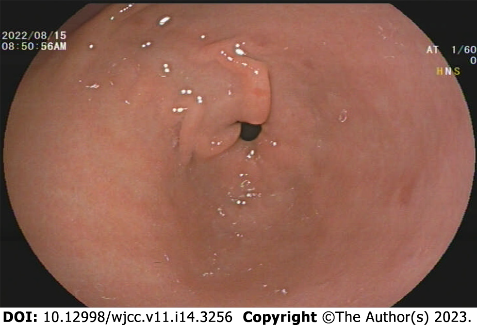
Abdominal computed tomography (CT) was performed after admission. It indicated a strip-shaped high-density focus in the stomach that had penetrated the caudate lobe of the liver. This was considered to be a foreign body in the stomach that had penetrated the liver (Figure 1). Emergency gastroscopy was performed to determine whether there were foreign bodies in the stomach. No obvious erosion or ulcers of the gastric mucosa was found during gastroscopy (Figure 2).
The patient was finally diagnosed with intrahepatic foreign body.
After receiving routine intravenous antibiotics treatment, his temperature decreased but did not return to normal. Abdominal CT demonstrated that the foreign body in the stomach had migrated into the liver. Emergency gastroscopy was carried out, and no residue of the foreign body was found in the stomach. Therefore, the patient underwent emergency laparoscopic surgery. During the operation, abscess formation was seen in the hepatogastric space, the abscess was cut, and one end of the fish bone was visible. The fish bone was completely removed. Following removal of the abscess, the gastric wall was examined, and no obvious damage was found (Video 1). The operation went smoothly, and the patient recovered.
Following surgery, the patient’s temperature gradually decreased to normal, without abdominal pain and other symptoms. He was discharged 1 wk after the operation and was followed up for 2 wk without experiencing obvious discomfort.
Chintamani et al[1] reported the world’s first case of foreign body in the digestive tract migrating into the liver, causing liver abscess in 1898. Since then only 59 cases have been reported in the literature[1,2]. However, with the development of digestive endoscopy technology[3], most foreign bodies in the digestive tract can be removed. A small number of foreign bodies, such as fish bones, toothpicks, iron wires, etc.[4], are thin and sharp. Most do not cause obvious symptoms when penetrating the gastrointestinal tract, making them difficult to find. The most common site of perforation is the stomach[5]. After penetrating the digestive tract, the foreign body often migrates into the left liver[6]. The patient still has no obvious symptoms at this time. With time, bacteria can undergo microbial replication and dissemination, causing liver abscess. The patient may develop fever, abdominal pain, and other symptoms, including serious infection, liver bleeding, etc., resulting in serious consequences[7].
Although intragastric foreign body migration into the liver is rare[8], it occurs occasionally. Most patients cannot recall the history of foreign body ingestion[3]. Clinicians need to be alert, and diagnosis depends on ultrasound, CT, etc. Ultrasound may be a convenient and radiation-free screening tool that can be used to identify abscesses and possible foreign bodies. On the other hand, CT is the first choice for diagnosis[7] due to its high resolution and accuracy in identifying foreign bodies. It can also be used to assess the depth of penetration and complications.
When it is suspected that a foreign body in the digestive tract has migrated into the liver, it is necessary to conduct timely digestive endoscopy. Some patients may have residues in the digestive tract. Foreign bodies can be removed by digestive endoscopy to avoid traumatic surgery. If necessary, before the foreign body is removed, endoscopic ultrasonography should be performed to determine the relationship between the foreign body and the surrounding blood vessels[9] to avoid massive bleeding and protect the safety of patients. In addition, surgical treatment should be carried out as early as possible for foreign bodies without residues in the gastrointestinal tract[10] rather than after the abscess has liquefied. Timely surgical[11] removal of foreign bodies can reduce the occurrence of complications and preserve the liver.
In the present case, the fish bone transferred from the stomach to the liver, and gastroscopy was performed in a timely manner. No obvious wound was found in the stomach; thus, it could not be removed under endoscopy. Therefore, laparoscopic foreign body removal was selected. Because it was a short amount of time that the foreign body had entered the liver, no obvious damage was found on the gastric wall, hepatogastric space, or liver during the operation. Following removal of the foreign body, complete debridement was conducted to ensure a good treatment effect. Therefore, for liver abscesses of unknown cause, clinicians should consider the possibility of foreign bodies[12], carefully observe the patient’s imaging findings, repeatedly ask about relevant medical history regarding ingestion of foreign bodies, carry out endoscopy as soon as possible when there is a high degree of suspicion of foreign bodies in the liver, and perform laparotomy if necessary. Surgery is the most effective method of treatment[13]. Early management is a prerequisite to ensure treatment efficacy.
We reported a case of intragastric foreign body that migrated into the liver. Although this is rare, it may cause serious infection and bleeding if not treated in time. This can lead to liver resection and can even be life-threatening, which should stimulate vigilance in clinicians.
Provenance and peer review: Unsolicited article; Externally peer reviewed.
Peer-review model: Single blind
Specialty type: Medicine, research and experimental
Country/Territory of origin: China
Peer-review report’s scientific quality classification
Grade A (Excellent): A
Grade B (Very good): 0
Grade C (Good): C, C
Grade D (Fair): D
Grade E (Poor): 0
P-Reviewer: Baryshnikova NV, Russia; Garbuzenko DV, Russia; Krishnan A, United States; Shelat VG, Singapore S-Editor: Liu GL L-Editor: Filipodia P-Editor: Yu HG
| 1. | Chintamani, Singhal V, Lubhana P, Durkhere R, Bhandari S. Liver abscess secondary to a broken needle migration--a case report. BMC Surg. 2003;3:8. [RCA] [PubMed] [DOI] [Full Text] [Full Text (PDF)] [Cited by in Crossref: 44] [Cited by in RCA: 61] [Article Influence: 2.8] [Reference Citation Analysis (0)] |
| 2. | Sobnach S, Castillo F, Blanco Vinent R, Kahn D, Bhyat A. Penetrating cardiac injury following sewing needle ingestion. Heart Lung Circ. 2011;20:479-481. [RCA] [PubMed] [DOI] [Full Text] [Cited by in Crossref: 6] [Cited by in RCA: 8] [Article Influence: 0.6] [Reference Citation Analysis (0)] |
| 3. | Burkholder R, Samant H. Management of Fish Bone-Induced Liver Abscess with Foreign Body Left In Situ. Case Reports Hepatol. 2019;2019:9075198. [RCA] [PubMed] [DOI] [Full Text] [Full Text (PDF)] [Cited by in Crossref: 2] [Cited by in RCA: 6] [Article Influence: 1.0] [Reference Citation Analysis (1)] |
| 4. | Subasinghe D, Jayasinghe R, Kodithuwakku U, Fernandopulle N. Hepatic abscess following foreign body perforation of the colon: A case report. SAGE Open Med Case Rep. 2022;10:2050313X221103357. [RCA] [PubMed] [DOI] [Full Text] [Full Text (PDF)] [Cited by in RCA: 2] [Reference Citation Analysis (0)] |
| 5. | Yan TD, Leung PHY, Zwirewich C, Harris A, Chartier-Plante S. An unusual cause of pericardial effusion: A case report of a hepatic abscess following foreign body migration and duodenal perforation. Int J Surg Case Rep. 2022;93:106931. [RCA] [PubMed] [DOI] [Full Text] [Full Text (PDF)] [Cited by in Crossref: 2] [Cited by in RCA: 4] [Article Influence: 1.3] [Reference Citation Analysis (0)] |
| 6. | Chong LW, Sun CK, Wu CC. Successful treatment of liver abscess secondary to foreign body penetration of the alimentary tract: a case report and literature review. World J Gastroenterol. 2014;20:3703-3711. [RCA] [PubMed] [DOI] [Full Text] [Full Text (PDF)] [Cited by in CrossRef: 35] [Cited by in RCA: 41] [Article Influence: 3.7] [Reference Citation Analysis (0)] |
| 7. | Pan W, Lin LJ, Meng ZW, Cai XR, Chen YL. Hepatic abscess caused by esophageal foreign body misdiagnosed as cystadenocarcinoma by magnetic resonance imaging: A case report. World J Clin Cases. 2021;9:6781-6788. [RCA] [PubMed] [DOI] [Full Text] [Full Text (PDF)] [Cited by in CrossRef: 1] [Cited by in RCA: 1] [Article Influence: 0.3] [Reference Citation Analysis (0)] |
| 8. | Sim GG, Sheth SK. Retained Foreign Body Causing a Liver Abscess. Case Rep Emerg Med. 2019;2019:4259646. [RCA] [PubMed] [DOI] [Full Text] [Full Text (PDF)] [Cited by in Crossref: 1] [Cited by in RCA: 1] [Article Influence: 0.2] [Reference Citation Analysis (0)] |
| 9. | Zhang F, Xu J, Zhu Y, Shi Y, Wu B, Wang H, Huang C. Endoscopic ultrasonography guided cutting scar of esophageal stricture after endoscopic injection sclerotherapy. BMC Gastroenterol. 2022;22:343. [RCA] [PubMed] [DOI] [Full Text] [Full Text (PDF)] [Reference Citation Analysis (0)] |
| 10. | Beckers G, Magema JP, Poncelet V, Nita T. Successful laparoscopic management of a hepatic abscess caused by a fish bone. Acta Chir Belg. 2021;121:135-138. [RCA] [PubMed] [DOI] [Full Text] [Cited by in Crossref: 2] [Cited by in RCA: 5] [Article Influence: 1.3] [Reference Citation Analysis (0)] |
| 11. | Nassif AT, Granella VH, Rucinski T, Cavassin BL, Bassani A, Nassif LT. Laparoscopy treatment of liver abscess secondary to an unusual foreign body (rosemary twig). Autops Case Rep. 2021;11:e2021317. [RCA] [PubMed] [DOI] [Full Text] [Full Text (PDF)] [Cited by in Crossref: 1] [Cited by in RCA: 1] [Article Influence: 0.3] [Reference Citation Analysis (0)] |
| 12. | Dangoisse C, Laterre PF. Tracking the foreign body, a rare cause of hepatic abscess. BMC Gastroenterol. 2014;14:167. [RCA] [PubMed] [DOI] [Full Text] [Full Text (PDF)] [Cited by in Crossref: 15] [Cited by in RCA: 17] [Article Influence: 1.5] [Reference Citation Analysis (0)] |
| 13. | Costa Almeida CE, Caroço T, Silva M, Baião JM, Guimarães A, Ângelo M. Hepatic resection due to a fish bone. Int J Surg Case Rep. 2021;81:105722. [RCA] [PubMed] [DOI] [Full Text] [Full Text (PDF)] [Cited by in RCA: 1] [Reference Citation Analysis (0)] |


