Published online Nov 26, 2022. doi: 10.12998/wjcc.v10.i33.12257
Peer-review started: May 29, 2022
First decision: June 27, 2022
Revised: July 18, 2022
Accepted: October 18, 2022
Article in press: October 18, 2022
Published online: November 26, 2022
Processing time: 178 Days and 0.9 Hours
Peroral endoscopic myotomy (POEM) is an established treatment option for eso
We performed POEM with an elastic ring for achalasia with obvious submucosal fibrosis. The short-term outcome was excellent, surgery time was significantly shorter, and success rate was higher with POEM for achalasia with obvious submucosal fibrosis.
POEM performed with an elastic ring is a feasible and effective endoscopic tre
Core Tip: Peroral endoscopic myotomy performed with an elastic ring is a feasible and effective endoscopic treatment modality for achalasia with obvious submucosal fibrosis.
- Citation: Wang BH, Li RY. Peroral endoscopic myotomy assisted with an elastic ring for achalasia with obvious submucosal fibrosis: A case report. World J Clin Cases 2022; 10(33): 12257-12260
- URL: https://www.wjgnet.com/2307-8960/full/v10/i33/12257.htm
- DOI: https://dx.doi.org/10.12998/wjcc.v10.i33.12257
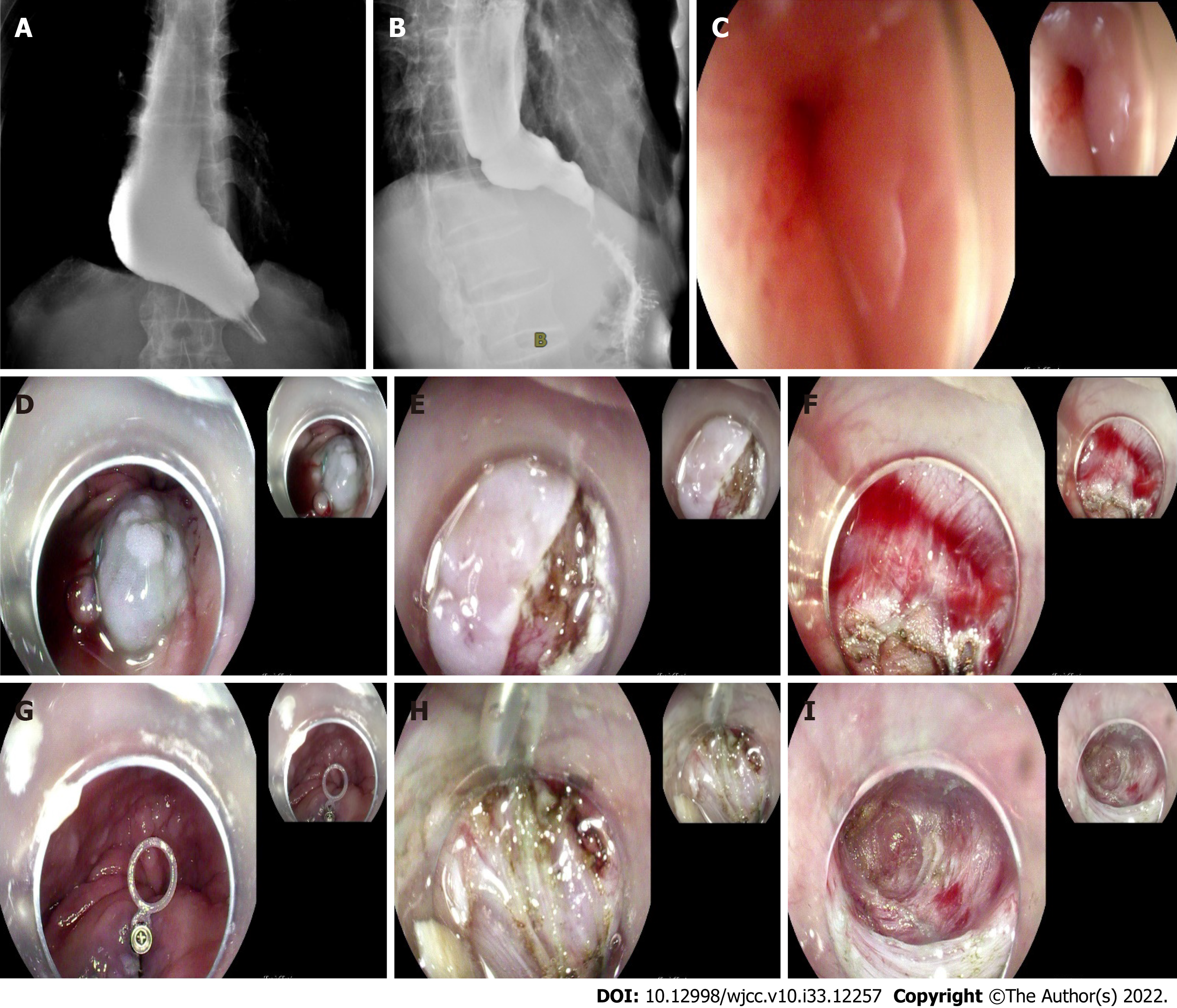
Submucosal fibrosis in achalasia patients is a rare cause of aborted peroral endoscopic myotomy (POEM) procedures. Implementation of new technology in achalasia is a challenge for endoscopic treatment when it comes to complex achalasia, because the dilated and tortuous esophageal lumen may make subsequent endoscopic dissection and separation of tissue planes difficult. Werner et al[1] reported peroral endoscopic dual myotomy (D-POEM) for achalasia with severe esophageal dilatation. Besides, he also reported open POEM (O-POEM) for achalasia with a sigmoid-shaped esophagus and achalasia with failed Heller myotomy. Wu et al[2] developed a modified version using a “Push and Pull” technique which reduced complications and accelerated wound recovery (Figure 1A).
A 54-year-old man who was unable to eat and drink for 1 wk presented at our hospital (Figure 1B).
The patient had choking sensation for 30 years (Figure 1C).
The patient had a history of ankylosing spondylitis. Physical examination on admission revealed malnutrition with a body mass index of 15.9. He had an Eckardt score of 12. A timed barium esophagram revealed that his esophagus was dilated and its lower end was narrow like a bird’s beak (Figure 1D). The contrast agent was not discharged after 10 min of observation. Preoperative gastroscopy revealed significant esophageal dilation and a large amount of food residue (Figure 1E). Esophageal stenosis was observed 38 cm away from the incisors, and the cardia was 52 cm away from them. Chest computed tomography ruled out external compression. This patient had never undergone any prior endoscopic intervention (Figure 1F).
The patient’s personal and family history was not remarkable.
The patient was thin. Abdominal examination was normal (Figure 1G).
Laboratory examinations showed no abnormality.
A timed barium esophagram revealed that the patient’s esophagus was dilated and its lower end was narrow like a bird’s beak. The contrast agent was not discharged after 10 min of observation (Figure 1H). Preoperative gastroscopy revealed significant esophageal dilation and a large amount of food residue. Esophageal stenosis was observed 38 cm away from the incisors, and the cardia was 52 cm away from them. Chest computed tomography ruled out external compression (Figure 1I).
The patient was diagnosed with achalasia.
Esophageal distortion was obvious in this patient, and the opening position of the tunnel was more suitable for the establishment of a complete tunnel. We used the endoscopic hood Olympus D201, and the dual knife had no water-jet function. Intraoperative submucosal injections were administered 10 cm above the cardia, resulting in a poor lift. There was obvious submucosal fibrosis. A 2-cm-long incision was made in the mucosa using the dual knife and submucosal adhesion was obvious. An elastic traction ring (patent number: ZL 2020 2 0016729.9) was used to tract the mucosa at the opening of the tunnel to the opposite side, and part of the circular muscle was cut to establish the submucosal tunnel. The elastic ring was deployed through the working channel. A clip was placed on the small ring at the initial incision site and a second clip was placed on the big ring attached to the opposing esophageal wall to create tension. The tunnel was built to 2 cm below the cardia. The muscle was incised 2 cm below the tunnel opening, and full-thickness myotomy was performed from 3 cm above the cardia to 2 cm below the cardia. Lastly, we removed the titanium clip holding the traction ring opposite the tunnel opening using a pair of biopsy forceps. The operation lasted 53 min.
Postoperatively, the cardia was obviously relaxed. The patient underwent gastrointestinal decompression for 48 h and fasted for 72 h. The timed barium esophagram was repeated 4 d after the operation, and the contrast agent passed through the cardia smoothly.
The symptoms of dysphagia were significantly relieved 3 wk after the operation, the Eckardt score was 2, and the patient’s weight gain was 5 kg.
Submucosal fibrosis in patients with achalasia is a rare cause of aborted POEM procedures. Over 5000 POEMs have been performed worldwide, with limited numbers of aborted procedures formally reported[1]. Zhou reported that 13 (0.77%) of 1693 POEMs (0.77%) were aborted, 12 (92.3%) of which were due to severe submucosal fibrosis that precluded tunneling[2,3]. The implementation of new technology in achalasia is a challenge for endoscopic treatment when it comes to complex achalasia, because the dilated and tortuous esophageal lumen may render subsequent endoscopic dissection and separation of tissue planes difficult. Werner et al[1] reported D-POEM for achalasia with severe esophageal dilatation. Besides, he also reported O-POEM for achalasia with a sigmoid-shaped esophagus and achalasia with failed Heller myotomy. Wu et al[2] developed a modified version using a “Push and Pull” technique which reduced complications and accelerated wound recovery. POEM with an elastic ring helps to clarify exposure levels and facilitate tunnel construction[4].
POEM with an elastic ring may be a feasible and effective endoscopic treatment modality for achalasia with obvious submucosal fibrosis[5-7]. We observed that the short-term outcome was excellent. This technique significantly shortened the operation time and improved the success rate of POEM for achalasia with obvious submucosal fibrosis.
Provenance and peer review: Unsolicited article; Externally peer reviewed.
Peer-review model: Single blind
Specialty type: Medicine, research and experimental
Country/Territory of origin: China
Peer-review report’s scientific quality classification
Grade A (Excellent): 0
Grade B (Very good): 0
Grade C (Good): C, C
Grade D (Fair): D
Grade E (Poor): E
P-Reviewer: Abe H, Japan; Hakimi T, Afghanistan; Sunjaya DB, United States; Viswanath YK, United Kingdom S-Editor: Wang LL L-Editor: Wang TQ P-Editor: Wang LL
| 1. | Werner YB, Costamagna G, Swanström LL. Clinical response to peroral endoscopic myotomy in patients with idiopathic achalasia at a minimum follow-up of 2 years. Gut. 2016;65:899-906. [RCA] [PubMed] [DOI] [Full Text] [Cited by in Crossref: 192] [Cited by in RCA: 168] [Article Influence: 18.7] [Reference Citation Analysis (0)] |
| 2. | Wu QN, Xu XY, Zhang XC, Xu MD, Zhang YQ, Chen WF, Cai MY, Qin WZ, Hu JW, Yao LQ, Li QL, Zhou PH. Submucosal fibrosis in achalasia patients is a rare cause of aborted peroral endoscopic myotomy procedures. Endoscopy. 2017;49:736-744. [RCA] [PubMed] [DOI] [Full Text] [Cited by in Crossref: 37] [Cited by in RCA: 40] [Article Influence: 5.0] [Reference Citation Analysis (0)] |
| 3. | Ramchandani M, Nageshwar Reddy D, Darisetty S, Kotla R, Chavan R, Kalpala R, Galasso D, Lakhtakia S, Rao GV. Peroral endoscopic myotomy for achalasia cardia: Treatment analysis and follow up of over 200 consecutive patients at a single center. Dig Endosc. 2016;28:19-26. [RCA] [PubMed] [DOI] [Full Text] [Cited by in Crossref: 80] [Cited by in RCA: 71] [Article Influence: 7.9] [Reference Citation Analysis (0)] |
| 4. | Yuan XL, Liu W, Ye LS, Yan P, Wang Y, Khan N, Hu B. Peroral endoscopic dual myotomy (dual POEM) for achalasia with severe esophageal dilatation. Endoscopy. 2018;50:E179-E180. [RCA] [PubMed] [DOI] [Full Text] [Cited by in Crossref: 5] [Cited by in RCA: 5] [Article Influence: 0.7] [Reference Citation Analysis (0)] |
| 5. | Liu W, Liu L, Chen HL, Zeng HZ, Wu CC, Ye LS, Hu B. Open peroral endoscopic myotomy for achalasia with sigmoid-shaped esophagus. Endoscopy. 2017;49:E311-E312. [RCA] [PubMed] [DOI] [Full Text] [Cited by in Crossref: 12] [Cited by in RCA: 11] [Article Influence: 1.4] [Reference Citation Analysis (0)] |
| 6. | Liu W, Wu CC, Hu B. Open peroral endoscopic myotomy for achalasia with failed Heller myotomy. Dig Endosc. 2018;30:268-269. [RCA] [PubMed] [DOI] [Full Text] [Cited by in Crossref: 7] [Cited by in RCA: 7] [Article Influence: 1.0] [Reference Citation Analysis (0)] |
| 7. | Zhang DF, Chen WF, Xu MD, Zhong YS, Zhang YQ, Li QL, Zhou PH. Modified peroral endoscopic myotomy: a "Push and Pull" technique. Surg Endosc. 2018;32:2165-2168. [RCA] [PubMed] [DOI] [Full Text] [Reference Citation Analysis (0)] |


