Published online Oct 16, 2022. doi: 10.12998/wjcc.v10.i29.10708
Peer-review started: May 9, 2022
First decision: May 30, 2022
Revised: June 10, 2022
Accepted: September 6, 2022
Article in press: September 6, 2022
Published online: October 16, 2022
Processing time: 142 Days and 20.1 Hours
Chondroid syringoma (CS) is a rare tumor of the apocrine or eccrine glands. CS of the lower back is rare, and its clinical manifestations are similar to those of lipoma, which is a common misdiagnosis for this disease.
A 39-year-old woman presented with a 2-year history of an asymptomatic subcutaneous mass on the lower back. The lesions increased progressively over time. The patient denied any history. Dermatological examination showed that there was a subcutaneous mass, ranging from 3-4 cm in diameter, with a clear boundary on the lower back. The surface of the skin was smooth without ulceration or scaling. Histopathologic examination was consistent with the diagnosis of CS.
CS is a rare tumor of the apocrine or eccrine glands. It usually presents as a wellcircumscribed and single subcutaneous masses. Histopathology showed the tumor was located in the dermis, with nests, sheets, and cords of basal-like cells, mucin deposition, and chondroid structures. We herein report a case of CS located in the lower back. CS of the lower back is rare, and its clinical manifestations are similar to those of lipoma, for which it is commonly misdiagnosed.
Core Tip: Chondroid syringoma (CS) is a rare tumor of the apocrine or eccrine glands. It usually occurs in the nose and surrounding areas, and it is rare in the lower back. It usually presents as a well-circumscribed, slow-growing, and single subcutaneous masses. It is easy to clinically misdiagnose CS as lipoma, but histopathological examination is helpful for the diagnosis and treatment of this disease. In our case, combined with the patient’s present illness, dermatological examination, and histopathology, the patient was diagnosed with CS. After surgical resection, no recurrence was found in follow-up visits.
- Citation: Huang QF, Shao Y, Yu B, Hu XP. Chondroid syringoma of the lower back simulating lipoma: A case report. World J Clin Cases 2022; 10(29): 10708-10712
- URL: https://www.wjgnet.com/2307-8960/full/v10/i29/10708.htm
- DOI: https://dx.doi.org/10.12998/wjcc.v10.i29.10708
Chondroid syringoma (CS), also known as mixed tumor of the skin (MTS), is a rare apocrine or eccrine tumor, accounting for 0.01% of primary skin tumors[1]. The etiology of CS is unknown, and it usually occurs in the head and neck, but is uncommon in the lower back. CS has no specific clinical manifestations and it is easily misdiagnosed as epidermoid cyst and lipoma[2]. In addition, CS has the potential for malignant transformation. The risk of clinical atypia and malignancy leads to delayed treatment[3]. Diagnosing CS is a challenge for clinicians. Therefore, we report a case of atypical CS and summarize its clinical manifestations, characteristic pathological findings, and treatment methods to raise clinicians’ awareness of the rare location of this rare disease.
A 39-year-old woman presented with an asymptomatic subcutaneous mass on the lower back which had been present for 2 years.
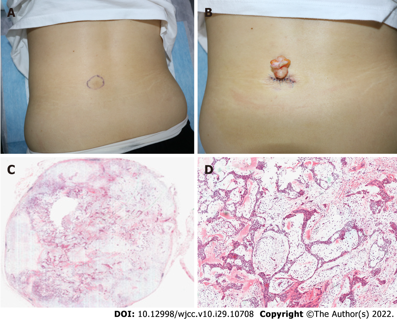
In 2020, a female patient presented with an asymptomatic subcutaneous mass on the lower back. Dermatological examination showed a subcutaneous mass, ranging from 3-4 cm in diameter, with clear boundaries. The surface of the skin was smooth without ulceration or scaling. We initially considered lipoma; however, histopathologic examination revealed a well-defined dermal tumor with nests, sheets, and cords of basal-like cells, glandular structures, interstitial mucin deposition, and chondroid structures in some areas. Therefore, our final diagnosis was CS.
The patient had no previous history.
The patient denied any family history of similar diseases or genetic history.
The physical examination indicated that the patient’s general condition was good, with no obvious abnormalities in the heart, lung, or abdomen, and superficial lymph nodes were not touched or enlarged. Upon dermatological examination, there was a subcutaneous mass, ranging from 3-4 cm in diameter, with a clear boundary on the lower back. The surface of the skin was smooth without ulceration or scaling (Figures 1A and 1B).
The patient’s laboratory tests were normal.
Imaging examination was not special.
The final diagnosis was CS.
The patient underwent surgical excision of the tumor, which revealed a yellow, smooth, tough mass with a clear boundary and a size of about 5 cm × 4 cm (Figure 1B).
There was no reoccurrence during the follow-up. No recurrence was found by palpation, and contrast-enhanced ultrasound was available for evaluation if necessary.
CS, also known as MTS, is a rare apocrine or eccrine tumor, accounting for 0.01% of primary skin tumors. CS was first described as a tumor located in the salivary gland by Billroth in 1895, and Virchow called it MTX a few years later because it appeared identical to mesenchymal neoplasm[1]. CS was first named in 1961[1]. Known for its chondroid sweat gland component, CS is mostly benign and has also been reported as malignant. The etiology of CS is unknown, and it usually occurs in the head and neck, especially in the nose and surrounding areas. It is rare in the external ear, lower lip, upper eyelid, scrotum, vulva, and other skin regions[1-3]. Lesions are mostly located in the dermis, with occasional occurrence up to the subcutaneous tissue. CS is more common in males between 20 and 40 years of age, while the malignant variant is more common in females[4]. The clinical features of CS are nonspecific and are characterized by isolated, raised, solid, asymptomatic nodules between 0.5 cm and 3.0 cm in diameter, with an average diameter of about 1 cm. Risk of malignancy increases in CS when lesions are greater than 3.0 cm in size[5]. Clinical diagnosis of CS is relatively difficult, and the diagnosis of CS is mainly based on histopathology. At present, the direction of CS differentiation is still controversial[6]. CS can differentiate either to eccrine or apocrine elements, with apocrine elements showing dominance. The resected tumor was comprised of epithelial and mesenchymal stromal derived elements. Histopathology showed that the tumor was located in the deep dermis or fat layer and differentiated into apocrine elements. It was characterized by irregular tubule-alveolar and ductal structures, which were composed of cuboid or polygonal cells in the shape of cords and nests, and embedded in myxoid and chondroid mesenchyma, with apocrine secretion[7]. The eccrine elements of CS differentiation showed a tubular structure, with epithelial cells scattered in chondroid and myxoid stroma, and without apocrine secretion[4,7]. CS contains acid mucopolysaccharide in cartilage and fibrous connective tissue, so Alcian blue staining can be positive. In addition, immunohistochemistry was helpful to understand the differentiation of CS, and the strong expression of CK15, EMA, carcinoembryonic antigen, and P63 suggested apocrine differentiation of the tumor. Histopathology can present obvious myoepithelial differentiation. Positive myoepithelial markers such as smooth muscle actin or calponin indicate myoepithelial differentiation, which can be diagnosed as myoepithelioma[8]. CS usually has a benign nature, but there is still a risk of malignancy. Histopathology showed cytological atypia, increased mitotic figures, infiltrative tumor margins, satellite nodules, and tumor liquefaction necrosis, which can be indicative of malignant transformation[2,9]. This patient would have typically been identified with lipoma, epidermoid cyst, dermoid cyst, etc. CS can be identified from other disease by histopathology. The clinical manifestations in this patient were similar to those of lipomas, with a subcutaneous active and tough mass. The histopathology of the lipoma revealed a well-defined dermal tumor with normal adipose cells. There are no fatty lobules separating the tumor tissue[9]. The histopathology of the epidermoid cyst showed a sharply defined cyst in the dermis with a wall composed of lamellar squamous epithelium. The contents of the cyst were horny material in the form of a net basket or plate layer[10]. Surgical resection is preferred for CS treatment. Incomplete resection of CS may lead to recurrence and malignant transformation. Therefore, the scope of surgical resection should be clearly defined before surgery and regular follow-up should be conducted after surgery[2].
The patient was an otherwise healthy middle-aged woman with a subcutaneous mass on the lower back for more than 2 years. The tumor gradually increased with a smooth skin surface and she was asymptomatic. The tumor was non-tender, slightly hard, and mobile, with a mass measuring 4 cm × 5 cm in size palpable on the lower back. Lipoma was considered in the initial diagnosis, but histopathological examination showed CS. Surgical resection was the first choice after diagnosis. The literature has reported that lesions with a diameter of more than 3 cm and occurring in females have a greater risk of malignant transformation. CS on the lower back is rare and easily misdiagnosed as lipoma or epidermoid cysts, resulting in delayed treatment and further increased risk of recurrence and malignant transformation.
This case is being reported for its rarity. A CS of the lower back is rare, and it is extremely easy to misdiagnose as lipoma. Even though CS is benign, it may become a malignant tumor. We report CS of the lower back mainly to improve clinicians’ understanding of this disease. Prompt diagnosis and treatment can reduce malignancy and recurrence.
Provenance and peer review: Unsolicited article; Externally peer reviewed.
Peer-review model: Single blind
Specialty type: Medicine, research and experimental
Country/Territory of origin: China
Peer-review report’s scientific quality classification
Grade A (Excellent): 0
Grade B (Very good): 0
Grade C (Good): C, C
Grade D (Fair): 0
Grade E (Poor): 0
P-Reviewer: Jeyaraman M, India; Taieb MAH, Tunisia S-Editor: Wang JJ L-Editor: Wang TQ P-Editor: Wang JJ
| 1. | Walvekar PV, Jakati S, Bothra N, Kaliki S. Isolated eyelid chondroid syringoma: a study of two cases. BMJ Case Rep. 2021;14. [RCA] [PubMed] [DOI] [Full Text] [Cited by in Crossref: 1] [Cited by in RCA: 4] [Article Influence: 1.0] [Reference Citation Analysis (0)] |
| 2. | Gotoh S, Ntege EH, Nakasone T, Matayoshi A, Miyamoto S, Shimizu Y, Nakamura H. Mixed tumour of the skin of the lower lip: A case report and review of the literature. Mol Clin Oncol. 2022;16:69. [RCA] [PubMed] [DOI] [Full Text] [Full Text (PDF)] [Cited by in Crossref: 1] [Cited by in RCA: 2] [Article Influence: 0.7] [Reference Citation Analysis (0)] |
| 3. | Bedir R, Yurdakul C, Sehitoglu I, Gucer H, Tunc S. Chondroid syringoma with extensive bone formation: a case report and review of the literature. J Clin Diagn Res. 2014;8:FD15-FD17. [RCA] [PubMed] [DOI] [Full Text] [Cited by in Crossref: 1] [Cited by in RCA: 2] [Article Influence: 0.2] [Reference Citation Analysis (0)] |
| 4. | Barnett MD, Wallack MK, Zuretti A, Mesia L, Emery RS, Berson AM. Recurrent malignant chondroid syringoma of the foot: a case report and review of the literature. Am J Clin Oncol. 2000;23:227-232. [RCA] [PubMed] [DOI] [Full Text] [Cited by in Crossref: 58] [Cited by in RCA: 65] [Article Influence: 2.6] [Reference Citation Analysis (0)] |
| 5. | Reddy PB, Nandini DB, Sreedevi R, Deepak BS. Benign chondroid syringoma affecting the upper lip: Report of a rare case and review of literature. J Oral Maxillofac Pathol. 2018;22:401-405. [RCA] [PubMed] [DOI] [Full Text] [Full Text (PDF)] [Cited by in Crossref: 6] [Cited by in RCA: 6] [Article Influence: 0.9] [Reference Citation Analysis (0)] |
| 6. | Agarwal R, Kulhria A, Singh K, Agarwal D. Cytodiagnosis of chondroid syringoma-Series of three cases. Diagn Cytopathol. 2021;49:E374-E377. [RCA] [PubMed] [DOI] [Full Text] [Cited by in Crossref: 1] [Cited by in RCA: 1] [Article Influence: 0.3] [Reference Citation Analysis (0)] |
| 7. | Jain A, Arava S. Chondroid syringoma with extensive cystic change and focal syringometaplasia: A rare histomorphological finding. Indian J Pathol Microbiol. 2018;61:143-144. [RCA] [PubMed] [DOI] [Full Text] [Cited by in Crossref: 1] [Cited by in RCA: 1] [Article Influence: 0.2] [Reference Citation Analysis (0)] |
| 8. | Bahrami A, Dalton JD, Krane JF, Fletcher CD. A subset of cutaneous and soft tissue mixed tumors are genetically linked to their salivary gland counterpart. Genes Chromosomes Cancer. 2012;51:140-148. [RCA] [PubMed] [DOI] [Full Text] [Cited by in Crossref: 83] [Cited by in RCA: 81] [Article Influence: 5.8] [Reference Citation Analysis (0)] |
| 9. | De Sanctis CM, Zara F, Sfasciotti GL. An Unusual Intraoral Lipoma: A Case Report and Literature Review. Am J Case Rep. 2020;21:e923503. [RCA] [PubMed] [DOI] [Full Text] [Full Text (PDF)] [Cited by in Crossref: 4] [Cited by in RCA: 4] [Article Influence: 0.8] [Reference Citation Analysis (0)] |
| 10. | Cui X, Wu X, Yao X. Surgical treatment for a giant epidermoid cyst on the buttock. Dermatol Ther. 2020;33:e13275. [RCA] [PubMed] [DOI] [Full Text] [Cited by in Crossref: 1] [Cited by in RCA: 2] [Article Influence: 0.4] [Reference Citation Analysis (0)] |


