Copyright
©The Author(s) 2022.
World J Clin Cases. Aug 16, 2022; 10(23): 8384-8391
Published online Aug 16, 2022. doi: 10.12998/wjcc.v10.i23.8384
Published online Aug 16, 2022. doi: 10.12998/wjcc.v10.i23.8384
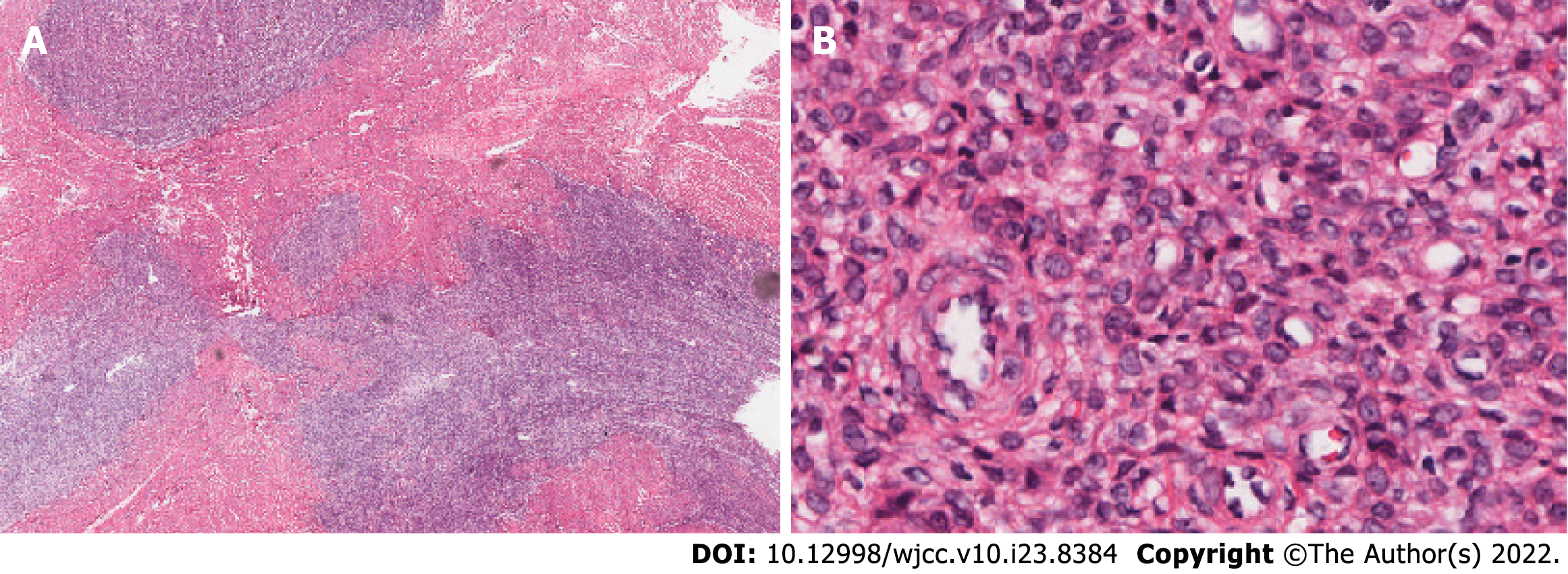
Figure 4 Biopsy material from the primary intrauterine tumor 20 years ago.
A: Light microscopy examination shows that the tumor had infiltrated the adjacent vaginal smooth muscular layer as tongues or islands (Original magnification: 40 ×; scale bar: 100 μm); B: Small oval to fusiform cells resemble the cells of proliferative endometrial stroma and whirl around spiral arteriole-like vessels (Original magnification: 400 ×; scale bar: 100 μm).
- Citation: Yu HY, Jin YL. Metastatic low-grade endometrial stromal sarcoma with variable morphologies in the ovaries and mesentery: A case report. World J Clin Cases 2022; 10(23): 8384-8391
- URL: https://www.wjgnet.com/2307-8960/full/v10/i23/8384.htm
- DOI: https://dx.doi.org/10.12998/wjcc.v10.i23.8384


