Published online Jul 6, 2022. doi: 10.12998/wjcc.v10.i19.6501
Peer-review started: August 2, 2021
First decision: November 6, 2021
Revised: November 15, 2021
Accepted: May 12, 2022
Article in press: May 12, 2022
Published online: July 6, 2022
Processing time: 326 Days and 2.5 Hours
In patients undergoing peritoneal dialysis (PD), catheter dysfunction is a common complication. A misplaced catheter is one of the reasons contributing to its dysfunction. The present study aimed to describe a case of misplaced PD catheter with an unusual location of the catheter tip.
A 61-year-old man undergoing PD for 4 years was investigated for progressive nausea and fatigue of 3 mo. Dialysis adequacy studies indicated inefficient dialysis. Imaging discovered that the PD catheter tip was mispositioned in the pelvic cavity with its tip outside the peritoneal cavity. Despite the dialysate accumulating outside the peritoneal cavity, the patient had not developed perineal or scrotal edema. The patient had experienced a sustainable prolonged dialysis efficacy in this case until the renal function deteriorated further in view of the poor dialysis outcome and worsening health condition. The patient was subsequently transitioned to hemodialysis.
Proper placement of the catheter in the peritoneal cavity should always be confirmed and re-checked when necessary in patients undergoing PD to ensure dialytic adequacy.
Core Tip: We present the case of a 61-year-old man undergoing prolonged peritoneal dialysis for 4 years who had the peritoneal dialysis catheter tip mispositioned outside the peritoneal cavity. Despite the prolonged dialysis with the misplaced catheter tip, the patient was able to sustain his dialysis adequacy without developing edema of the perineal, scrotal or abdominal muscles. This is the first case report describing this unique location of the catheter tip between the peritoneum and transversalis fascia that prevented early complication of dialysate migrating into the muscle causing edema while simultaneously allowing a sustainable dialysis through the available peritoneal surface.
- Citation: Shen QQ, Behera TR, Chen LL, Attia D, Han F. Sustained dialysis with misplaced peritoneal dialysis catheter outside peritoneum: A case report. World J Clin Cases 2022; 10(19): 6501-6506
- URL: https://www.wjgnet.com/2307-8960/full/v10/i19/6501.htm
- DOI: https://dx.doi.org/10.12998/wjcc.v10.i19.6501
Patients with renal dysfunction require renal replacement therapy that may include hemodialysis and peritoneal dialysis. In peritoneal dialysis (PD), the dialysate is introduced to the peritoneal cavity through the PD catheter, the tip of which stays positioned within in the peritoneal cavity for introduction and evacuation of the dialysate fluid. Though PD is a relatively safe procedure, there could be complications associated to its use. The complications of PD are commonly classified as infectious and noninfectious. Although infectious complications such as peritonitis leading to morbidity and mortality remain the main cause of PD failure[1], non-infectious causes also result in patient morbidity, which could be prevented by early recognition and management[2]. The various non-infectious complications are due to erroneous PD catheter insertion technique, failure to retain the catheter in the peritoneal cavity or a dialysate-induced rise in intra-abdominal pressure and the resulting metabolic effects. Here we report a case of a patient with end-stage renal disease undergoing PD without any incidence of significant deterioration over 4 years until he presented with inadequate small solute clearance as a complication of the misplaced extra-peritoneal catheter.
A 61-year-old man with end-stage renal disease undergoing PD was referred to our hospital with progressive nausea and fatigue for 3 mo.
The patient had developed renal failure secondary to uric acid nephropathy and was implanted with straight catheter with double polyester cuff. He was undergoing PD for 4 years. He had been undergoing continuous ambulatory PD with 3 exchanges per day; one with 1.5% solution and the remaining two with 2.5% solution, thereby achieving around 500 mL of peritoneal ultrafiltrate.
Other than the uric acid nephropathy, the patient had chronic hypertension.
There was no family history of renal failure or any relevant nephropathy in the family.
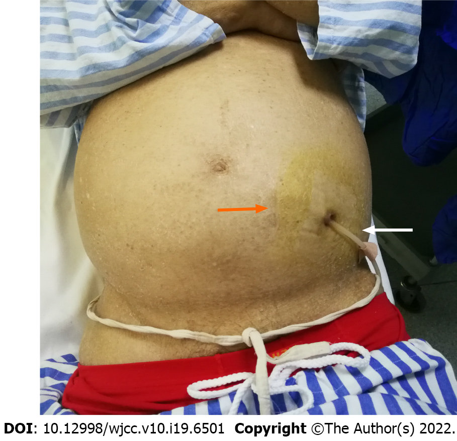
On admission, physical examination showed a tense distended abdomen without signs of edema in the perineum or lower extremities (Figure 1).
Laboratory assessment revealed severe anemia in the patient, with blood urea nitrogen 19.66 mmol/L, serum creatinine 884 µmol/L, uric acid 611 µmol/L, albumin 21.2 g/L, calcium 2.03 mmol/L, phosphate 2.10 mmol/L and parathyroid hormone level of 204 pmol/L. Peritoneal equilibration test determined his 4-hour equilibration between dialysate and plasma creatine to be 0.571, classifying him as a low-average transporter. The peritoneal dialysis adequacy parameters measured 5 mo prior to hospitalization as peritoneal kt/Vurea and dialysis creatinine clearance were 1.314 and 42.706 L/wk, respectively; the renal kt/Vurea and creatinine clearance at that time were 0.22 and 8.741 L/wk, respectively.
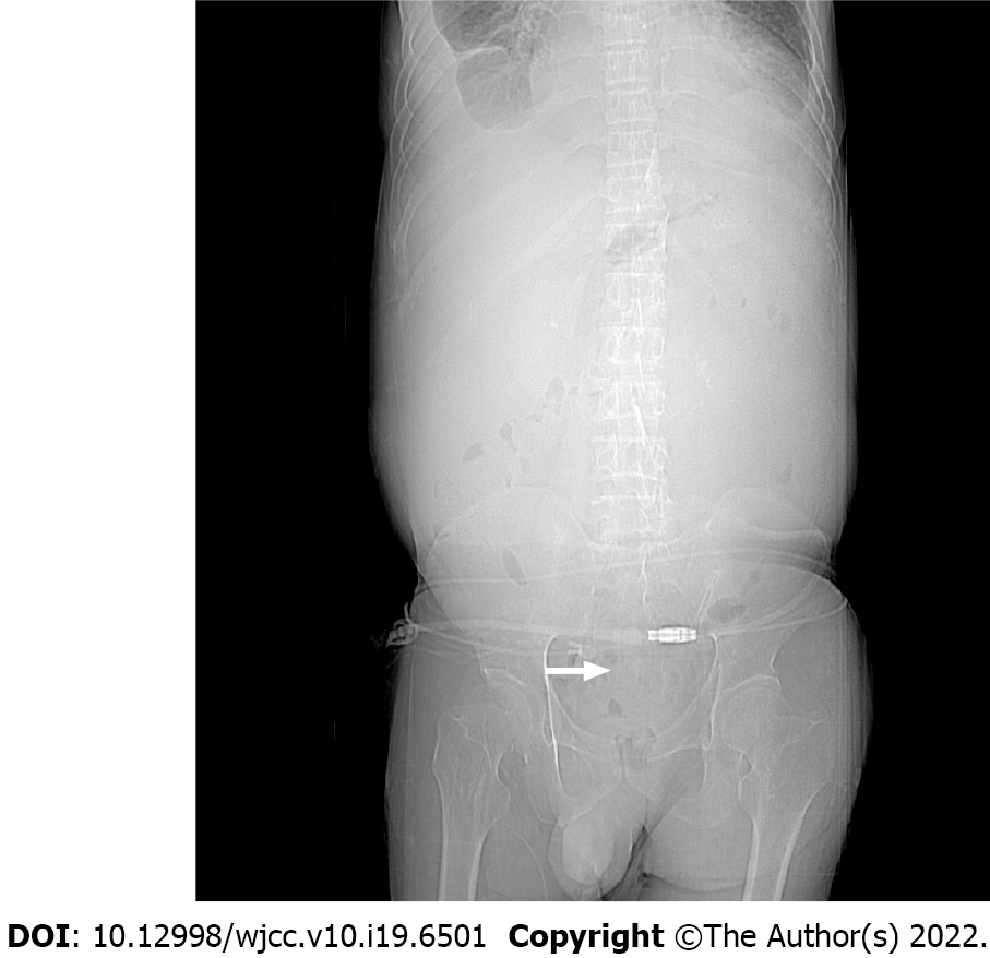
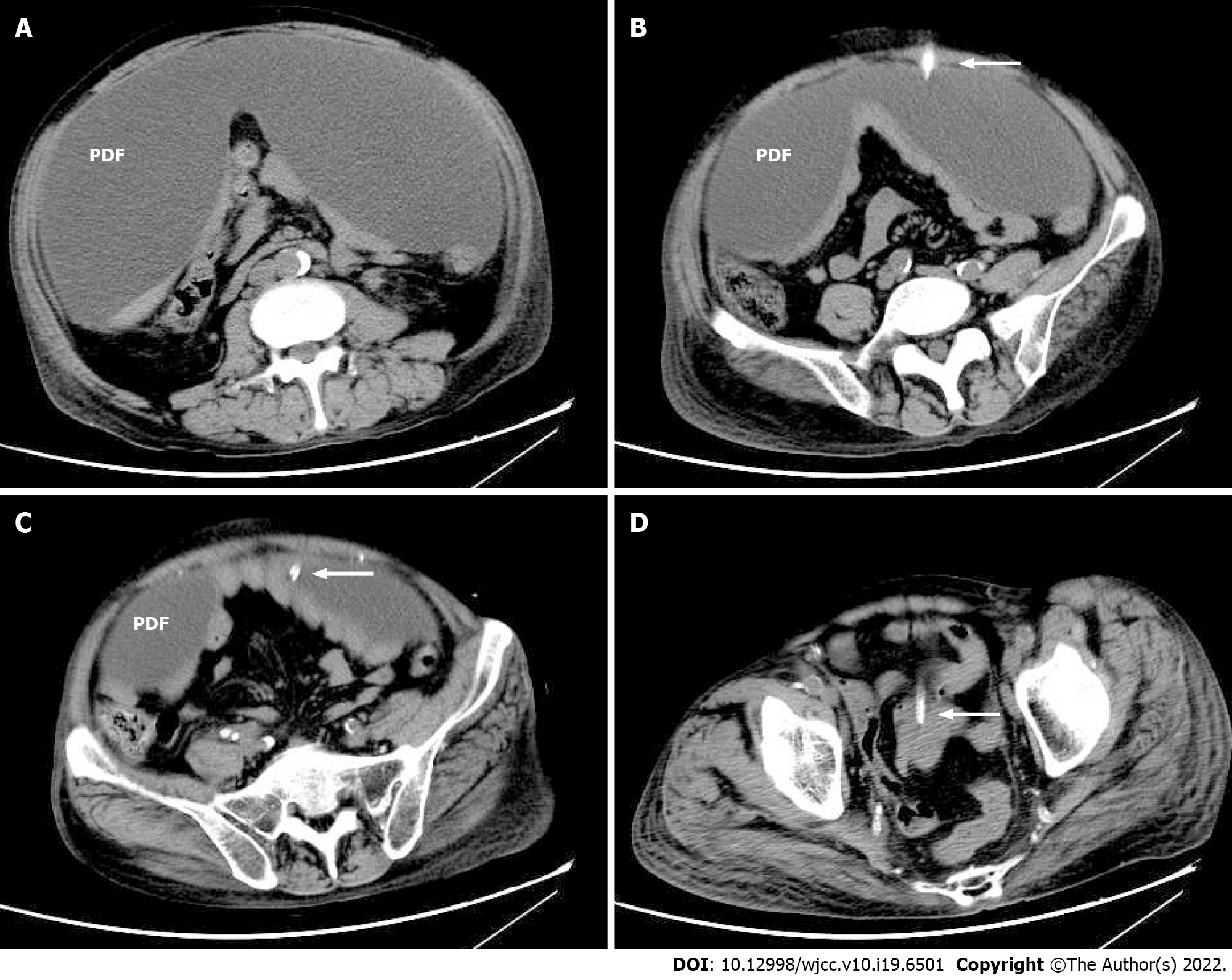
Imaging studies were performed to evaluate anatomical etiology. The abdominal plain film showed the tip of peritoneal catheter positioned in the true pelvis (Figure 2). The abdominal computed tomography confirmed the peritoneal dialysis catheter and the dialysis fluid to be outside the abdominal cavity (Figure 3).
Misplaced PD catheter tip was identified to be the reason of his deteriorating dialysis efficacy leading to his presentation.
In view of the worsening health condition due to poor dialysis outcome, the PD solution was drained, and the catheter was removed, transferring the patient to hemodialysis mode of renal replacement therapy.
The patient’s laboratory parameters and symptoms improved after the hemodialysis. The patient was followed up for surgical creation of fistula for future hemodialysis, which was continued at the local hospital.
Techniques for PD catheter insertion include placement by percutaneous needle-guidewire with or without image guidance, open surgical dissection, peritoneoscopic and laparoscopic implantation[3]. Irrespective of catheter insertion approach, the placement of catheter entry inside the peritoneal cavity must always be confirmed. The small pelvis below the pelvic brim is the ideal site for catheter tip location; positioning of the tip outside of this is considered a malposition of PD catheter, which has been attributed to contribute to catheter dysfunction. Several techniques of catheter insertion have been investigated including catheter tip fixation, in order to prevent its migration[4].
The catheter insertion in this patient was done by open dissection as evidenced by the surgical scar of the vertical paramedian incision (Figure 1). The catheter was located in the pelvis; however, the catheter tip was outside the peritoneal cavity. Despite the unintended location of the dialysis catheter over a period of 4 years of ongoing PD, the patient did not develop perineal or scrotal edema nor did the dialysate enter into the abdominal muscle. This could be possible if the catheter entered through the posterior rectus sheath and transversalis fascia without passing through the peritoneum, thus the catheter tip was located between the transversalis fascia and peritoneum. Another possible explanation of the position of the catheter tip could be catheter extrusion from its normal position.
PD relies on the use of the peritoneum as a semipermeable membrane for the exchange of solutes and water. The host-related factors, including peritoneal membrane surface area and peritoneal blood flow, influence transcapillary movement of solute and water[5]. In this case the patient remained sustainable through the PD for a prolonged period due to the residual renal reserves until the limited effective surface area of the peritoneum could not support adequate dialysis. The low PD adequacy parameters along with the worsening health condition prompted imaging to detect PD catheter failure. In patients not achieving the minimal delivered clearance goal, it is reasonable to ensure that the PD catheter is located within the peritoneal cavity.
This is the first case report describing a sustained PD for over 4 years with a misplaced extra-PD catheter in a patient undergoing PD. The unique location of the catheter tip between the peritoneum and transversalis fascia prevented early complication of dialysate migration into the muscle causing edema while allowing filtration through the available peritoneal surface area. To prevent complications of PD it is important to confirm and re-check when necessary the placement of catheter tip inside the peritoneal cavity.
Provenance and peer review: Unsolicited article; Externally peer reviewed.
Peer-review model: Single blind
Specialty type: Urology and nephrology
Country/Territory of origin: United States
Peer-review report’s scientific quality classification
Grade A (Excellent): 0
Grade B (Very good): B
Grade C (Good): C, C, C
Grade D (Fair): 0
Grade E (Poor): 0
P-Reviewer: Moyses Neto M, Brazil; Sachdeva S, India; Yang WY, China S-Editor: Liu JH L-Editor: Filipodia P-Editor: Liu JH
| 1. | Li PK, Szeto CC, Piraino B, de Arteaga J, Fan S, Figueiredo AE, Fish DN, Goffin E, Kim YL, Salzer W, Struijk DG, Teitelbaum I, Johnson DW. ISPD Peritonitis Recommendations: 2016 Update on Prevention and Treatment. Perit Dial Int. 2016;36:481-508. [RCA] [PubMed] [DOI] [Full Text] [Full Text (PDF)] [Cited by in Crossref: 589] [Cited by in RCA: 635] [Article Influence: 70.6] [Reference Citation Analysis (0)] |
| 2. | McCormick BB, Bargman JM. Noninfectious complications of peritoneal dialysis: implications for patient and technique survival. J Am Soc Nephrol. 2007;18:3023-3025. [RCA] [PubMed] [DOI] [Full Text] [Cited by in Crossref: 63] [Cited by in RCA: 60] [Article Influence: 3.3] [Reference Citation Analysis (0)] |
| 3. | Crabtree JH, Shrestha BM, Chow KM, Figueiredo AE, Povlsen JV, Wilkie M, Abdel-Aal A, Cullis B, Goh BL, Briggs VR, Brown EA, Dor FJMF. Creating and Maintaining Optimal Peritoneal Dialysis Access in the Adult Patient: 2019 Update. Perit Dial Int. 2019;39:414-436. [RCA] [PubMed] [DOI] [Full Text] [Cited by in Crossref: 254] [Cited by in RCA: 201] [Article Influence: 33.5] [Reference Citation Analysis (0)] |
| 4. | Chen JC, Lee WJ, Liu TP. Modified laparoscopic technique for fixation of peritoneal dialysis catheter. Surg Laparosc Endosc Percutan Tech. 2014;24:e146-e150. [RCA] [PubMed] [DOI] [Full Text] [Cited by in Crossref: 4] [Cited by in RCA: 4] [Article Influence: 0.4] [Reference Citation Analysis (0)] |
| 5. | Waniewski J, Werynski A, Lindholm B. Effect of blood perfusion on diffusive transport in peritoneal dialysis. Kidney Int. 1999;56:707-713. [RCA] [PubMed] [DOI] [Full Text] [Cited by in Crossref: 38] [Cited by in RCA: 39] [Article Influence: 1.5] [Reference Citation Analysis (0)] |


