Copyright
©The Author(s) 2021.
World J Clin Cases. Jan 26, 2021; 9(3): 651-658
Published online Jan 26, 2021. doi: 10.12998/wjcc.v9.i3.651
Published online Jan 26, 2021. doi: 10.12998/wjcc.v9.i3.651
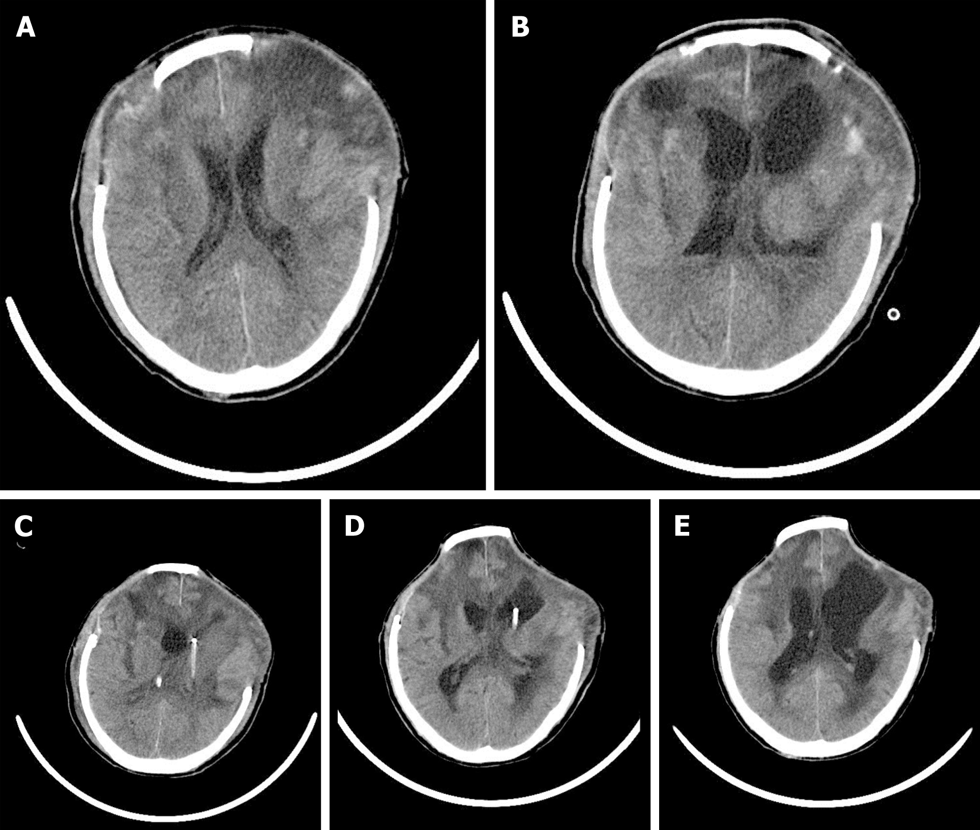
Figure 1 Computed tomography images of the patient.
A: No ventricle pus was detected by head computed tomography (CT) on day 18; B: Head CT image obtained on day 24 showing ventricle pus; C: Head CT image after bilateral ventricular drainage; D: Head CT image obtained on day 30 showing that the ventricle pus had disappeared; E: Head CT image after removing the left ventricular drainage tube.
Figure 2 The timeline and antibiotics usage of the patient.
IVT: Intraventricular; CVI: Continuous ventricular irrigation; CSF: Cerebral spinal fluid.
- Citation: Li W, Li DD, Yin B, Lin DD, Sheng HS, Zhang N. Successful treatment of pyogenic ventriculitis caused by extensively drug-resistant Acinetobacter baumannii with multi-route tigecycline: A case report. World J Clin Cases 2021; 9(3): 651-658
- URL: https://www.wjgnet.com/2307-8960/full/v9/i3/651.htm
- DOI: https://dx.doi.org/10.12998/wjcc.v9.i3.651


