Copyright
©The Author(s) 2021.
World J Clin Cases. Jul 26, 2021; 9(21): 5948-5954
Published online Jul 26, 2021. doi: 10.12998/wjcc.v9.i21.5948
Published online Jul 26, 2021. doi: 10.12998/wjcc.v9.i21.5948
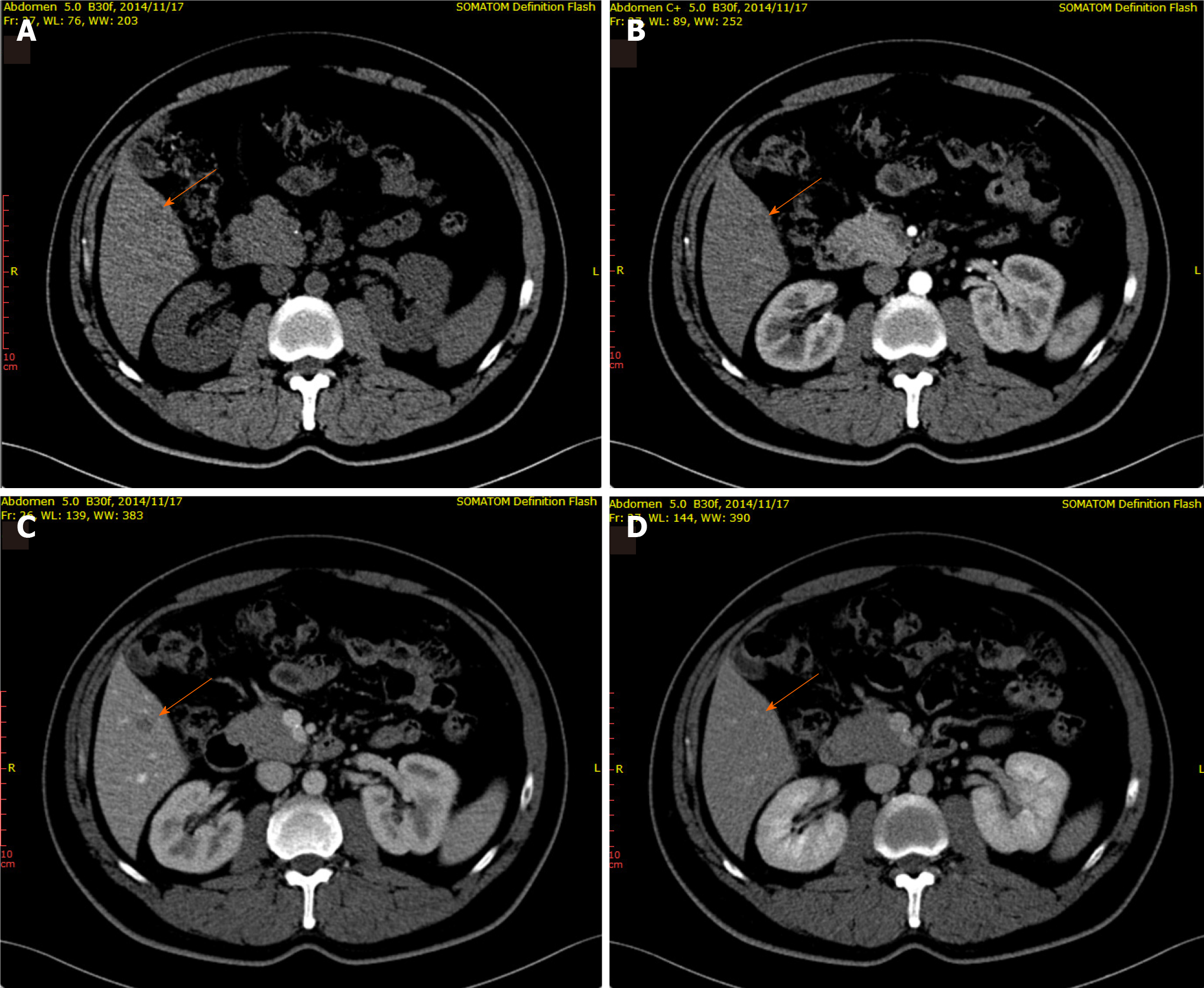
Figure 1 A 46-year-old male patient with focal liver lesions involving the right lobe of the liver.
A: Routine computerized tomography (CT) scan revealed an indistinct hypodense lesion (arrow) located in segment V of the liver; B: In the arterial phase, contrast enhanced CT revealed a homogeneous mass with no enhancement (arrow); C and D: A well-defined round mass (arrow), measured about 10 mm × 9 mm, was observed in the portal venous phase (C) and the mass showed iso-density in the delayed phase (D).
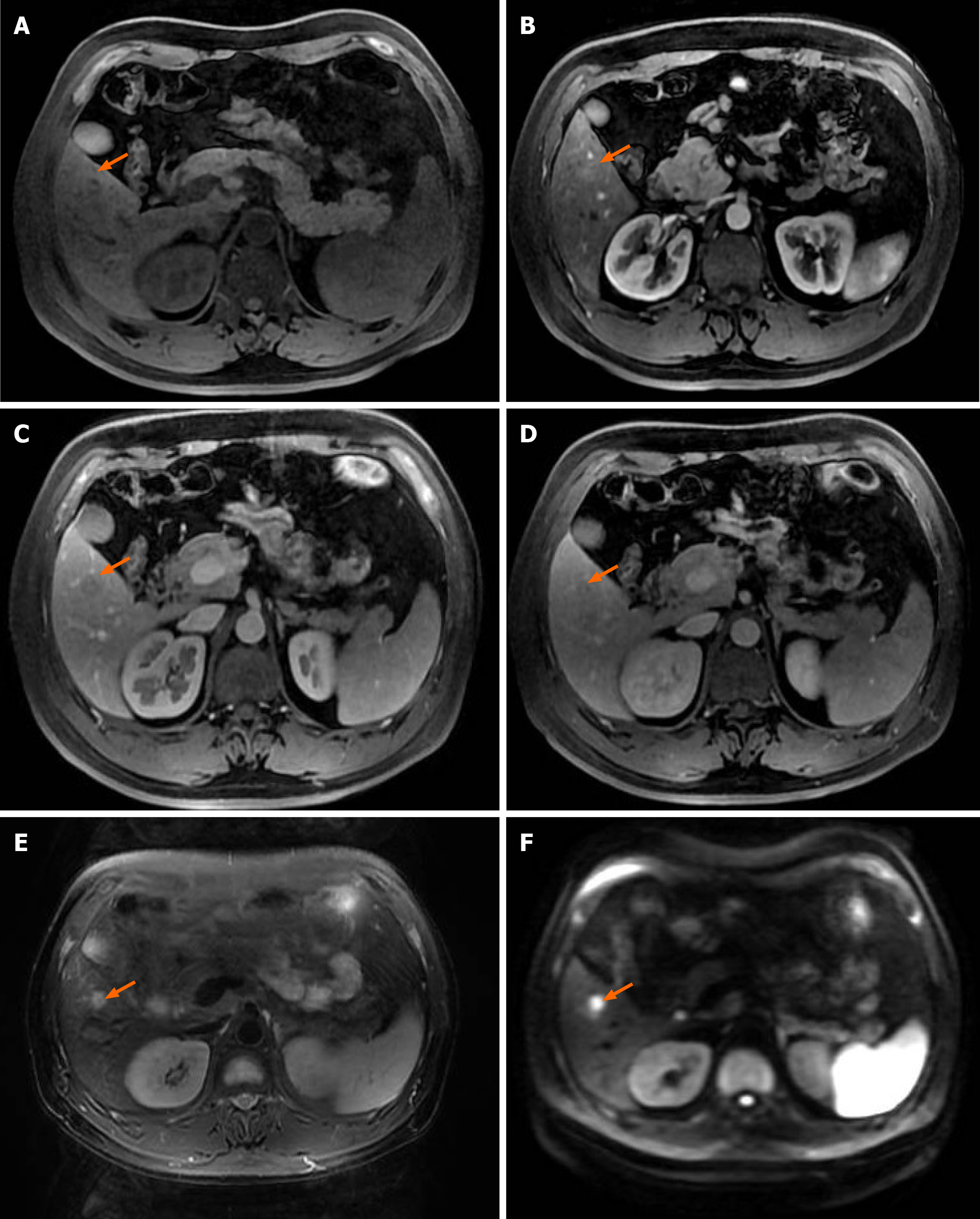
Figure 2 Magnetic resonance imaging of upper abdomen.
Magnetic resonance imaging scan revealed an iso T1 and long T2 signals, lesion only showed slightly enhancement in arterial phase, relatively low signal in portal and delayed phase hyperintense signal was observed in diffusion-weighted imaging. A: Iso T1 signal; B: Lesion only showed slightly enhancement in arterial phase; C and D: Relatively low signal in portal and delayed phase; E: Long T2 signal; F: Hyperintense signal was observed in diffusion-weighted imaging.
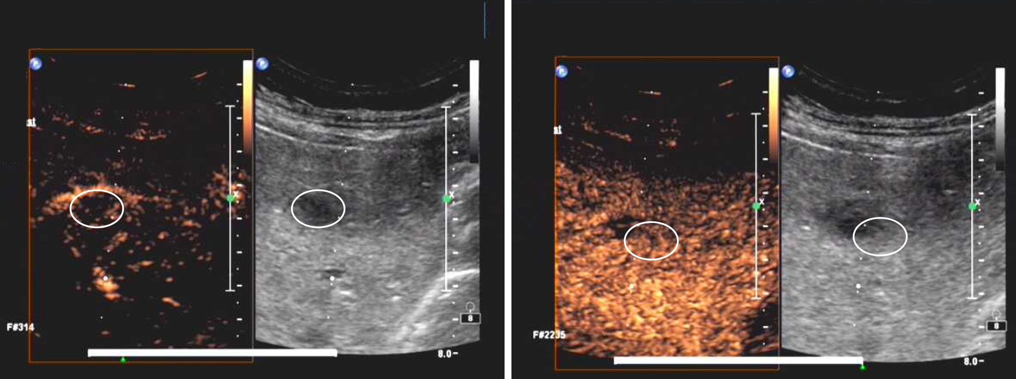
Figure 3 Realtime contrast-enhanced ultrasound of the patient.
The lesion identified by B-mode ultrasound and contrast agent SonoVue (Bracco, Italy) was then injected through the cubital vein with a fixed body position. However, no microbubble contrast agent filling was observed during the 5 min consecutive examination.
- Citation: Wang ZD, Haitham S, Gong JP, Pen ZL. Contrast enhanced ultrasound in diagnosing liver lesion that spontaneously disappeared: A case report . World J Clin Cases 2021; 9(21): 5948-5954
- URL: https://www.wjgnet.com/2307-8960/full/v9/i21/5948.htm
- DOI: https://dx.doi.org/10.12998/wjcc.v9.i21.5948


