Copyright
©The Author(s) 2021.
World J Clin Cases. Jul 26, 2021; 9(21): 5948-5954
Published online Jul 26, 2021. doi: 10.12998/wjcc.v9.i21.5948
Published online Jul 26, 2021. doi: 10.12998/wjcc.v9.i21.5948
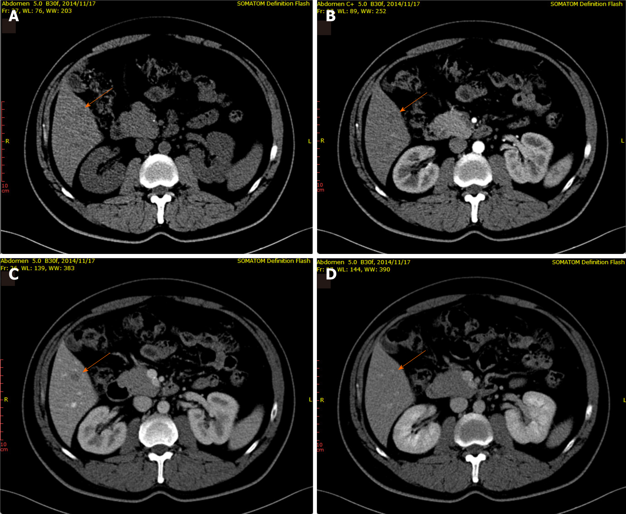
Figure 1 A 46-year-old male patient with focal liver lesions involving the right lobe of the liver.
A: Routine computerized tomography (CT) scan revealed an indistinct hypodense lesion (arrow) located in segment V of the liver; B: In the arterial phase, contrast enhanced CT revealed a homogeneous mass with no enhancement (arrow); C and D: A well-defined round mass (arrow), measured about 10 mm × 9 mm, was observed in the portal venous phase (C) and the mass showed iso-density in the delayed phase (D).
- Citation: Wang ZD, Haitham S, Gong JP, Pen ZL. Contrast enhanced ultrasound in diagnosing liver lesion that spontaneously disappeared: A case report . World J Clin Cases 2021; 9(21): 5948-5954
- URL: https://www.wjgnet.com/2307-8960/full/v9/i21/5948.htm
- DOI: https://dx.doi.org/10.12998/wjcc.v9.i21.5948


