Copyright
©The Author(s) 2021.
World J Clin Cases. Jun 26, 2021; 9(18): 4852-4858
Published online Jun 26, 2021. doi: 10.12998/wjcc.v9.i18.4852
Published online Jun 26, 2021. doi: 10.12998/wjcc.v9.i18.4852
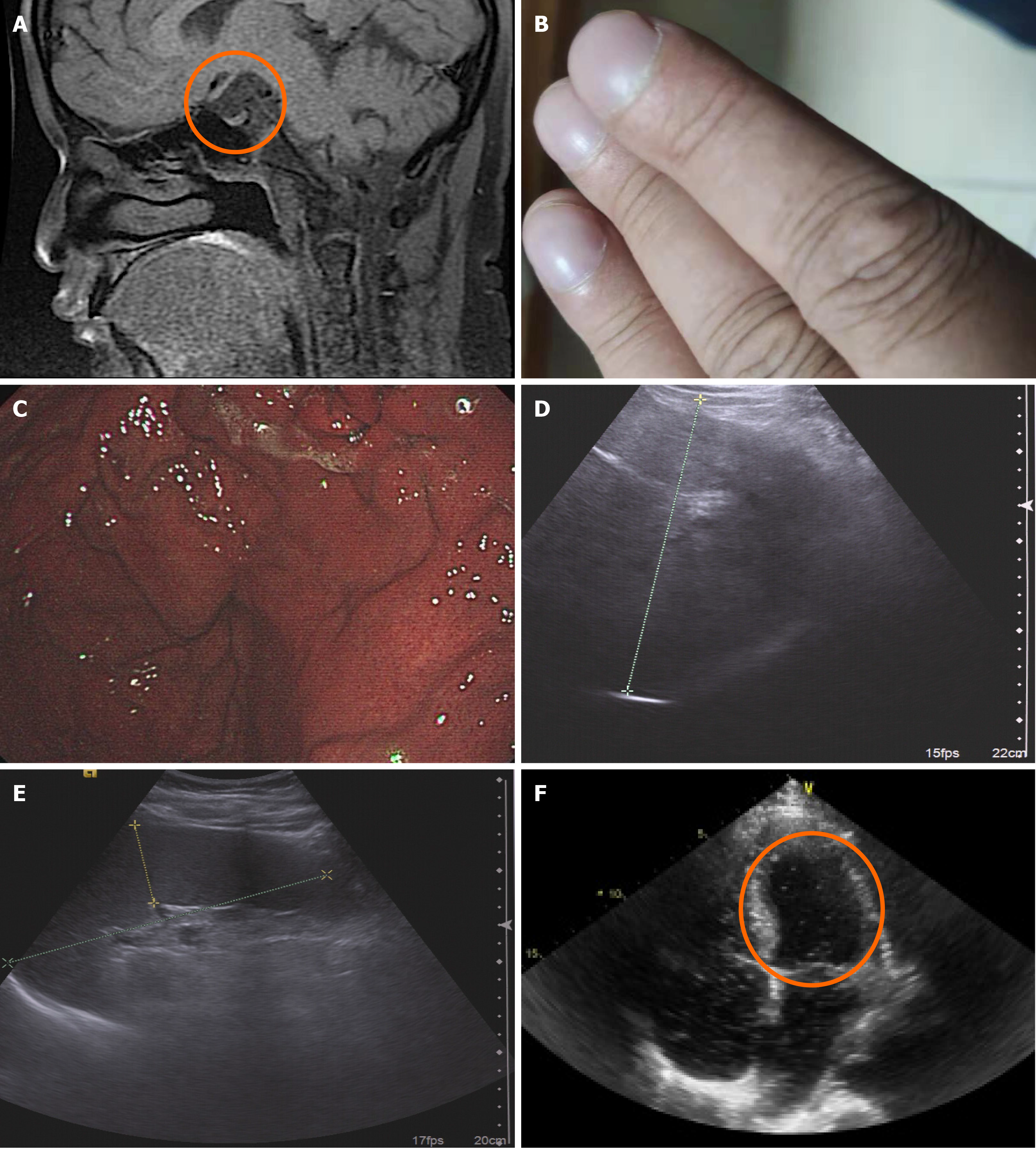
Figure 1 Imaging examination on admission.
A: Magnetic resonance imaging of the Sellar region. Orange circle shows the hallmarks of pituitary stalk interruption syndrome, including invisible pituitary stalk, and hypoplastic anterior pituitary gland combined with disappeared hyperintense signal in the posterior pituitary; B: Clubbed fingers; C: Prominent gastric varices under gastroscopy; D: Ultrasonic examination of the liver: Coarse texture with an oblique diameter of 16.6 cm and more echo compared to the right renal cortex, in keeping with liver cirrhosis and diffuse fatty liver; E: Enlarged spleen (15.9 cm × 4.4 cm); F: Transthoracic contrast echocardiography showed opacification in the left chamber of the heart by micro-bubbles five heartbeats after the appearance of microbubbles in the right atrium (orange circle).
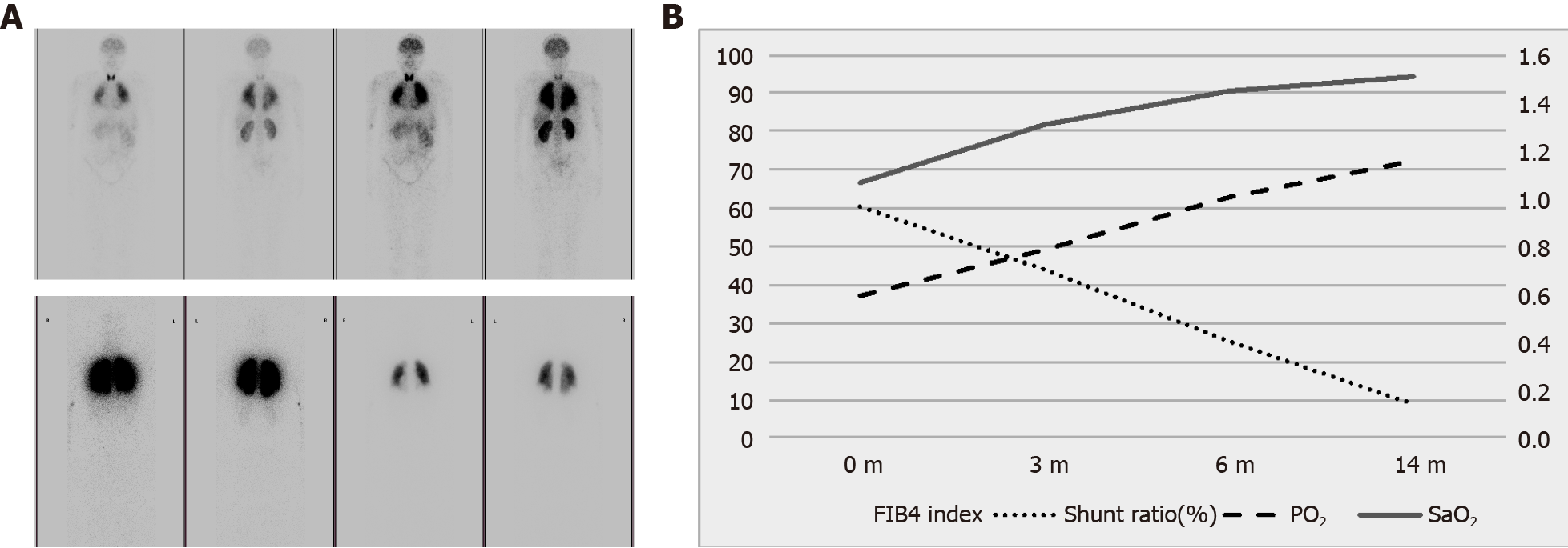
Figure 2 Response to hormone treatment.
A: Uptake ratio of radionuclides 99mTc macroaggregated albumin of the whole body. Intrapulmonary shunting returned to normal (bottom) from 64.4% (top). These images are from department of nuclear medicine, Peking Union Medical College Hospital; B: The right Y-axis represents fibrosis 4 (FIB-4) index, and the left Y-axis represents the intrapulmonary shunt ratio in percentage items, with PO2 and SaO2 in mmHg units. The PO2 and SaO2 levels markedly increased along with declination of intrapulmonary shunt ratio and FIB-4 index. FIB-4: Fibrosis 4.
- Citation: Ji W, Nie M, Mao JF, Zhang HB, Wang X, Wu XY. Growth hormone cocktail improves hepatopulmonary syndrome secondary to hypopituitarism: A case report. World J Clin Cases 2021; 9(18): 4852-4858
- URL: https://www.wjgnet.com/2307-8960/full/v9/i18/4852.htm
- DOI: https://dx.doi.org/10.12998/wjcc.v9.i18.4852


