Copyright
©The Author(s) 2021.
World J Clin Cases. Jun 26, 2021; 9(18): 4765-4771
Published online Jun 26, 2021. doi: 10.12998/wjcc.v9.i18.4765
Published online Jun 26, 2021. doi: 10.12998/wjcc.v9.i18.4765
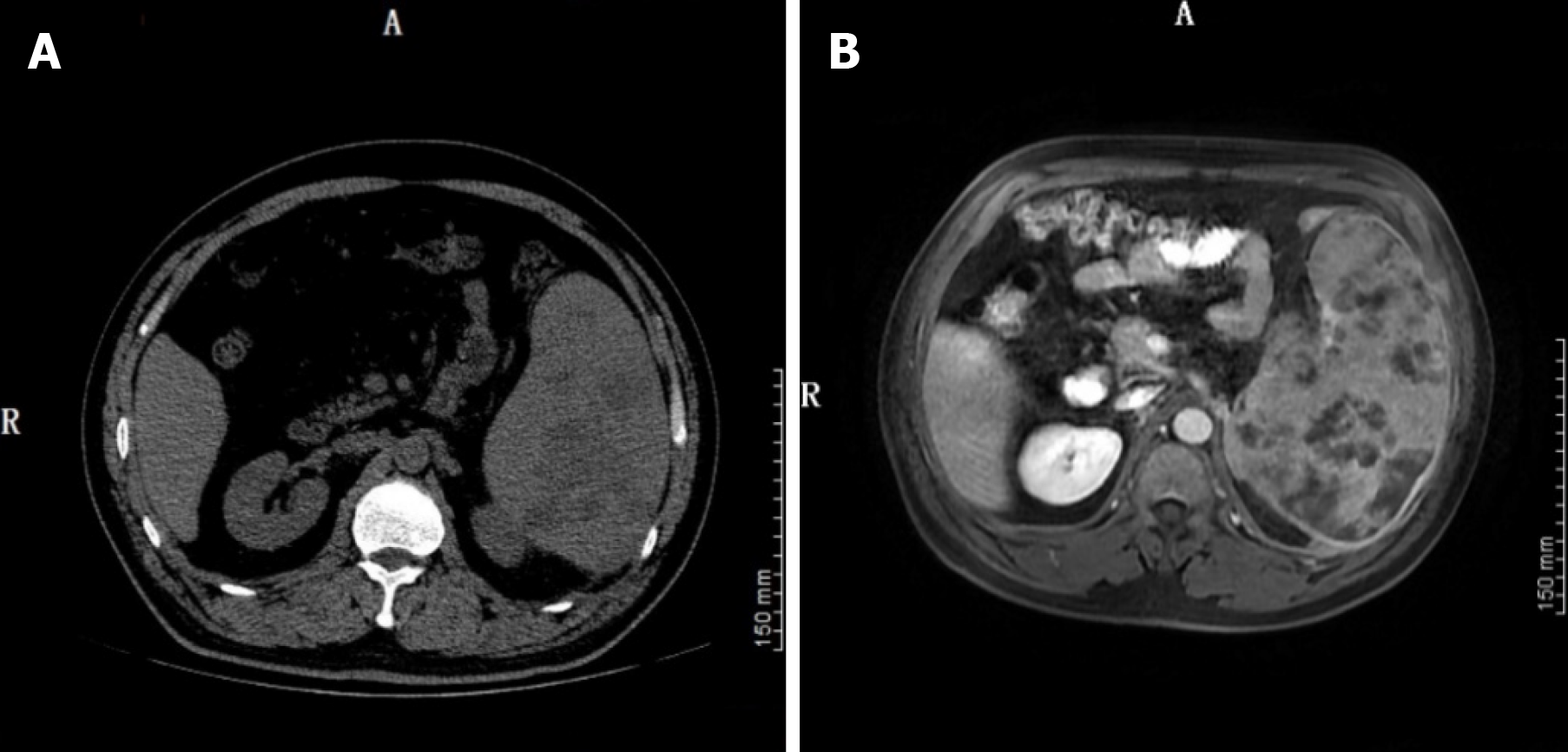
Figure 1 Abdominal computed tomography scan and magnetic resonance imaging before surgery.
A: The abdominal computed tomography performed on July 20, 2020 revealed splenomegaly, with multiple low density shadows and mixed density shadows; B: The magnetic resonance imaging on July 25, 2020 revealed multiple space-occupying lesions in the spleen, which was considered to be lymphoma.
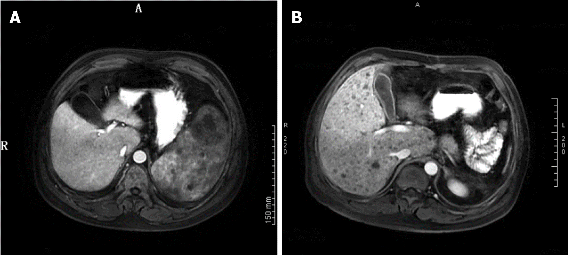
Figure 2 Abdominal magnetic resonance imaging images of the liver pre- and post-operation.
A: The magnetic resonance imaging (MRI) performed before surgery on July 25, 2020 showed no Kaposi’s sarcoma (KS) lesions in the liver; B: The MRI on September 5, 2020, when the patient was readmitted to the hospital after surgery, showed multiple KS metastatic lesions in the liver (black spots).
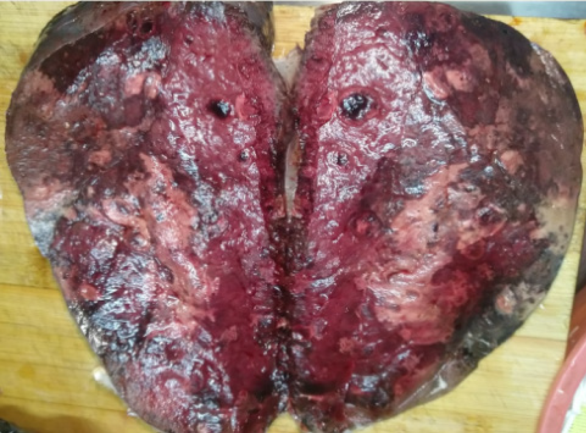
Figure 3 The excised spleen.
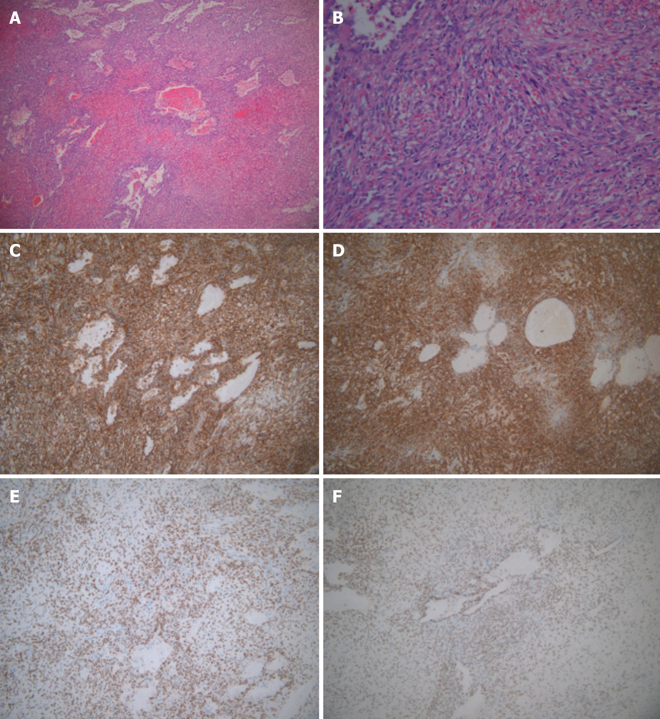
Figure 4 Hematoxylin and eosin staining and immunohistochemistry.
A: Multinodular tumors with a clear boundary and composed of proliferative spindle cells with interstitial hemorrhage (× 10); B: Proliferating spindle cells divided by slit-like spaces containing red blood cells, which were sieve-like or beehive in shape, and eosinophilic hyaline bodies scattered in the cytoplasm or extracellular of spindle cells (× 200); C: Immunohistochemical staining was positive for CD31, revealing splenic Kaposi’s sarcoma (KS) (× 200); D: Immunohistochemical staining was positive for CD34, revealing splenic KS (× 200); E: Immunohistochemical staining was positive for ERG (× 200); F: Immunohistochemical staining was positive for FLI-1 (× 200).
- Citation: Zhao CJ, Ma GZ, Wang YJ, Wang JH. Splenic Kaposi’s sarcoma in a human immunodeficiency virus-negative patient: A case report. World J Clin Cases 2021; 9(18): 4765-4771
- URL: https://www.wjgnet.com/2307-8960/full/v9/i18/4765.htm
- DOI: https://dx.doi.org/10.12998/wjcc.v9.i18.4765


