Copyright
©The Author(s) 2021.
World J Clin Cases. Jun 6, 2021; 9(16): 4001-4006
Published online Jun 6, 2021. doi: 10.12998/wjcc.v9.i16.4001
Published online Jun 6, 2021. doi: 10.12998/wjcc.v9.i16.4001
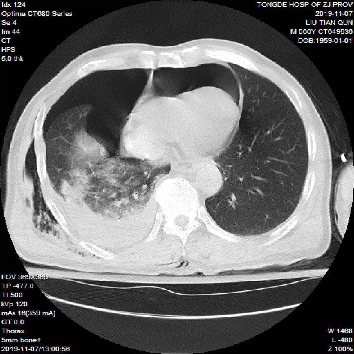
Figure 1 Chest computed tomography showed pneumopericardium, right hemopneumothorax, and lung contusion.
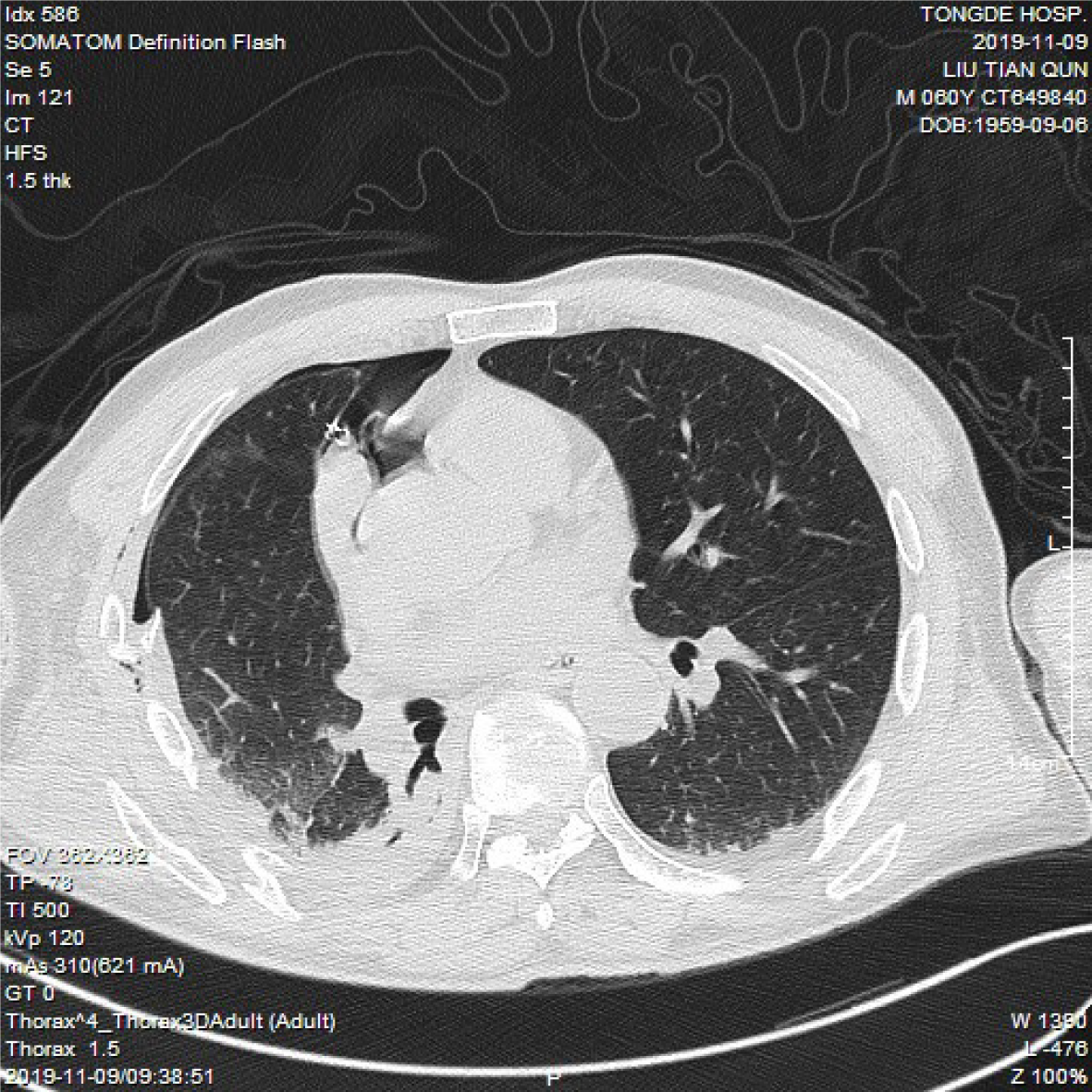
Figure 2 The pneumopericardium and right hemopneumothorax were obviously absorbed after tube thoracostomy was performed.
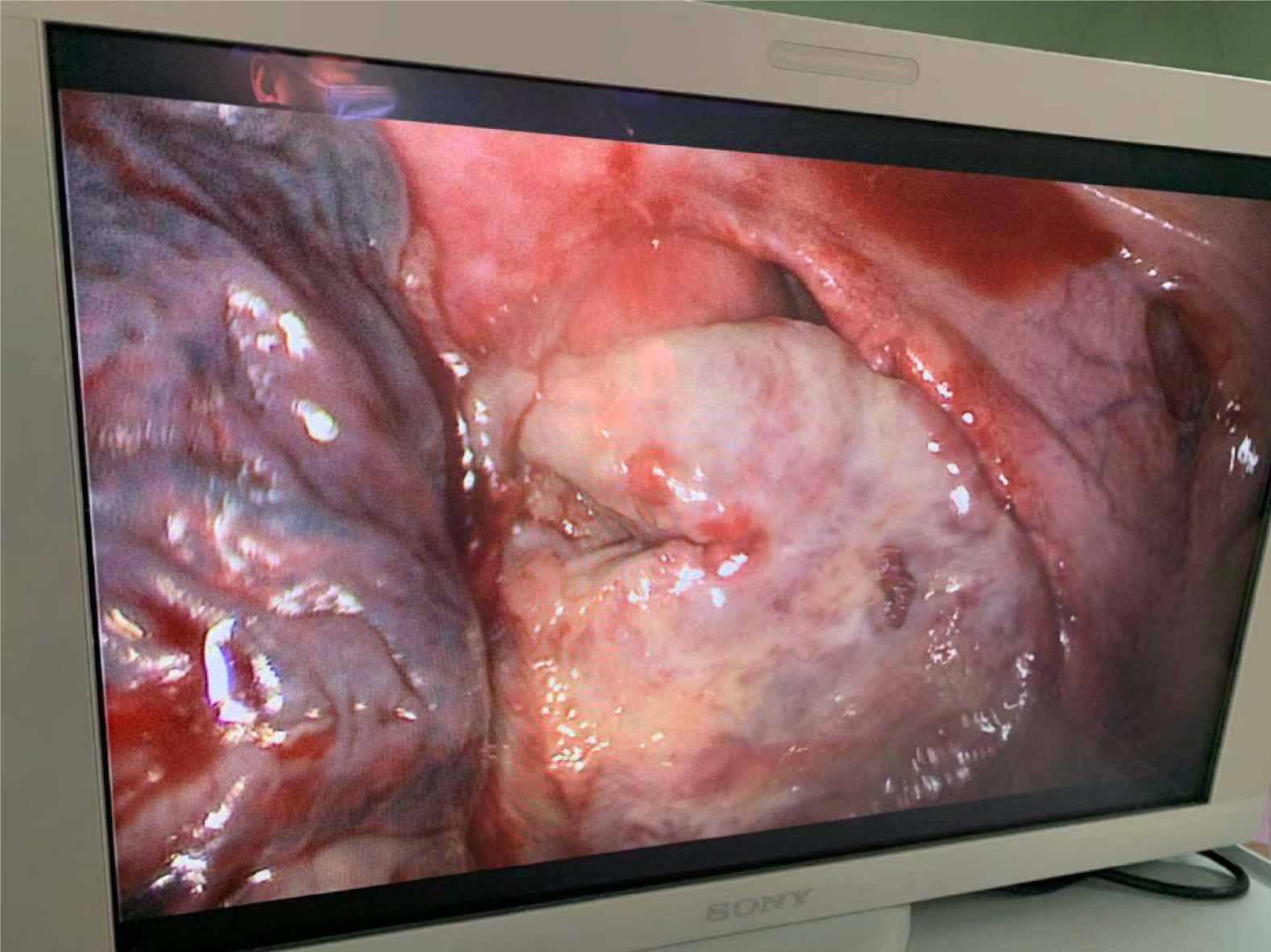
Figure 3 The ruptured pericardium, exposed right atrial appendage, right atrium, and superior vena cava under thoracoscopy.
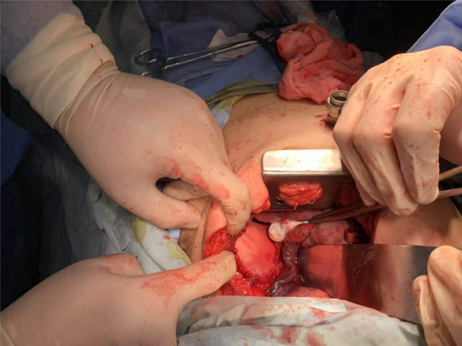
Figure 4 The ruptured pericardium was repaired with a heart Dacron patch.
- Citation: Wu YY, He ZL, Lu ZY. Thoracoscopic diagnosis of traumatic pericardial rupture with cardiac hernia: A case report. World J Clin Cases 2021; 9(16): 4001-4006
- URL: https://www.wjgnet.com/2307-8960/full/v9/i16/4001.htm
- DOI: https://dx.doi.org/10.12998/wjcc.v9.i16.4001


