Copyright
©The Author(s) 2021.
World J Clin Cases. Jun 6, 2021; 9(16): 3988-3995
Published online Jun 6, 2021. doi: 10.12998/wjcc.v9.i16.3988
Published online Jun 6, 2021. doi: 10.12998/wjcc.v9.i16.3988
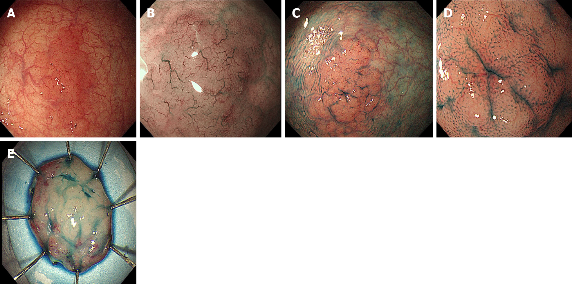
Figure 1 Endoscopic findings.
A: A laterally spreading tumor-like elevated lesion was observed on white light endoscopy in the rectum; B: Narrow band imaging showed enlarged branch-like vessels on the surface of the lesion; C and D: Indigo carmine staining made the lesion margin clearer (C), and pit pattern II was observed on magnifying endoscopy (D); E: On assessment of the endoscopic submucosal dissection specimen, a slightly elevated, laterally spreading tumor measuring 25 mm × 20 mm was identified in the mucosal layer.
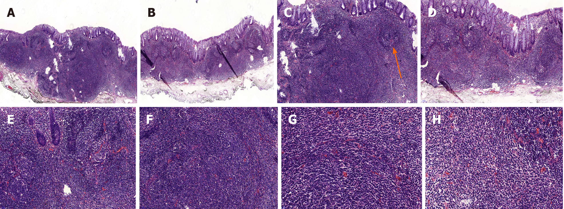
Figure 2 Histopathological findings (hematoxylin and eosin staining).
Diffusely hyperplastic lymphoid tissue could be observed in the lamina propria with visible lymphoid follicle structures (orange arrow, C). There were a large number of lymphoid cells around the lymphoid follicles that had clear cytoplasm and similar sizes. A and B: Magnification 50 ×; C and D: Magnification 100 ×; E and F: Magnification 200 ×; G and H Magnification 400 ×.
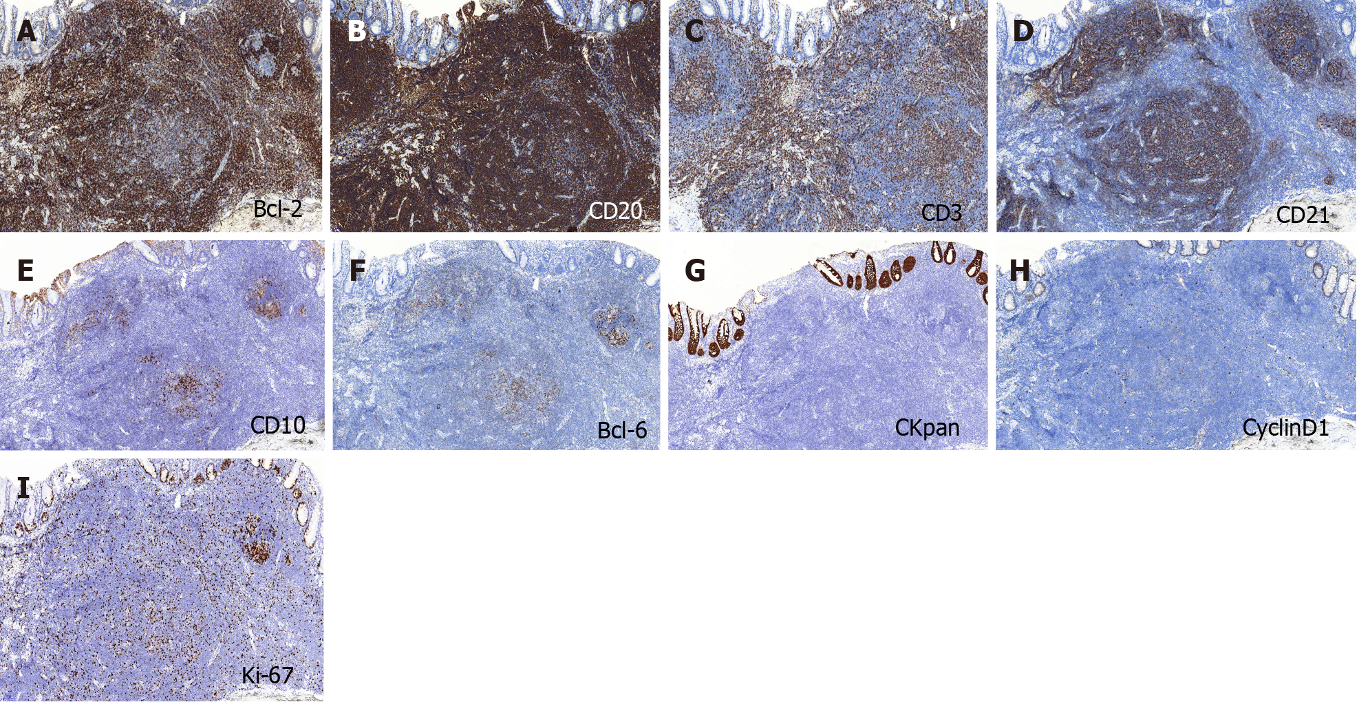
Figure 3 Immunohistochemical findings.
A and B: BCL-2 and CD20 were strongly expressed in the tumor; C and D: CD3 and CD21 were positive; E and F: CD10 and BCL-6 showed scattered positivity in the tumor; G: CKpan was expressed in the residual epithelium; H: CyclinD1 was negative in the tumor; I: The Ki-67 labeling index was 20%. Magnification of all photographs is 100 ×.
Figure 4 Immunohistochemical findings (CD31 marker).
Hyperplastic capillaries can be seen in the mucosa, but the branch-like structures were not obvious. A: Magnification 100 ×; B: Magnification 200 ×.
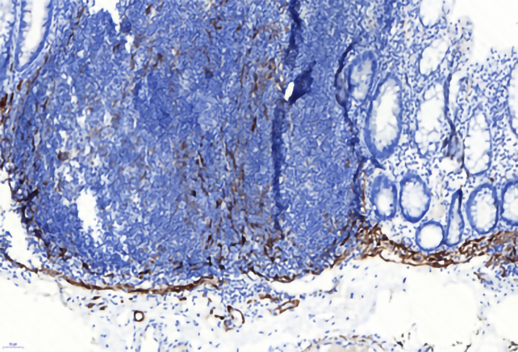
Figure 5 Immunohistochemical findings (Desmin marker).
The tumor was located above the muscularis mucosa and did not involve the submucosa, and the expansive growth of the tumor made the muscularis mucosa thinner.
- Citation: Wei YL, Min CC, Ren LL, Xu S, Chen YQ, Zhang Q, Zhao WJ, Zhang CP, Yin XY. Laterally spreading tumor-like primary rectal mucosa-associated lymphoid tissue lymphoma: A case report. World J Clin Cases 2021; 9(16): 3988-3995
- URL: https://www.wjgnet.com/2307-8960/full/v9/i16/3988.htm
- DOI: https://dx.doi.org/10.12998/wjcc.v9.i16.3988


