Copyright
©The Author(s) 2021.
World J Clin Cases. Jun 6, 2021; 9(16): 3988-3995
Published online Jun 6, 2021. doi: 10.12998/wjcc.v9.i16.3988
Published online Jun 6, 2021. doi: 10.12998/wjcc.v9.i16.3988
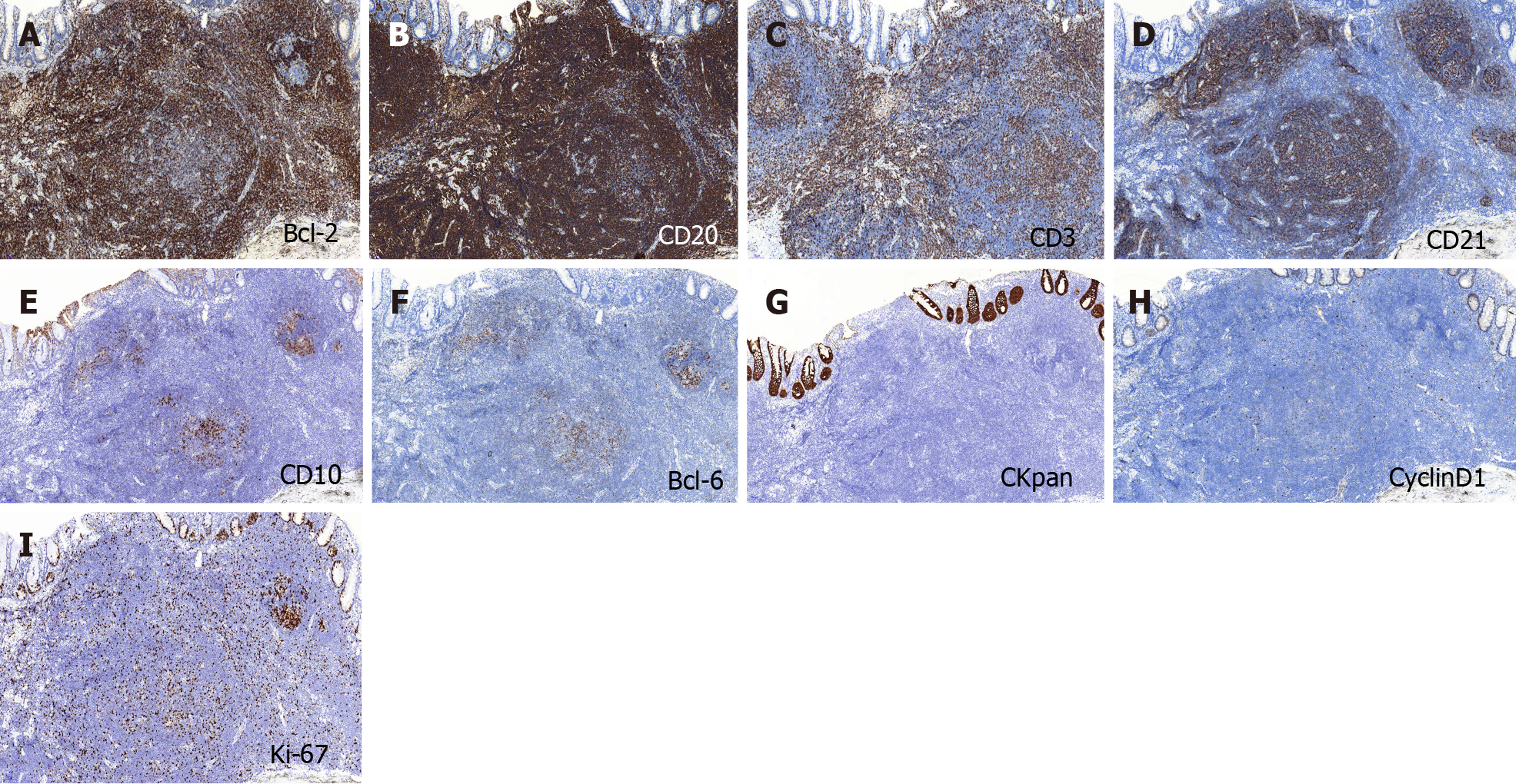
Figure 3 Immunohistochemical findings.
A and B: BCL-2 and CD20 were strongly expressed in the tumor; C and D: CD3 and CD21 were positive; E and F: CD10 and BCL-6 showed scattered positivity in the tumor; G: CKpan was expressed in the residual epithelium; H: CyclinD1 was negative in the tumor; I: The Ki-67 labeling index was 20%. Magnification of all photographs is 100 ×.
- Citation: Wei YL, Min CC, Ren LL, Xu S, Chen YQ, Zhang Q, Zhao WJ, Zhang CP, Yin XY. Laterally spreading tumor-like primary rectal mucosa-associated lymphoid tissue lymphoma: A case report. World J Clin Cases 2021; 9(16): 3988-3995
- URL: https://www.wjgnet.com/2307-8960/full/v9/i16/3988.htm
- DOI: https://dx.doi.org/10.12998/wjcc.v9.i16.3988


