Copyright
©The Author(s) 2021.
World J Clin Cases. May 6, 2021; 9(13): 3114-3119
Published online May 6, 2021. doi: 10.12998/wjcc.v9.i13.3114
Published online May 6, 2021. doi: 10.12998/wjcc.v9.i13.3114
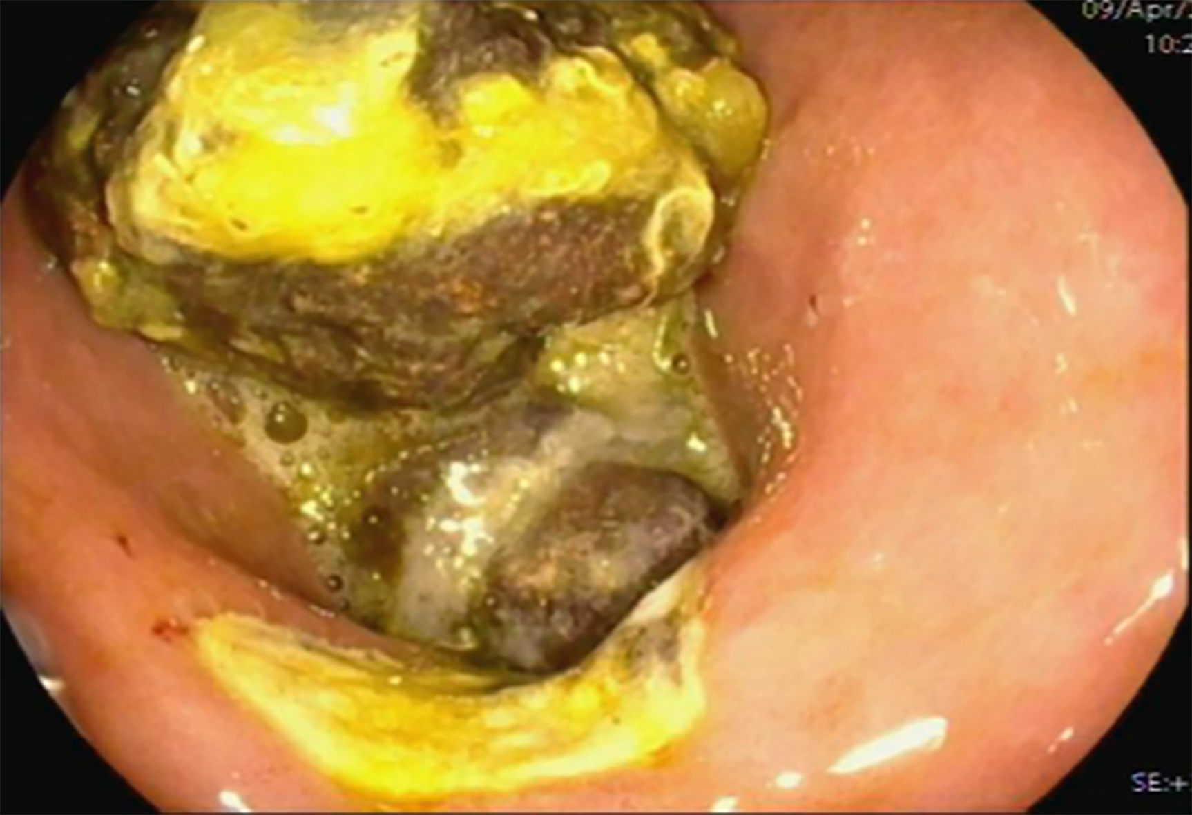
Figure 1 Gastroscopy before admission revealed a gastric ulcer and a large bezoar in the stomach.
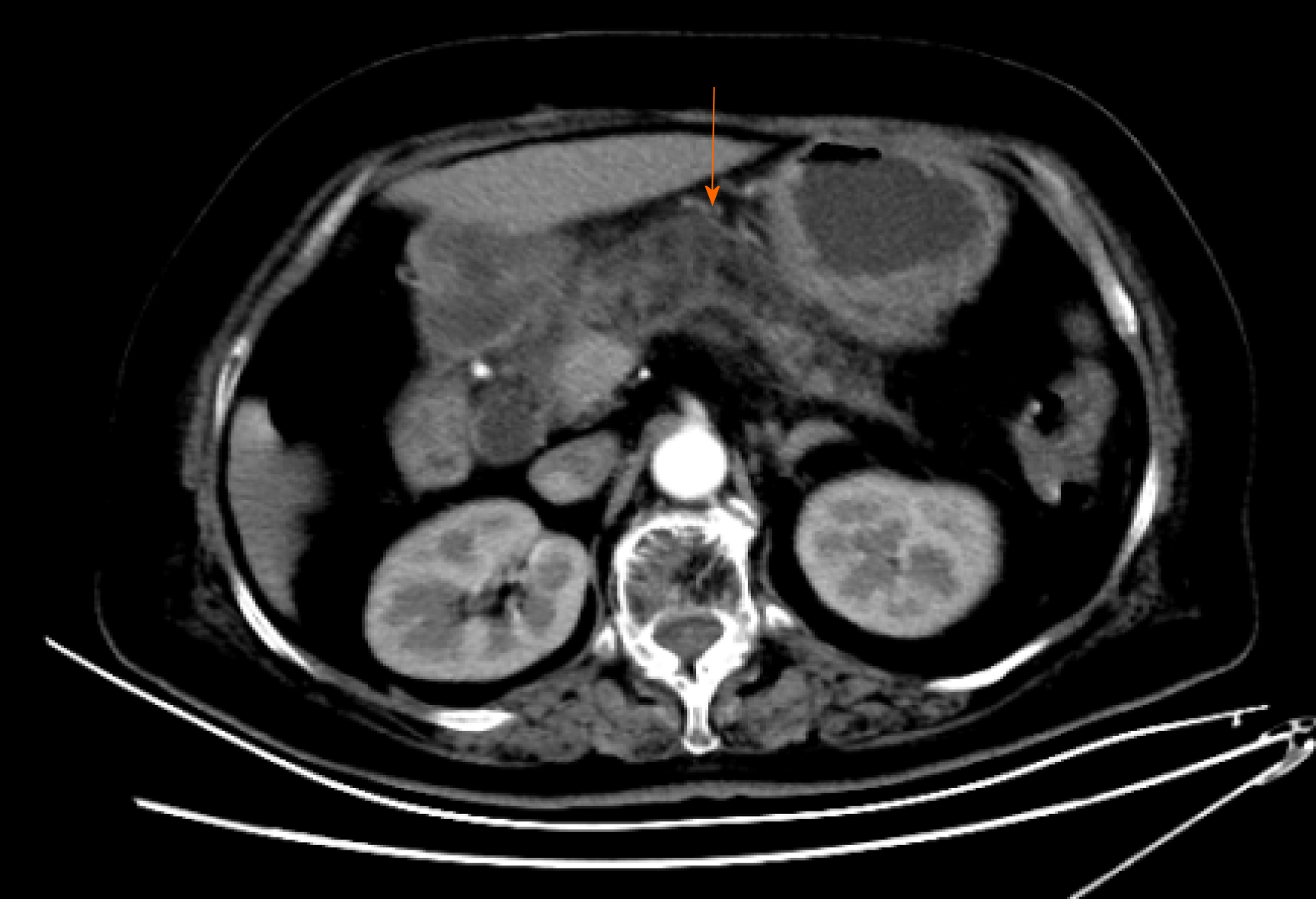
Figure 2 Abdominal computed tomography revealed mild inflammation of the pancreas.
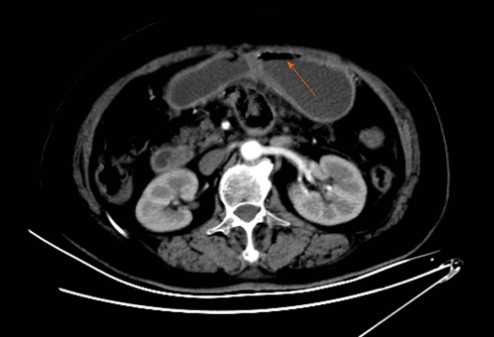
Figure 3 Abdominal computed tomography re-examination revealed gastric distension and loops of small intestine with multiple gas-fluid levels, which suggested intestinal obstruction.
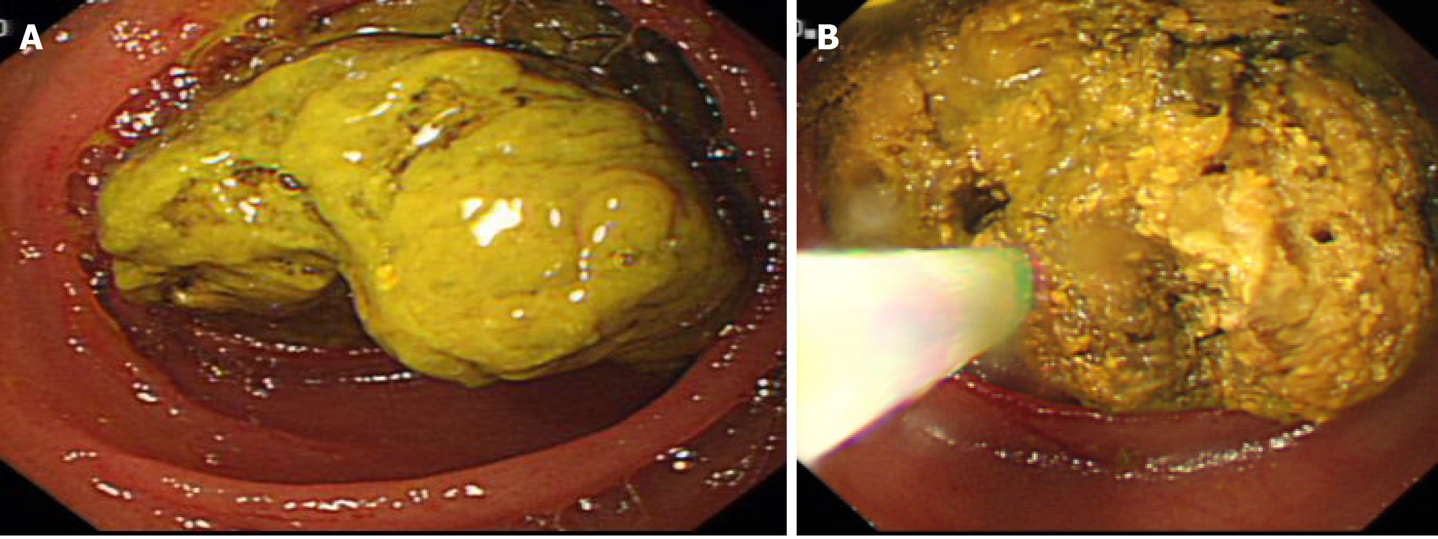
Figure 4 Esophagogastroduodenoscopy.
A: Large yellowish hard mass in the jejunal lumen; B: Forward-viewing endoscope was urgently used to fragment the bezoar with the help of a basket and net.
- Citation: Wang TT, He JJ, Liu J, Chen WW, Chen CW. Acute pancreatitis and small bowel obstruction caused by a migratory gastric bezoar after dissolution therapy: A case report. World J Clin Cases 2021; 9(13): 3114-3119
- URL: https://www.wjgnet.com/2307-8960/full/v9/i13/3114.htm
- DOI: https://dx.doi.org/10.12998/wjcc.v9.i13.3114


