Copyright
©The Author(s) 2020.
World J Clin Cases. Apr 26, 2020; 8(8): 1471-1476
Published online Apr 26, 2020. doi: 10.12998/wjcc.v8.i8.1471
Published online Apr 26, 2020. doi: 10.12998/wjcc.v8.i8.1471
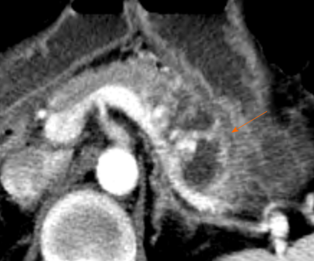
Figure 1 Contrast enhanced computed tomography scan before embolization.
Contrast-enhanced computed tomography scan before angiogram reveals swelling of pancreas with pseudocyst in the involved area of arteriovenous malformation (arrow) at the pancreas body and tail.
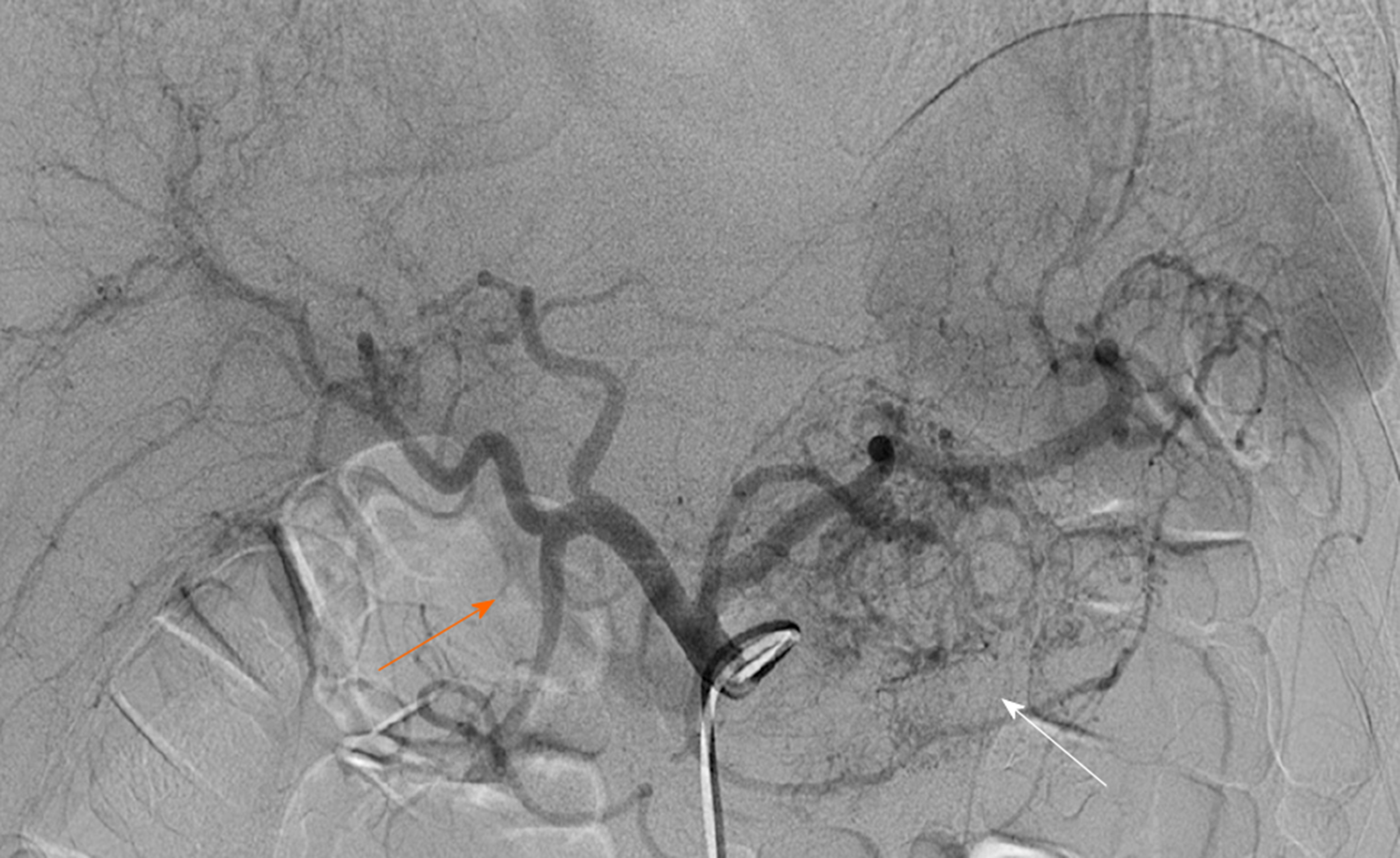
Figure 2 Initial celiac arteriography.
Celiac trunk angiography shows numerous fine feeders (white arrow) mainly originating from the left gastric artery, splenic artery and dorsal pancreatic artery, as well as early portal vein opacification (orange arrow).
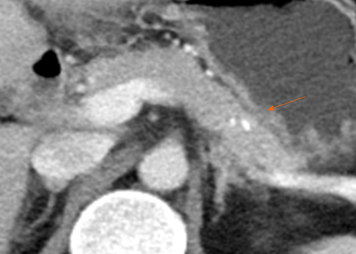
Figure 3 One-year follow-up contrast enhanced computed tomography scan.
Computed tomography scan shows regression of most vascular networks except for a few small residual vessels in the pancreas proximal body. There was no evidence of pancreatitis. A high-density spot (orange arrow) in the pancreas was embolic agent (histoacryl glue).
- Citation: Yoon SY, Jeon GS, Lee SJ, Kim DJ, Kwon CI, Park MH. Embolization of pancreatic arteriovenous malformation: A case report. World J Clin Cases 2020; 8(8): 1471-1476
- URL: https://www.wjgnet.com/2307-8960/full/v8/i8/1471.htm
- DOI: https://dx.doi.org/10.12998/wjcc.v8.i8.1471


