Copyright
©The Author(s) 2020.
World J Clin Cases. Apr 26, 2020; 8(8): 1471-1476
Published online Apr 26, 2020. doi: 10.12998/wjcc.v8.i8.1471
Published online Apr 26, 2020. doi: 10.12998/wjcc.v8.i8.1471
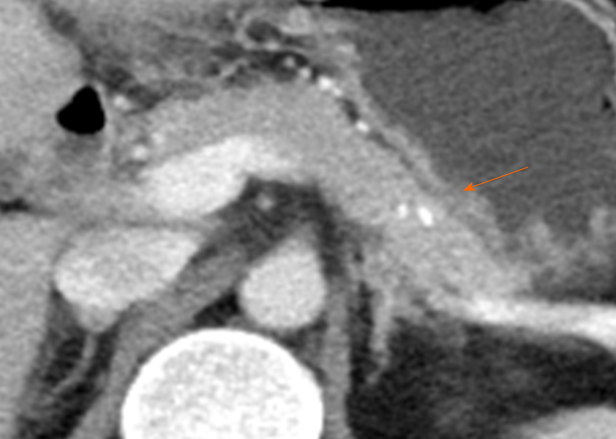
Figure 3 One-year follow-up contrast enhanced computed tomography scan.
Computed tomography scan shows regression of most vascular networks except for a few small residual vessels in the pancreas proximal body. There was no evidence of pancreatitis. A high-density spot (orange arrow) in the pancreas was embolic agent (histoacryl glue).
- Citation: Yoon SY, Jeon GS, Lee SJ, Kim DJ, Kwon CI, Park MH. Embolization of pancreatic arteriovenous malformation: A case report. World J Clin Cases 2020; 8(8): 1471-1476
- URL: https://www.wjgnet.com/2307-8960/full/v8/i8/1471.htm
- DOI: https://dx.doi.org/10.12998/wjcc.v8.i8.1471


