Copyright
©The Author(s) 2020.
World J Clin Cases. Apr 6, 2020; 8(7): 1251-1256
Published online Apr 6, 2020. doi: 10.12998/wjcc.v8.i7.1251
Published online Apr 6, 2020. doi: 10.12998/wjcc.v8.i7.1251
Figure 1 Four-phase computed tomography images.
A: Computed tomography on arterial phase performed in May 2012 showing a 2.0-cm mass with central necrosis and peripheral solid portions; B: Computed tomography on arterial phase performed in November 2015 showing a partial response of the mass.
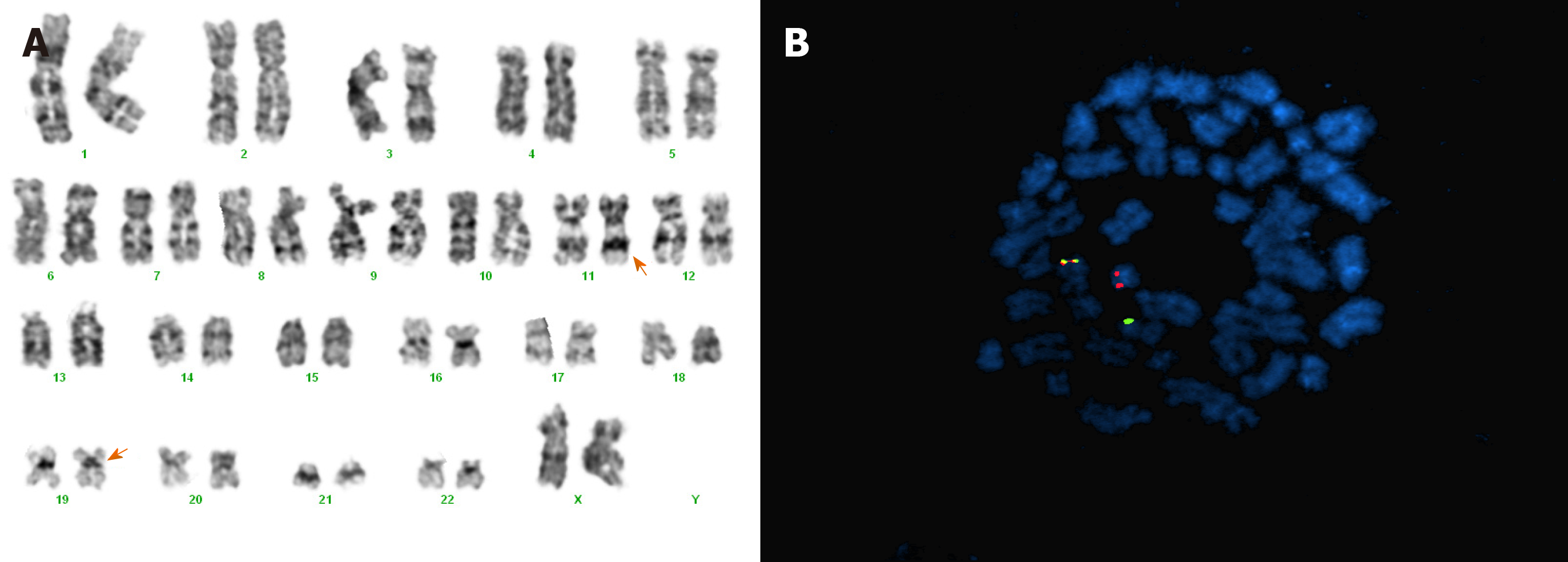
Figure 2 A chromosome study and fluorescence in situ hybridization study.
A: Giemsa-banding karyogram at initial diagnosis: 46, XX, t(11;19)(q23;p13.1). The arrows indicate the involved chromosomes; B: Metaphase fluorescence in situ hybridization with the KMT2A probe revealed a red signal on the derivative chromosome 19, a green signal on the derivative chromosome 11, and a fusion signal on the normal chromosome 11.
- Citation: Kim HJ, Baek SK, Maeng CH, Kim SY, Park TS, Han JJ. Acute myeloid leukemia with t(11;19)(q23;p13.1) in a patient with a gastrointestinal stromal tumor undergoing imatinib therapy: A case report. World J Clin Cases 2020; 8(7): 1251-1256
- URL: https://www.wjgnet.com/2307-8960/full/v8/i7/1251.htm
- DOI: https://dx.doi.org/10.12998/wjcc.v8.i7.1251


