Copyright
©The Author(s) 2020.
World J Clin Cases. Jan 26, 2020; 8(2): 382-389
Published online Jan 26, 2020. doi: 10.12998/wjcc.v8.i2.382
Published online Jan 26, 2020. doi: 10.12998/wjcc.v8.i2.382
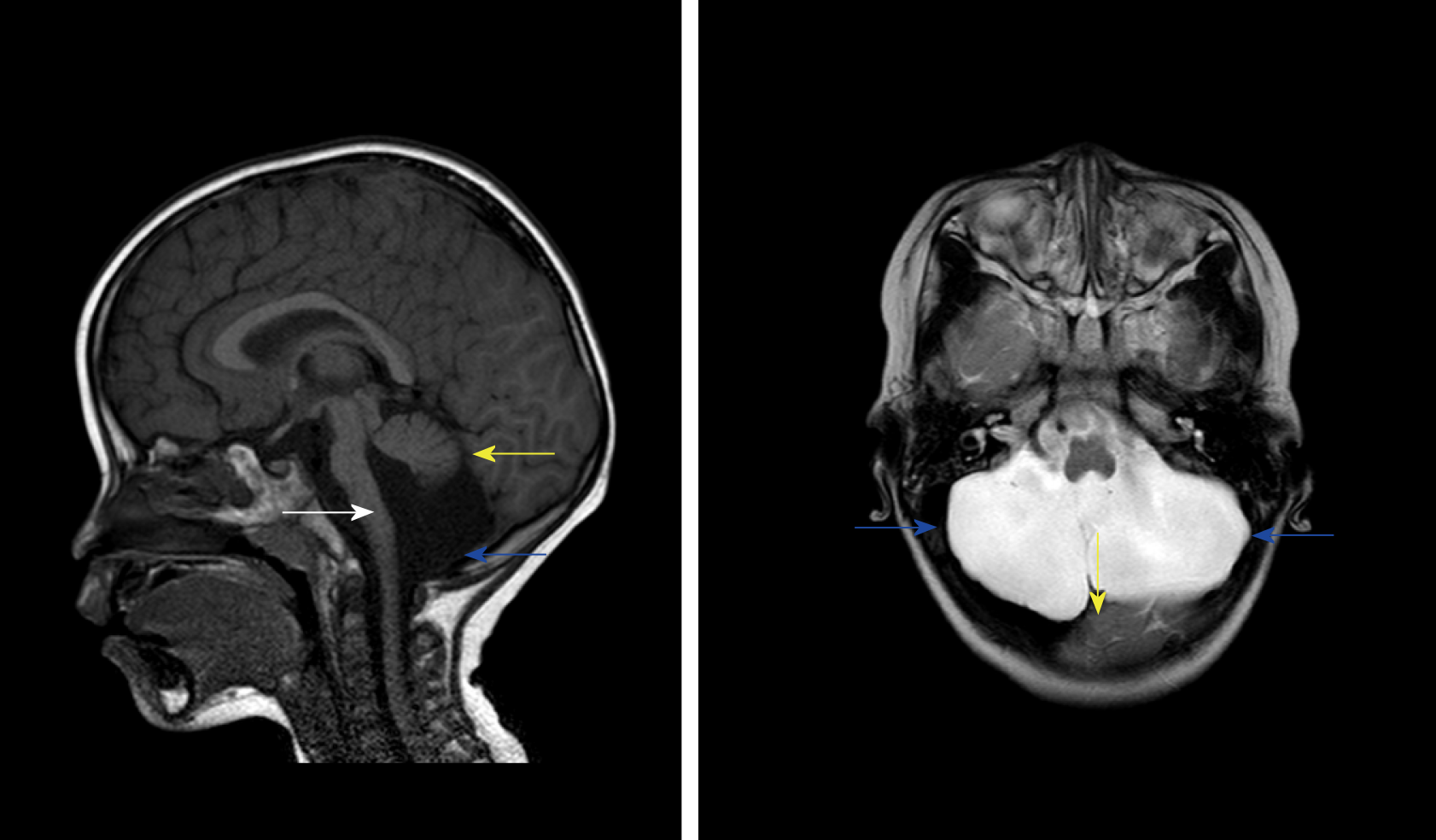
Figure 1 Dandy-Walker variant and pontocerebellar hypoplasia showed in the patient’s first magnetic resonance imaging.
Sagittal T1WI magnetic resonance imaging showed posterior fossa cystic lesions, cyst-like dilatation of the fourth ventricle and communication with the cistern magnum (blue arrow), and hypoplasia of the inferior vermis of the cerebellum. The developed superior vermis was elevated upwardly and rotated by posterior fossa cystic lesions (yellow arrow) and caused the death. The pons was smaller, and the medulla was not compressed (white arrow). Axial T2WI magnetic resonance imaging showed that most of the posterior fossa was occupied by cystic lesions (blue arrow), the cyst had a large volume, the superior vermis was compressed upward, and the pons and medulla were not compressed (white arrow).
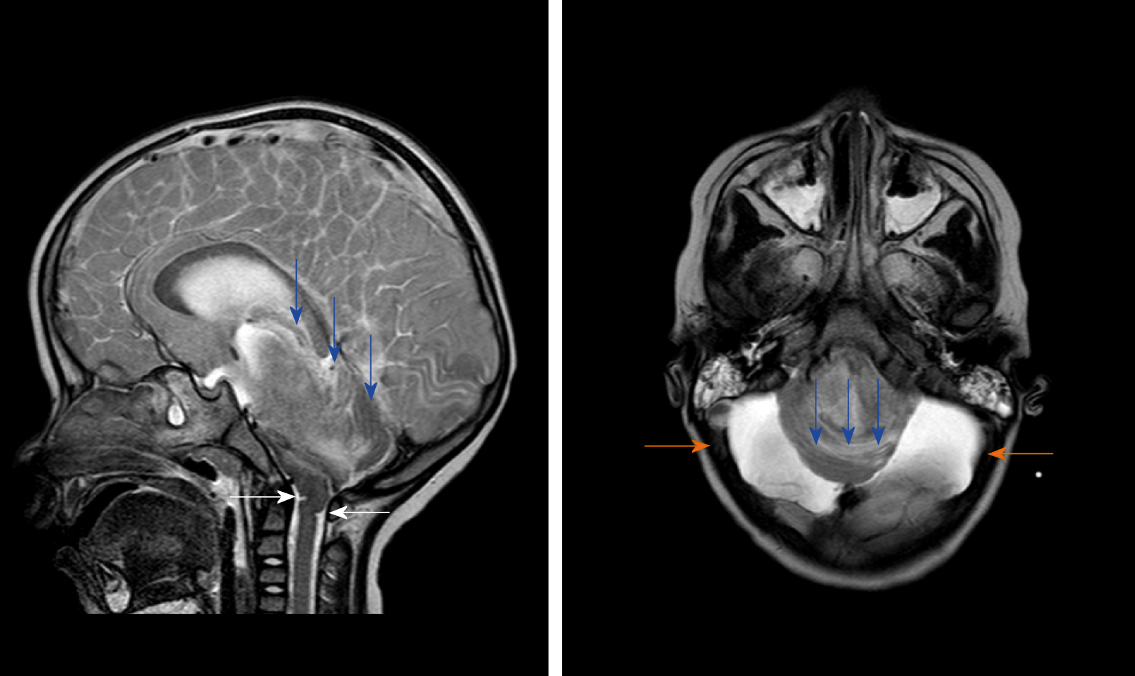
Figure 2 T2WI magnetic resonance examination after the condition suddenly deteriorated.
The gyri were swelling, the sulcus became superficial (arrow), the supratentorial cranial pressure was increased, the pressure gradient pushed the supratentorial structure down (blue arrow), resulting in compression of posterior fossa cystic lesions and volume decrease, and the medulla moved down and folded into a “Z-like” shape (white arrow). Axial T2WI magnetic resonance imaging showed that the supratentorial structure pushed down the curtain, the pons and medulla were obviously compressed by the hernia tissue (blue arrow), and the surrounding cerebrospinal fluid gap disappeared. Posterior fossa cystic lesions shrank in volume under compression, with residual bilateral cystic areas (orange arrow).
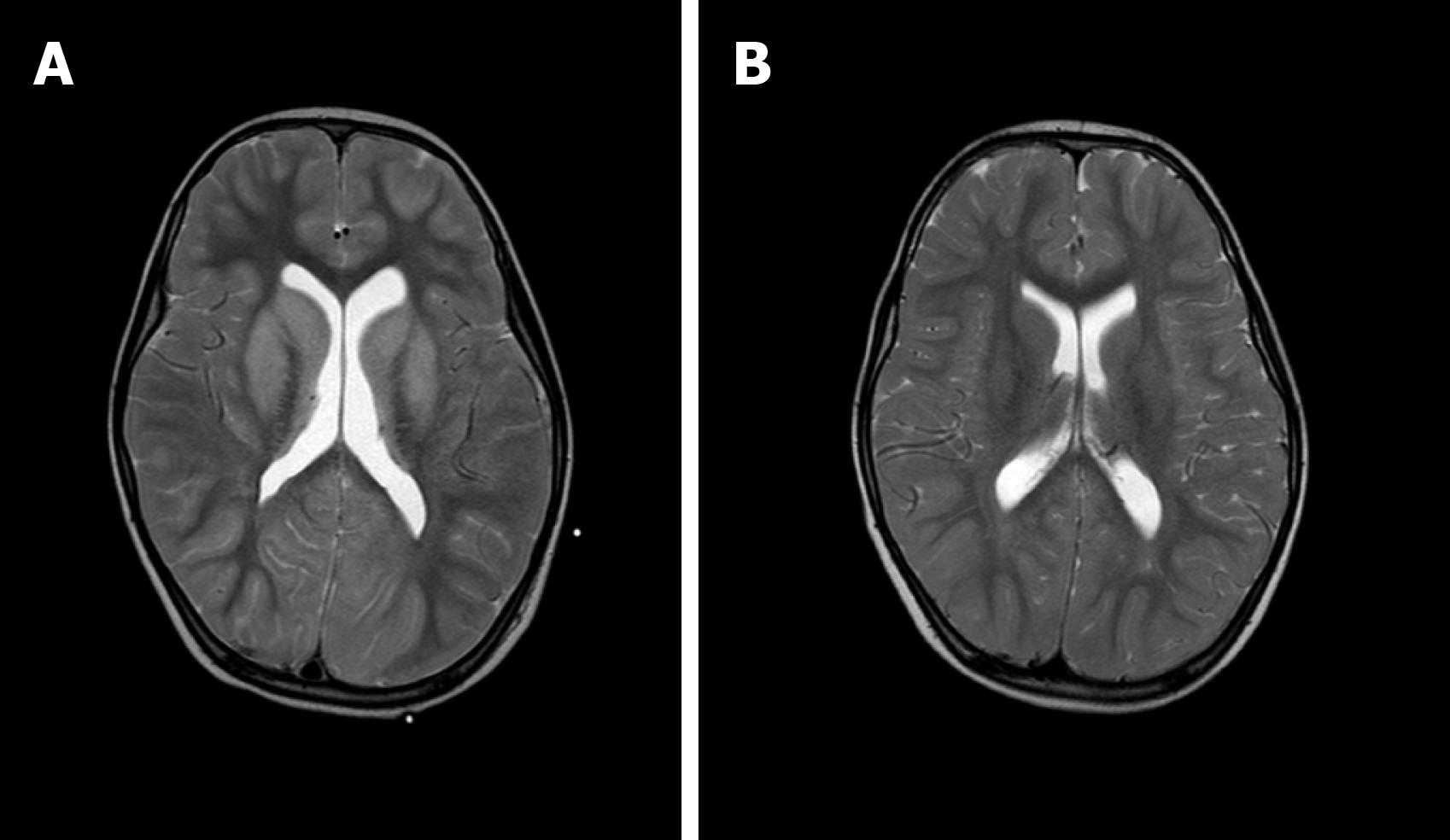
Figure 3 Imaging of the basal ganglia after and before brainstem folding.
A: After brain stem folding, swelling of the bilateral putamen, cauda nucleus, frontal lobe, parietal lobe, and occipital cortex, increased T2WI signals, and slightly enlarged bilateral lateral ventricle can be seen; B: Three days before brain stem folding, no swelling was observed in the bilateral putamen, caudate nucleus, frontal lobe, parietal lobe, or occipital cortex, with normal signals, and no expansion was observed in the bilateral lateral ventricles.
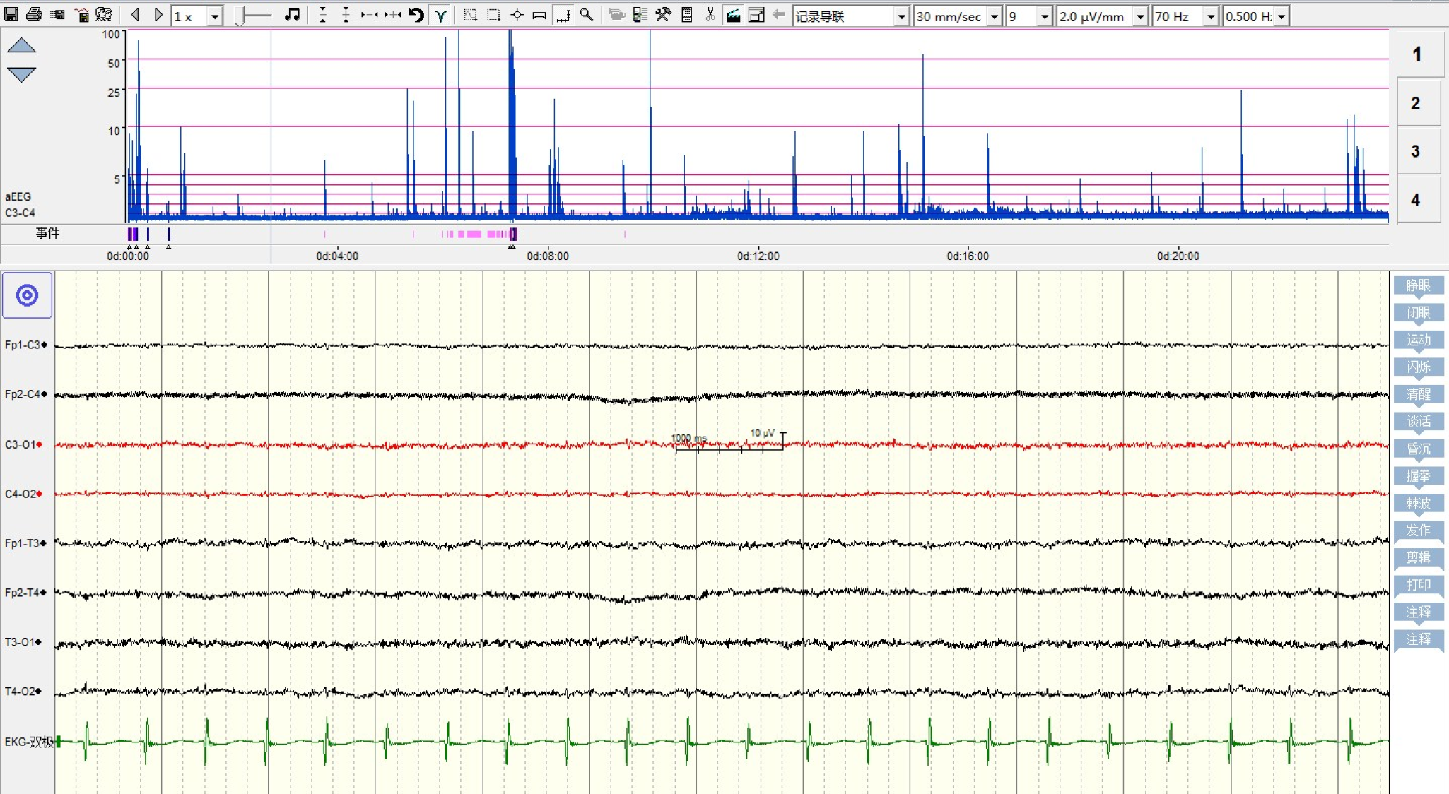
Figure 4 Electric silence in electroencephalograph.
- Citation: Li SY, Li PQ, Xiao WQ, Liu HS, Yang SD. Brainstem folding in an influenza child with Dandy-Walker variant. World J Clin Cases 2020; 8(2): 382-389
- URL: https://www.wjgnet.com/2307-8960/full/v8/i2/382.htm
- DOI: https://dx.doi.org/10.12998/wjcc.v8.i2.382


