Copyright
©The Author(s) 2020.
World J Clin Cases. Jan 26, 2020; 8(2): 382-389
Published online Jan 26, 2020. doi: 10.12998/wjcc.v8.i2.382
Published online Jan 26, 2020. doi: 10.12998/wjcc.v8.i2.382
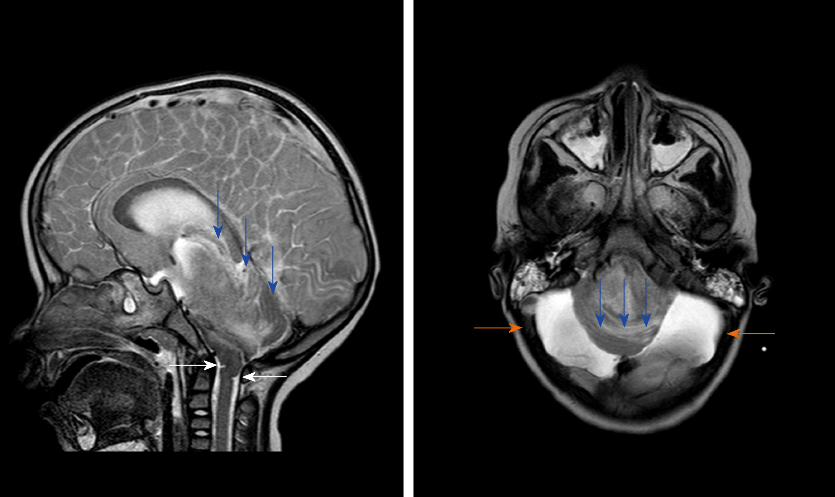
Figure 2 T2WI magnetic resonance examination after the condition suddenly deteriorated.
The gyri were swelling, the sulcus became superficial (arrow), the supratentorial cranial pressure was increased, the pressure gradient pushed the supratentorial structure down (blue arrow), resulting in compression of posterior fossa cystic lesions and volume decrease, and the medulla moved down and folded into a “Z-like” shape (white arrow). Axial T2WI magnetic resonance imaging showed that the supratentorial structure pushed down the curtain, the pons and medulla were obviously compressed by the hernia tissue (blue arrow), and the surrounding cerebrospinal fluid gap disappeared. Posterior fossa cystic lesions shrank in volume under compression, with residual bilateral cystic areas (orange arrow).
- Citation: Li SY, Li PQ, Xiao WQ, Liu HS, Yang SD. Brainstem folding in an influenza child with Dandy-Walker variant. World J Clin Cases 2020; 8(2): 382-389
- URL: https://www.wjgnet.com/2307-8960/full/v8/i2/382.htm
- DOI: https://dx.doi.org/10.12998/wjcc.v8.i2.382


