Copyright
©The Author(s) 2020.
World J Clin Cases. Jan 26, 2020; 8(2): 362-369
Published online Jan 26, 2020. doi: 10.12998/wjcc.v8.i2.362
Published online Jan 26, 2020. doi: 10.12998/wjcc.v8.i2.362
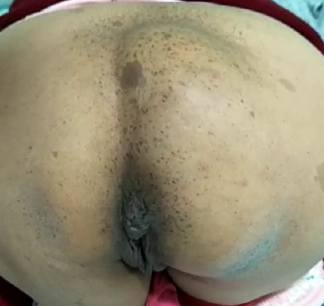
Figure 1 Preoperative physical examination.
A bulge in the right sacrococcygeal region, which was approximately 8 cm × 8 cm in size and soft in palpation with no tenderness, was revealed upon standing.
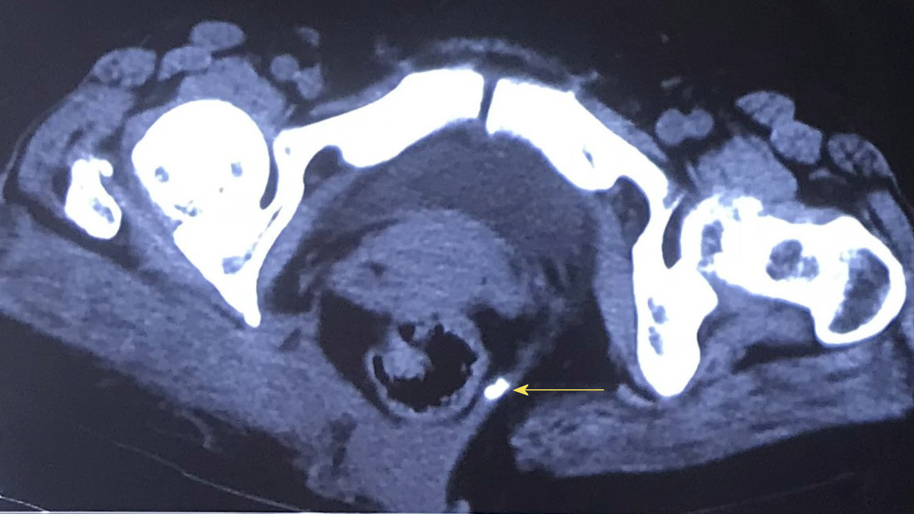
Figure 2 Computed tomography image.
Computed tomography revealed rectal herniation and an abnormality in the structure of the tissues between the sacrococcyx (arrow) and the right gluteus muscle.
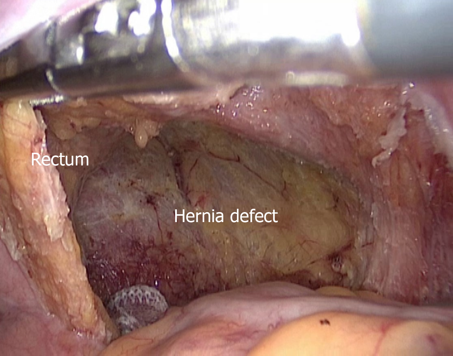
Figure 3 Laparoscopic findings.
In the right anterior sacrococcygeal region, a defect approximately 6 cm × 6 cm in size was identified, through which part of the rectum protruded.
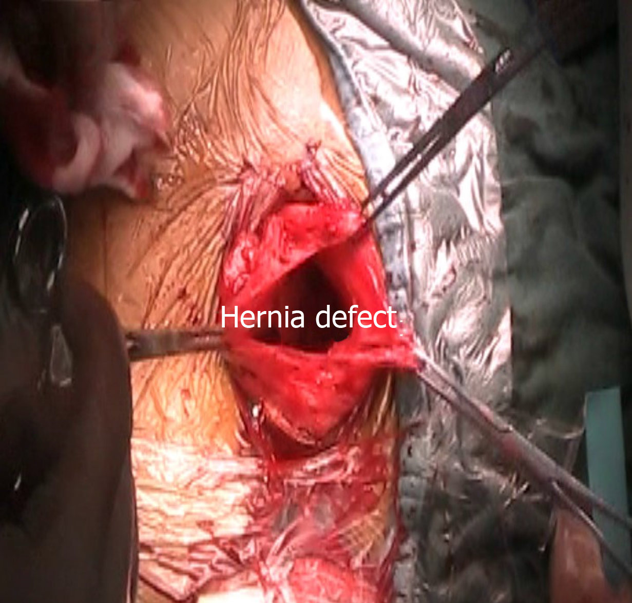
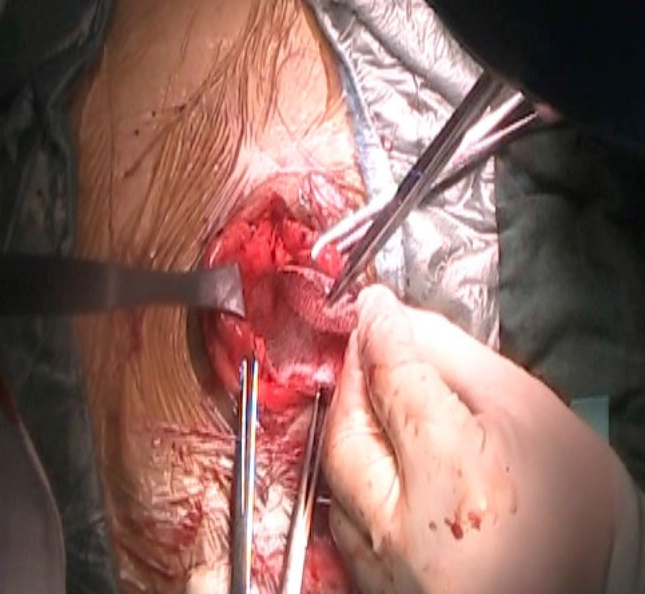
Figure 4 Sacrococcygeal findings.
The hernia defect was visualized after opening the hernia sac.
Figure 5 Mesh placement via the sacrococcygeal approach.
A properly sized self-gripping polyester mesh was placed deep into the defect and intermittently sutured to the edges.
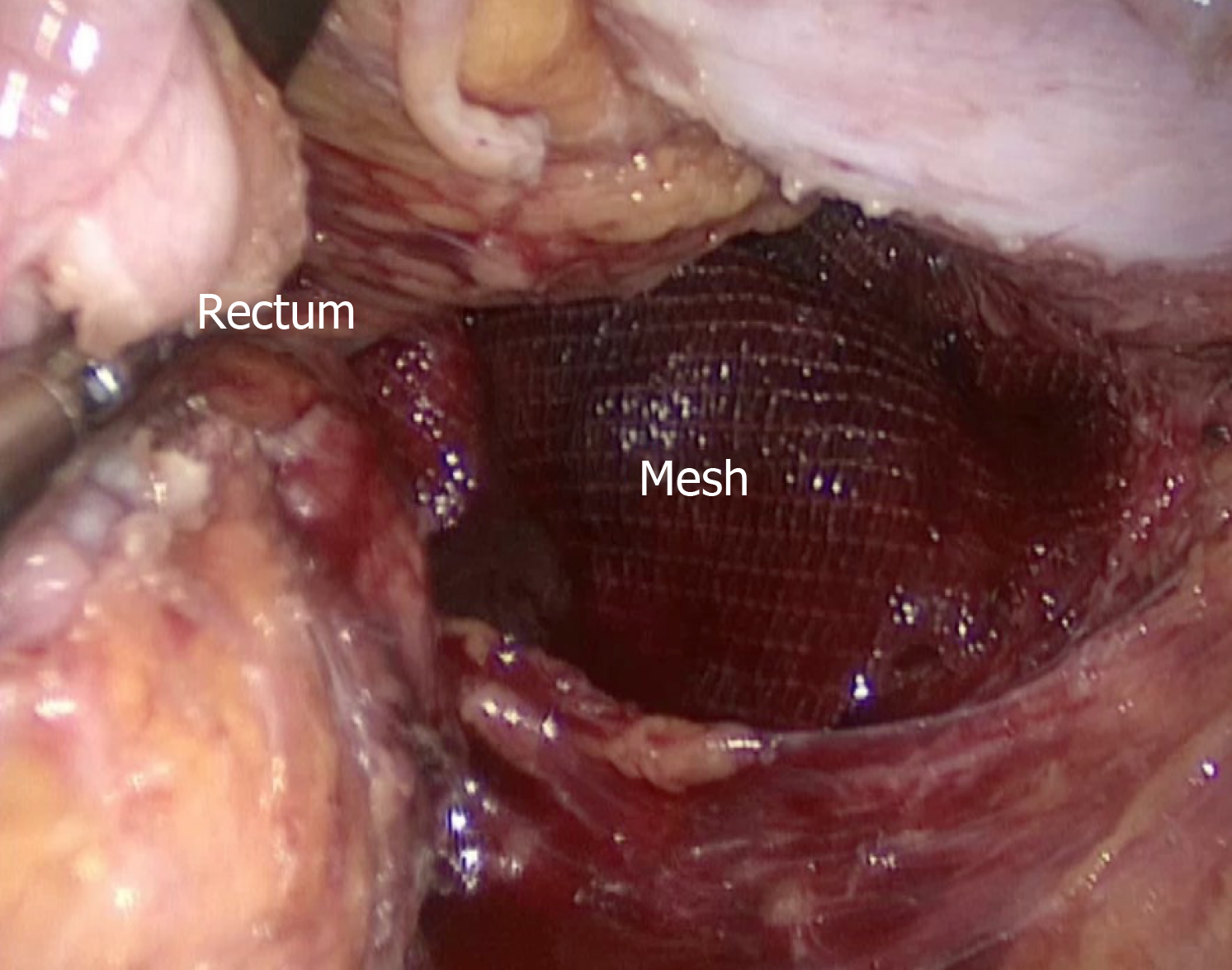
Figure 6 Mesh accommodation under laparoscopy.
The implanted mesh was inspected for accommodation in a good location.
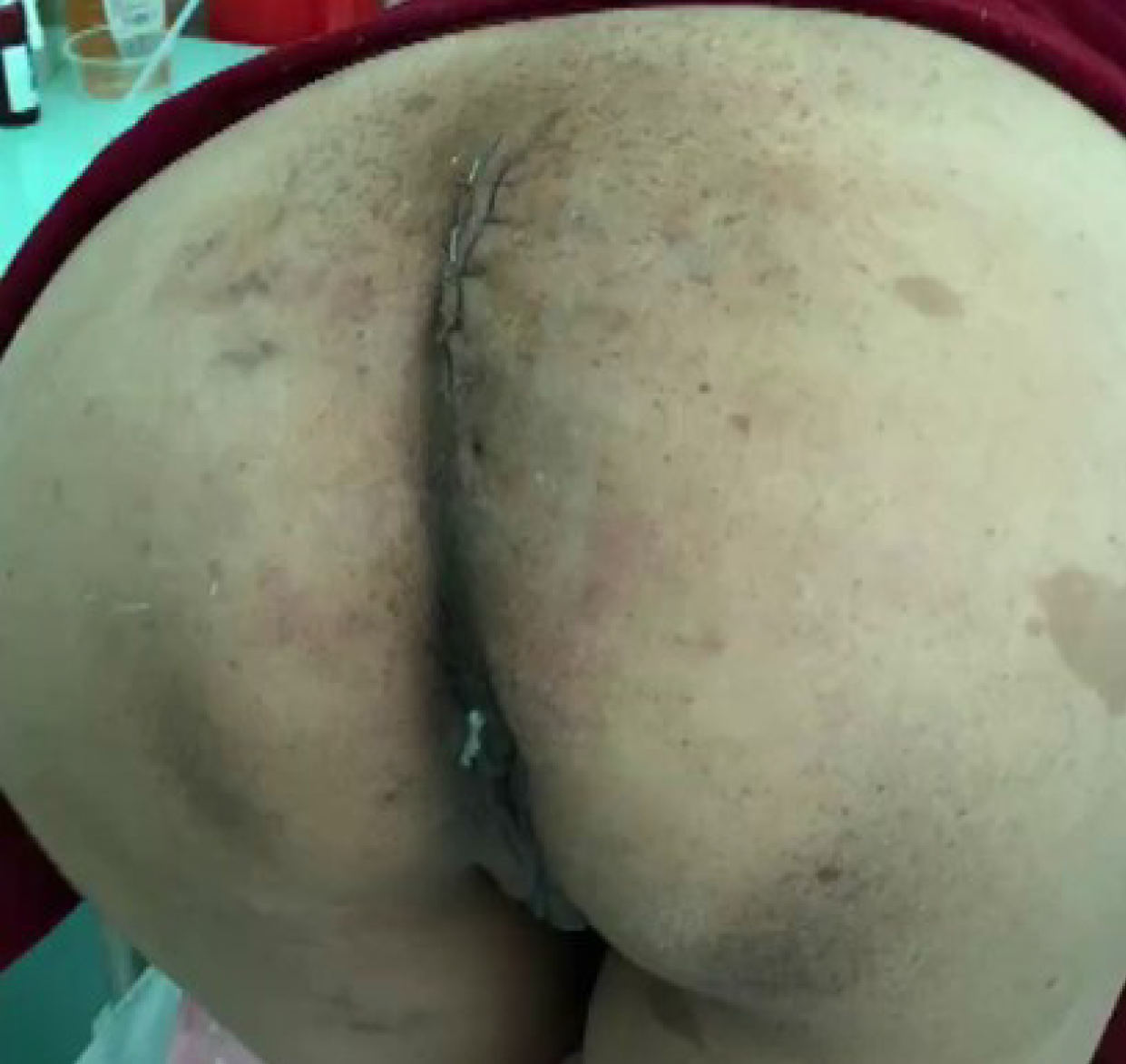
Figure 7 Postoperative physical examination.
At the time of discharge, no relapsing bulge was found when performing the Valsalva manoeuvre.
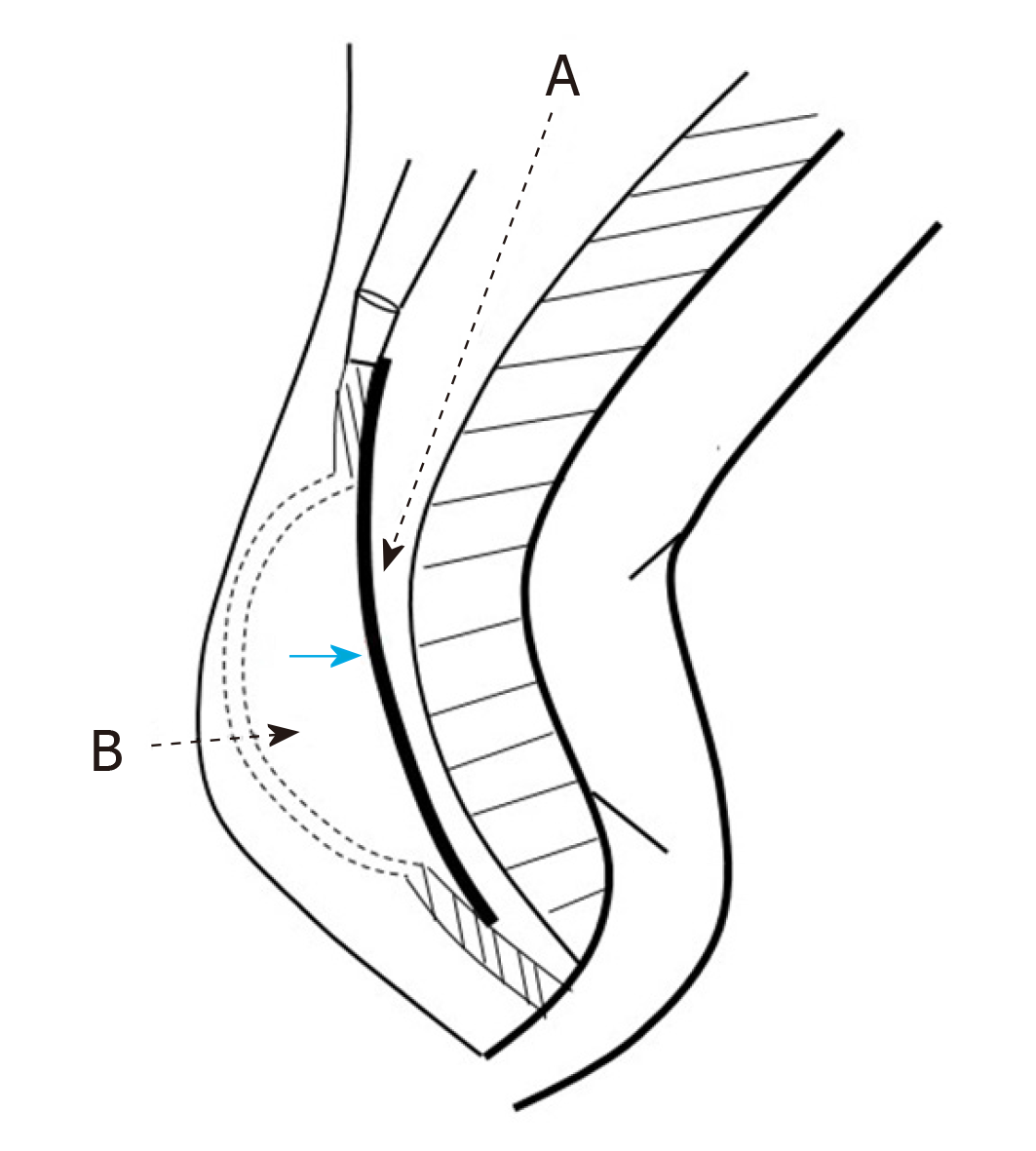
Figure 8 Diagram of the anatomy of the repair.
Line A is the surgical plane for laparoscopic approach, and it is posterior to the mesorectum; line B is the surgical path for sacrococcygeal approach, and it extends through the skin and subcutaneous fat to the level of the defect, and allows the placement of the mesh (blue arrow).
- Citation: Dong YQ, Liu LJ, Fu Z, Chen SM. Mesh repair of sacrococcygeal hernia via a combined laparoscopic and sacrococcygeal approach: A case report. World J Clin Cases 2020; 8(2): 362-369
- URL: https://www.wjgnet.com/2307-8960/full/v8/i2/362.htm
- DOI: https://dx.doi.org/10.12998/wjcc.v8.i2.362


